Full Papers

Surface-Enhanced Raman Spectroscopy of Carbon Nanotubes in Aqueous Solution

  • Niu Yang ,
  • Liu Qinghai ,
  • Yang Juan ,
  • Gao Dongliang ,
  • Qin Xiaojun ,
  • Luo Da ,
  • Zhang Zhenyu ,
  • Li Yan
Expand
  • Beijing National Laboratory for Molecular Sciences, Key Laboratory for the Physics and Chemistry of Nanodevices, State Key Laboratory of Rare Earth Materials Chemistry and Applications, Peking University, Beijing 100871, China

Received date: 2012-05-11

  Online published: 2012-06-19

Supported by

Project supported by the Ministry of Science and Technology of China (No. 2011CB933003) and the National Natural Science Foundation of China (Nos. 21125103, 11179011).

Abstract

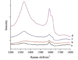
Carbon nanotubes (CNTs) exhibit intrinsic spectroscopic properties and are potential Raman and NIR florescence probes for bioimaging. However, the weak Raman intensity greatly obstructs such applications. Surface-enhanced Raman scattering (SERS) has shown to be an effective way to increase the Raman signal. SERS has been widely studied for solid samples. However, solid state is distinctly different from the environment in biosystems. Herein, we studied the SERS of Au- nanoparticle-decorated CNTs in aqueous solution, which offers a similar environment to biosystems. We found that the functionalization of CNTs with-SH groups can improve the attachment of Au nanoparticles on tube surfaces thus benefit the SERS effect. The particle size is another important issue for SERS. Particles of 50 nm show much stronger enhancement than those of 12 nm. The Raman intensity of CNTs increases with the increase of the concentration of Au nanoparticles. Hexamethylene diamine molecules can act as the bi-linkers between Au nanoparticles, compressing the interparticle distance. This was proved by the red-shift of the band at ca. 540 nm and the appearance of a broad band around 700 nm in the absorption spectra of Au nanoparticles. Therefore the addition of hexamethylene diamine can further increase the Raman signal of CNTs by the strong coupling of the surface plasma from the Au nanoparticles with very small interparticle distances. Two kinds of commonly used small molecule Raman probes p-aminothiophenol and Rhodamine B both show remarkably enhanced Raman intensity when added to the aqueous dispersion of Au/CNT hybrids. This shows that these Raman probe molecules can absorb onto the Au/CNT hybrids and their Raman spectra are able to be greatly enhanced by Au nanoparticles decorated on CNTs. Because various Raman bands from either CNTs or Raman probe molecules can be used for Raman imaging, this kind of Raman probe molecule/Au/CNT tri-component hybrid systems may be used as a potential nanostructured platform for multiplexed Raman imaging based on SERS effect.

Cite this article

Niu Yang , Liu Qinghai , Yang Juan , Gao Dongliang , Qin Xiaojun , Luo Da , Zhang Zhenyu , Li Yan . Surface-Enhanced Raman Spectroscopy of Carbon Nanotubes in Aqueous Solution[J]. Acta Chimica Sinica, 2012 , 70(14) : 1533 -1537 . DOI: 10.6023/A12050222

References

[1] Lacerda, L.; Raffa, S.; Prato, M.; Bianco, A.; Kostarelos, K. Nano Today 2007, 2, 38.

[2] Wang, J.; Cui, R.; Liu, Y.; Zhou, W.; Jin, Z.; Li, Y. J. Phys. Chem. C 2009, 113, 5075.

[3] Dresselhaus, M. S.; Dresselhaus, G.; Saito, R.; Jorio, A. Phys. Rep. 2005, 409, 47.

[4] Dresselhaus, M. S.; Dresselhaus, G.; Jorio, A.; Souza, A. G.; Saito, R. Carbon 2002, 40, 2043.

[5] Wang, X.; Wang, C.; Cheng, L.; Lee, S.-T.; Liu, Z. J. Am. Chem. Soc. 2012, 134, 7414.

[6] Kneipp, K.; Wang, Y.; Kneipp, H.; Perelman, L. T.; Itzkan, I.; Dasari, R. R.; Feld, M. S. Phys. Rev. Lett. 1997, 78, 1667.

[7] Chu, H. B.; Wang, J. Y.; Ding, L.; Yuan, D. N.; Zhang, Y.; Liu, J.; Li, Y. J. Am. Chem. Soc. 2009, 131, 14310.

[8] Chu, H. B.; Wei, L.; Cui, R. L.; Wang, J. Y.; Li, Y. Coord. Chem. Rev. 2010, 254, 1117.

[9] Chu, H. B.; Cui, R. L.; Wang, J. Y.; Yang, J. A.; Li, Y. Carbon 2011, 49, 1182.

[10] Zavaleta, C. L.; Smith, B. R.; Walton, I.; Doering, W.; Davis, G.; Shojaei, B.; Natan, M. J.; Gambhir, S. S. Proc. Natl. Acad. Sci. U. S. A. 2009, 106, 13511.

[11] Jorio, A.; Pimenta, M. A.; Filho, A. G. S.; Saito, R.; Dresselhaus, G.; Dresselhaus, M. S. New J. Phys. 2003, 5, 139.1~139.17.

[12] Pimenta, M. A.; Dresselhaus, G.; Dresselhaus, M. S.; Cancado, L. G.; Jorio, A.; Saito, R. PCCP 2007, 9, 1276.

[13] Zhao, W.; Brook, M. A.; Li, Y. ChemBioChem 2008, 9, 2363.

[14] Zheng, J.; Li, X.; Gu, R.; Lu, T. J. Phys. Chem. B 2003, 106, 1019.

[15] Zheng, J.; Zhou, Y.; Li, X.; Ji, Y.; Lu, T.; Gu, R. Langmuir 2003, 19, 632.

[16] Wang, Y.; Chen, H.; Dong, S.; Wang, E. J. Chem. Phys. 2006, 124, 074709.

[17] Kim, K.; Kim, K. L.; Shin, D.; Choi, J.-Y.; Shin, K. S. J. Phys. Chem. C 2012, 116, 4774.

[18] Zhang, J.; Li, X.; Sun, X.; Li, Y. J. Phys. Chem. B 2005, 109, 12544.

[19] Hildebrandt, P.; Stockburger, M. J. Phys. Chem. 1984, 88, 5935.

[20] Volny, M.; Sengupta, A.; Wilson, C. B.; Swanson, B. D.; Davis, E. J.; Turecek, F. Anal. Chem. 2007, 79, 4543.

[21] Datsyuk, V.; Kalyva, M.; Papagelis, K.; Parthenios, J.; Tasis, D.; Siokou, A.; Kallitsis, I.; Galiotis, C. Carbon 2008, 46, 833.

[22] Peng, Y.; Liu, H. W. Ind. Eng. Chem. Res. 2006, 45, 6483.

[23] Chiang, I. W.; Brinson, B. E.; Huang, A. Y.; Willis, P. A.; Bronikowski, M. J.; Margrave, J. L.; Smalley, R. E.; Hauge, R. H. J. Phys. Chem. B 2001, 105, 8297.

[24] Xiao, Y. H.; Ye, X. X.; He, L.; Che, J. F. Polym. Int. 2012, 61, 190.

[25] Cai, K. Y.; Yao, K. D.; Cui, Y. L.; Yang, Z. M.; Li, X. Q.; Xie, H. Q.; Qing, T. W.; Gao, L. B. Biomaterials 2002, 23, 1603.

[26] Kam, N. W. S.; O'Connell, M.; Wisdom, J. A.; Dai, H. J. Proc. Natl. Acad. Sci. U. S. A. 2005, 102, 11600.

[27] Jana, N. R.; Gearheart, L.; Murphy, C. J. Chem. Mater. 2001, 13, 2313.
Outlines

/