Article

A Two-Photon Fluorescent Probe for Specific Imaging of Furin Activity in Living Cells and Tissues

  • Liu Hongwen ,
  • Zhu Longmin ,
  • Lou Xiaofeng ,
  • Yuan Lin ,
  • Zhang Xiao-Bing
Expand
  • Molecular Science and Biomedicine Laboratory(MBL), College of Chemistry and Chemical Engineering, State Key Laboratory of Chemo/Biosensing and Chemometrics, Hunan University, Changsha 410082, China

Received date: 2020-07-22

  Online published: 2020-07-28

Supported by

Project supported by the National Natural Science Foundation of China (Nos. 21890744 and 21877029), National Key R&D Program of China (No. 2019YFA0210103), and the National Postdoctoral Program for Innovative Talents (No. BX20180093).

Abstract

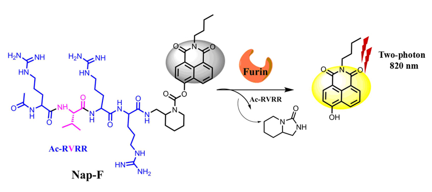
Furin, the most characteristic member of the proprotein convertase (PCs), has important biological functions. The expression level of furin is related to many diseases, for example, the occurrence and development of cancer is closely related to the expression level of furin. Although several small-molecule fluorescent probes for furin have been developed, which were designed based on near-infrared dye or one-photon dye. These probes exhibit low Stocks' shift or shallow penetration depth, which leading to self-quenching and strong interference. Two-photon fluorescent probes, which utilize two near-infrared photons as the excitation source, can overcome these problems. Herein, a furin-activatable two-photon fluorescent probe (Nap-F) was developed firstly that allowed for detection and imaging of furin in live cells and tumor tissues. Nap-F consists of a classical two-photon fluorophore (1,8-naphthalimide), a furin-particular polypeptide sequence RVRR and a self-eliminating linker. Nap-F is water-soluble and in a fluorescence-off state itself due to the inhibited intramolecular charge transfer (ICT). In the absence of furin, no noticeable fluorescence enhancement was detected, even over 3 days in buffer solution, indicating its good stability. Upon the conversion by furin, it displayed a dramatically fluorescence enhancement at 545 nm, and exhibits high specificity and sensitivity to furin. Nap-F was applied for visualizing the difference in the expression level of furin in various cells, demonstrating its capacity of distinguishing some cancer cells from normal cells. Furthermore, Nap-F was utilized to visualize the variation of furin expression level efficiently after immobilization of hypoxia-inducible factor-1 (HIF-1) by CoCl2, with the results indicating that there is a positive correlation between the expression level of furin and the degree of hypoxia in tumor cells. Owing to the excellent property of Nap-F, the probe was also successful utilized to imaging furin activity in tumor tissues. Thus, Nap-F is able to serve as a potential tool for better exploring the intrinsic link between hypoxic physiological environment and cellular carcinogenesis and detecting cancer in preclinical applications.

Cite this article

Liu Hongwen , Zhu Longmin , Lou Xiaofeng , Yuan Lin , Zhang Xiao-Bing . A Two-Photon Fluorescent Probe for Specific Imaging of Furin Activity in Living Cells and Tissues[J]. Acta Chimica Sinica, 2020 , 78(11) : 1240 -1245 . DOI: 10.6023/A20070323

References

[1] Steiner, D. F. Curr. Opin. Chem. Biol. 1998, 2, 31.
[2] Seidah, N. G.; Mayer, G.; Zaid, A.; Rousselet, E.; Nassoury, N.; Poirier, S.; Essalmani, R.; Prat, A. Int. J. Biochem. Cell Biol. 2008, 40, 1111.
[3] Bassi, D. E.; Fu, J.; de Cicco, R. L.; Klein-Szanto, A. J. P. Mol. Carcinogen 2005, 44, 151.
[4] McMahon, S.; Grondin, F.; McDonal, P. P.; Richard, D. E.; Dubois, C. M. J. Biol. Chem. 2005, 280, 6561.
[5] Zhang, J.; Liu, H.-W.; Hu, X.-X.; Li, J.; Liang, L.-H.; Zhang, X.-B.; Tan, W. Anal. Chem. 2015, 87, 11832.
[6] Li, X.; Gao, X.; Shi, W.; Ma, H. Chem. Rev. 2014, 114, 590.
[7] Yang, L.; Liu, B.; Li, N.; Tang, B. Acta Chim. Sinica, 2017, 75, 1047. (in Chinese). (杨立敏, 刘波, 李娜, 唐波. 化学学报, 2017, 75, 1047.)
[8] Yang, Z.; He, Y.; Dai, B.; Dou, B.; Wang, J.; Peng, X. Acta Chim. Sinica, 2011, 69, 445(in Chinese). (杨志刚, 和艳霞, 戴彬, 窦柏蕊, 王静云, 彭孝军. 化学学报, 2011, 69, 445.)
[9] Guan, X.; Wang, L.; Li, Z.; Liu, M.; Wang, K.; Lin, B.; Yang, X.; Lai, S.; Lei, Z.. Acta Chim. Sinica, 2019, 77, 1036(in Chinese). (关晓琳, 王林, 李志飞, 刘美娜, 王凯龙, 林斌, 杨学琴, 来守军, 雷自强. 化学学报, 2019, 77, 1036.)
[10] Hou, J.; Li, K.; Qin, C. Yu, X.; Chin. J. Org. Chem. 2018, 38, 612(in Chinese). (后际挺, 李坤, 覃彩芹, 余孝其. 有机化学, 2018, 38, 612.)
[11] Liu, H.-W.; Chen, L.; Xu, C.; Li, Z.; Zhang, H.; Zhang, X.-B.; Tan, W. Chem. Soc. Rev. 2018, 47, 7140.
[12] Zhu, L.; Liu, H.-W.; Yang, Y.; Hu, X.-X.; Li, K.; Xu, S.; Li, J.-B.; Ke, G.; Zhang, X.-B. Anal. Chem. 2019, 91, 9682.
[13] Li, K.; Hu, X.-X.; Liu, H.-W.; Xu, S.; Huan, S.-Y.; Li, J.-B.; Deng, T.-G.; Zhang, X.-B. Anal. Chem. 2018, 90, 11680.
[14] Zhao, X.; Lv, G.; Peng, Y.; Liu, Q.; Li, X.; Wang, S.; Li, K.; Qiu, L.; Lin, J. ChemBioChem 2018, 19, 1060.
[15] Mu, J.; Liu, F.; Rajab, M. S.; Shi, M.; Li, S.; Goh, C.; Lu, L.; Xu, Q.-H.; Liu, B.; Ng, L. G., Xing B. Angew. Chem. Int. Ed. 2014, 53, 14357.
[16] Liu, X.; Liang, G. Chem. Commun. 2017, 53, 1037.
[17] Liu, H.-W.; Liu, Y.; Wang, P.; Zhang, X.-B. Methods Appl. Fluoresc. 2017, 5, 012003.
[18] Kim, H.; Cho, B. Chem. Rev. 2015, 115, 5014.
[19] Kim, H.; Cho, B. Acc. Chem. Res. 2009, 42, 863.
[20] Liu, H.-W.; Zhang, X.-B.; Zhang, J.; Wang, Q.-Q.; Hu, X.-X.; Wang, P.; Tan, W. Anal. Chem. 2015, 87, 8896.
[21] Huang, C.; Chen, H.; Li, F. An, S. Chin. J. Org. Chem. 2019, 39, 2467(in Chinese). (黄池宝, 陈会, 李福琴, 安思雅. 有机化学, 2019, 39, 2467.)
[22] Dragulescu-Andrasi, A.; Kothapalli, S.-R.; Tikhomirov, G. A.; Rao, J.; Gambhir, S. S. J. Am. Chem. Soc. 2013, 135, 11015.
[23] Yuan, Y.; Zhang, J.; Cao, Q.; An, L.; Liang, G. Anal. Chem. 2015, 87, 6180.
[24] Xu, S.; Liu, H.-W.; Hu, X.-X.; Huan, S.-Y.; Zhang, J.; Liu, Y.-C.; Yuan, L.; Qu, F.-L.; Zhang, X.-B.; Tan, W. Anal. Chem. 2017, 89, 7641.
[25] Thorn-Seshold, O.; Vargas-Sanchez, M.; McKeon, S.; Hasserodt, J. Chem. Commun. 2012, 48, 6253.
[26] Ou-Yang, J.; Li, Y.-F.; Wu, P.; Jiang, W.-L.; Liu, H.-W.; Li, C.-Y. ACS Sens. 2018, 3, 1354.
[27] Ma, J.; Evrard, S.; Badiola, I.; Siegfried, G.; Khatib, A. M. Eur. J. Cell Biol., 2017, 96, 457.
[28] Feng, L.; Li, P.; Hou, J.; Cui, Y.-L.; Tian, X.-G.; Yu, Z.-L.; Cui, J.-N.; Wang, C.; Huo, X.-K.; Ning, J., Ma X.-C. Anal. Chem. 2018, 90, 13341.
Outlines

/