


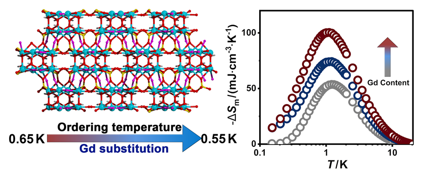
1 Introduction
Sub-kelvin temperatures are indispensable for frontier research areas ranging from quantum information processing and superconducting electronics to precision metrology and deep-space exploration.[1-2] At present, the principal technologies used to access the sub-kelvin regime include 3He-4He dilution refrigeration, 3He adsorption refrigeration and adiabatic demagnetization refrigeration (ADR). While the former two technologies rely on scarce 3He resources, efficient and helium-independent ADR is regarded as a complementary and potentially more accessible cooling strategy.[3-4]
The performance of ADR is ultimately governed by the magnetocaloric refrigerant. In an ideal paramagnet, the application of an external magnetic field aligns magnetic moments and reduces magnetic entropy. Subsequent adiabatic removal of the magnetic field induces spin disordering under entropy-conserving conditions, thereby leading to a decrease in temperature.[5] Accordingly, the cooling performance of an ADR refrigerant is closely associated with its magnetic entropy content and the absence of magnetic ordering above the targeted operating temperature. Therefore, ideal sub-kelvin magnetic refrigerants should possess a high density of magnetic moments while maintaining a low magnetic ordering temperature (T0).
Despite substantial progress over the past decades,[6-13] lowering T0 often requires dilution of the magnetic ions, resulting in an inherent trade-off between suppressing magnetic ordering and maintaining high magnetic entropy density. As a result, commercial refrigerants such as CrK(SO4)2•12H2O (CPA) and NH4Fe(SO4)2•12H2O (FAA) continue to play pivotal roles in ADR technology; however, they suffer from intrinsic limitations. The abundant lattice water molecules in these salts not only reduce their magnetocaloric effect (MCE), but also compromise their chemical and thermal stability, making them susceptible to dehydration and structural degradation during practical operation under vacuum or even mild heating conditions.[14] More recently, inorganic magnetic refrigerants have shown improved stability; however, their relatively low magnetic ion density still limits their achievable magnetocaloric performance.[4,14-15] These challenges underscore the urgent need to develop magnetic materials that combine large magnetic entropy changes (-ΔSm) with a suppressed T0.
Owing to spin-orbit coupling and crystal-field effects, only the lowest Kramers doublet of Yb3+ is thermally populated at low temperatures. Consequently, Yb3+ can often be described as an effective spin-1/2 ground state in the sub-kelvin regime.[16-17] Compared with conventional Gd-based refrigerants, Yb3+ systems commonly exhibit weaker exchange and dipolar interactions, resulting in lower magnetic ordering temperatures that are favorable for ADR. Beyond the selection of suitable magnetic ions, however, enhancing magnetic entropy while preserving a low T0 remains a critical challenge in refrigerant design. Recently, we proposed a magnetic-density optimization strategy based on trace Gd3+ doping, in which a small amount of isotropic Gd3+ ions is introduced into a weakly interacting rare-earth lattice. This approach effectively increases the available magnetic entropy without significantly strengthening magnetic exchange interactions, thereby improving magnetocaloric performance in the sub-kelvin region.[18-20] These findings suggest that combining a weakly coupled Yb3+ framework with controlled Gd3+ substitution may provide an effective route toward next-generation ADR materials.
Herein, we report the synthesis, crystal structure, magnetic properties, and sub-kelvin magnetocaloric effect of a new ytterbium fluorohydroxysulfate framework, Yb2SO4(OH)2F2. This compound adopts a dense three-dimensional framework constructed from interconnected Yb-centered polyhedra and sulfate bridges, affording a high magnetic-ion density while maintaining weak magnetic interactions. Furthermore, partial substitution of Yb3+ with Gd3+ was employed to optimize the magnetic entropy content following our previously established strategy. Magnetic and thermodynamic investigations reveal weak antiferromagnetic interactions and a pronounced magnetocaloric effect at sub-kelvin temperatures. These results demonstrate that magnetic-density optimization through Gd incorporation is an effective strategy for enhancing sub-kelvin magnetocaloric performance and identify fluorohydroxysulfate frameworks as a promising platform for adiabatic demagnetization refrigeration.
2 Results and Discussion
2.1 Structural Information
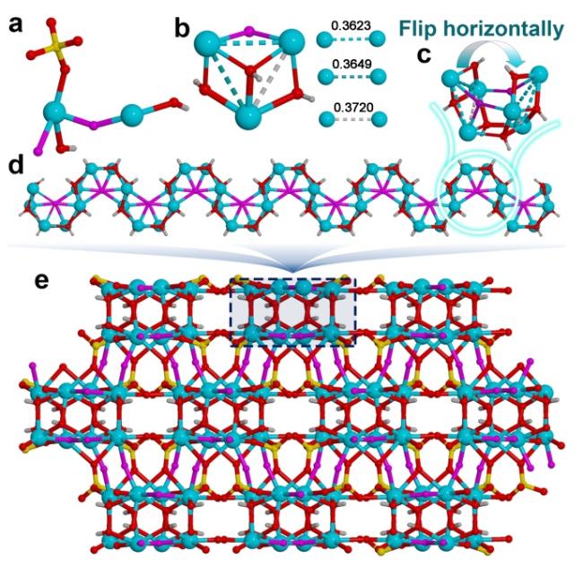
Single-crystal X-ray diffraction analysis revealed that (Yb1-xGdx)2SO4(OH)2F2 crystallizes in the monoclinic space group (Supporting Information (SI), Table S1). The powder X-ray diffraction patterns of 1—3 are in good agreement with the simulated pattern, confirming the phase purity of the samples (SI, Figure S1). The ratio of Gd to Yb in 2 and 3 were determined by inductively coupled plasma mass spectrometry (ICP-MS) (SI, Table S2). In addition, elemental mapping of 2 and 3 (SI, Figure S2) demonstrates the homogeneous distribution of Gd and Yb throughout the samples. Given their structural similarities, the structure of Yb2SO4(OH)2F2 is discussed in detail below. The asymmetric unit in 1 contains two crystallographically independent Yb3+ ions, one SO42− group, two OH− ligands, and two F− ligands (Figure 1a). One Yb3+ center is eight-coordinated by two F−, three OH−, and three O atoms from two SO42− groups, giving a slightly distorted triangular dodecahedron (SI, Figure S3a) with continuous shape measure (CShM) values of 1.018—1.098 (SI, Table S3). In contrast, the other Yb3+ center is eight-coordinated by three F−, three OH−, and two O atoms from two SO42− groups, giving a slightly distorted square antiprism (Figure S3b) with CShM values of 0.953—1.016. Three neighboring Yb3+ ions are interconnected by three OH− ligands and one F− ligand to form a triangular trinuclear motif, with Yb…Yb separations ranging from 0.3623 to 0.3720 nm (Figure 1b). Two such triangular units are related by a horizontal flip (Figure 1c) and are further connected to generate a hexanuclear {Yb6} unit. These {Yb6} units are subsequently linked in a zigzag, or “Z”-shaped, manner along the a axis through shared OH− ligands, forming an infinite one-dimensional chain structure (Figure 1d). Furthermore, neighboring zigzag chains are interconnected by F− and SO42− groups, which act as interchain bridges and extend the structure into a three-dimensional framework (Figure 1e). The Yb—F and Yb—O bond lengths are 0.2121(3)—0.2465(7) and 0.2242(7)—0.2687(7) nm, respectively, while the Yb—F—Yb and Yb—O—Yb bond angles range from 99.4(3)° to 151.1(3)° and 90.0(3)° to 108.0(3)°, respectively. These parameters remain essentially unchanged upon Gd3+ doping, with corresponding bond lengths of 0.2121(5)—0.2479(7) and 0.2253(8)—0.2683(8) nm and bond angles of 100.1(3)°—150.7(3)° and 90.3(3)°—108.0(4)°, respectively.
2.2 Magnetic properties
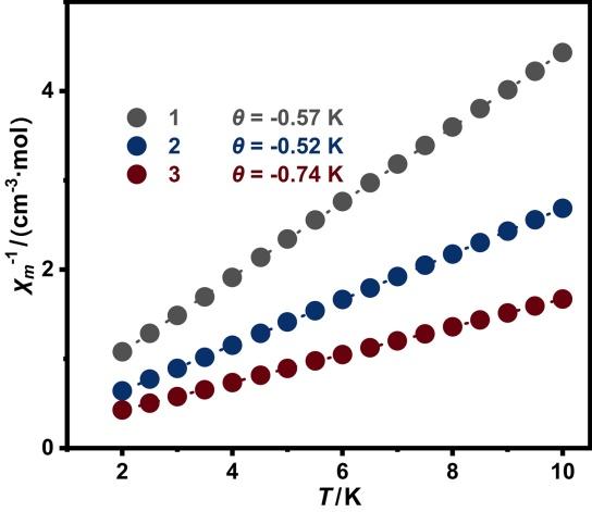
Temperature-dependent magnetic susceptibility measurements were performed on polycrystalline samples of 1—3 under an applied DC field of 0.1 T (SI, Figure S4). At 300 K, the χmT values are consistent with those expected for the corresponding concentrations of magnetically isolated Yb3+ and Gd3+ ions (SI, Table S4). Upon cooling, χmT decreases gradually for all three compounds. Although this behavior may suggest antiferro- magnetic interactions, the magnetic properties of Yb3+- based systems are often strongly influenced by crystal-field splitting, which can also contribute significantly to the temperature dependence of χmT. To minimize the influence of thermally populated crystal electric field excited states and thereby better reflect the intrinsic low-temperature magnetic interactions, Curie-Weiss fitting was performed over the 2—10 K range, following common practice for rare-earth magnetic systems. This analysis yielded Weiss constants (θ) of -0.57, -0.52, and -0.74 K for 1—3, respectively (Figure 2). These small negative θ values indicate the presence of weak antiferromagnetic interactions in all three compounds. Although Gd substitution slightly increases the magnitude of θ at higher doping levels, the magnetic interactions remain weak across the series. Notably, Gd incorporation increases the spin density without introducing significant magnetic exchange, thereby achieving a favorable balance between large magnetic entropy and suppressed magnetic ordering, both of which are desirable characteristics for efficient adiabatic demagnetization refrigeration at sub-kelvin temperatures.[21]
2.3 Magnetocaloric Effect
Considering the preference for low magnetic fields in practical applications, heat-capacity measurements were performed for 1—3 between 0.1 and 20 K under zero field and an applied magnetic field of 2 T to evaluate their low-temperature magnetocaloric effects (Figures 3a and S5). At higher temperatures, the heat capacity is dominated by lattice contributions, whereas magnetic contributions become increasingly significant upon cooling. To isolate the magnetic contribution from the total heat capacity, the lattice contribution (Clattice) was estimated using the Debye model, and the magnetic heat capacity (Cm) was then obtained by subtracting Clattice from the total heat capacity according to Cm=Cp-Clattice. As shown in Figure 3b, the magnetic heat capacities exhibit pronounced λ-type anomalies characteristic of long-range magnetic ordering. The corresponding T0 values are 0.65, 0.60, and 0.55 K for 1—3, respectively, revealing a gradual suppression of magnetic ordering upon Gd substitution. Notably, although Gd incorporation slightly strengthens the overall magnetic interactions, as indicated by the increased θ values, the ordering temperature is continuously reduced. This apparent contrast highlights the difference between local magnetic exchange interactions and the development of long-range magnetic order. In this system, Gd-induced exchange randomness, together with competing ferro- and antiferromagnetic interactions, likely frustrates long-range magnetic ordering and thus lowers T0.[18-20] While increasing the Gd3+ content can enhance the magnetic entropy, it also strengthens exchange and dipolar interactions, which tend to raise T0. In principle, dilution of a pure Gd3+ lattice with diamagnetic Y3+ can also suppress magnetic interactions and reduce the magnetic ordering temperature. However, due to the strong dipolar interactions associated with the large magnetic moment of Gd3+, achieving sufficiently low ordering temperatures generally requires extensive dilution, which inevitably reduces the magnetic ion density and thus significantly limits the achievable magnetic entropy change. This highlights the intrinsic trade-off between suppressing magnetic ordering and maintaining high magnetic density in designing efficient sub-Kelvin magnetocaloric materials. Herein, the combination of enhanced magnetic entropy (SI, Figure S6) and suppressed magnetic ordering is highly desirable for sub-kelvin magnetic refrigeration.
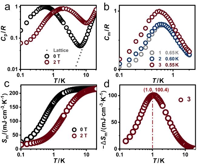


Figure 3 (a) Experimental heat capacity (Cp) normalized to the gas constant (R) of 3; (b) temperature dependence of the zero-field magnetic heat capacity (Cm) in 1—3; (c) temperature dependence of the experimental magnetic entropy Sm in 3 under 0 T and 2 T; (d) the calculated -ΔSm values of 3 under 2 T. |
To further evaluate the effect of Gd substitution on the thermodynamic properties, the magnetic entropy (Sm) was calculated by integrating the magnetic heat capacity according to Sm(T)=∫(Cm/T)dT. As shown in Figure S7 (SI), the magnetic entropy increases systematically with increasing Gd content over the entire temperature range. Given its highest magnetic entropy and lowest ordering temperature among the three compounds (Figure S6b), 3 was selected as a representative system for detailed magnetocaloric investigation. As shown in Figures 3c and 3d, compound 3 exhibits a maximum magnetic entropy change of approximately 100 mJ•cm-3•K-1 under ΔH=2 T, which is substantially higher than those of hydrated paramagnetic salts such as CPA and FAA (Table 1). Although the magnetocaloric performance of 3 does not exceed that of the best-performing Yb-based fluoride refrigerants, which benefit from high magnetic-ion densities and minimal nonmagnetic components, its maximum entropy change remains highly competitive among reported Yb-based refrigerants. In particular, the -ΔSm values of 3 surpass those of most pure Yb-based framework materials reported to date, demonstrating the effectiveness of the present fluorohydroxysulfate framework in preserving large magnetic entropy at sub-kelvin temperatures. More importantly, the enhanced magnetocaloric performance is achieved without compromising either structural stability or magnetic-ion density. In contrast to many Yb-based fluoride refrigerants, in which increasing Gd content typically strengthens magnetic correlations and raises the magnetic ordering temperature, Gd incorporation in the present fluorohydroxysulfate framework does not induce stronger magnetic ordering. Instead, T0 is slightly suppressed with increasing Gd content. Consequently, the introduction of Gd3+ simultaneously increases the magnetic-ion density and preserves, or even enhances, the accessibility of magnetic entropy in the sub-kelvin regime. This unusual combination effectively mitigates the conventional trade-off between magnetic entropy density and magnetic ordering, leading to a substantially enhanced magnetocaloric response.
Table 1 Magnetic ordering temperature (T0) and maximum magnetic entropy changes (-ΔSmax) under 2 T of selected Yb-based refrigerants |
| Refrigerants | T0/K | -ΔSmax (2 T)/(mJ•cm-3•K-1) | Ref. |
|---|---|---|---|
| KYb3F10 | <0.05 | 133 | [22] |
| Gd0.1Yb0.9F3 | 0.18 | 197 | [18] |
| K(Gd0.1Yb0.9)3F10 | <0.05 | 142 | [20] |
| LiGd0.1Yb0.9F4 | 0.085 | 136 | [19] |
| Yb3Ga5O12 | 0.054, 0.18 | 116 | [23] |
| 1 | 0.65 | 54 | This work |
| 2 | 0.60 | 74 | |
| 3 | 0.55 | 100 | |
| KBaYb(BO3)2 | 0.009 | 61 | [14] |
| NaYbP2O7 | — | 57 | [15] |
| YbPt2Sn | 0.25 | 93 | [5] |
| YbNi4Mg | 0.3 | 71 | [24] |
| YbNi1.6Sn(8T) | 0.14 | 131 | [25] |
| YbCu4.6Au0.4 | — | 43 | [25] |
| (NH4)Fe(SO4)2•12H2O | 26 | 52 | [26] |
| CrK(SO4)2•12H2O | 9 | 41 | [26] |
3 Conclusion
In summary, a new ytterbium fluorohydroxysulfate framework, Yb2SO4(OH)2F2, and its Gd-doped analogues have been successfully synthesized and characterized. The dense three-dimensional framework combines high magnetic-ion density with weak magnetic interactions, resulting in magnetic ordering temperatures below 1 K. Notably, Gd incorporation increases the magnetic entropy while slightly suppressing the ordering temperature, thereby mitigating the conventional trade-off between magnetic density and magnetic ordering. Benefiting from this synergistic effect, the optimized compound exhibits a maximum magnetic entropy change of approximately 100 mJ•cm−3•K−1 under ΔH=2 T, nearly twice that of FAA and more than twice that of CPA. These results demonstrate that magnetic-density optimization through Gd substitution is an effective strategy for enhancing sub-kelvin magnetocaloric performance and establish fluorohydroxysulfate frameworks as promising candidates for sub-kelvin magnetic refrigeration.
4 Experimental Section
4.1 Synthesis of Yb2SO4(OH)2F2 (1)
A mixture of Yb2O3 (1 mmol), concentrated H2SO4 (98%, 1 mmol) and hydrofluoric acid (≈40%, 2 mmol) was dissolved in 5 mL of H2O and stirred for 20 min at room temperature. The resulting solution was transferred into a 23 mL Teflon-lined stainless-steel autoclave and heated at 230 ℃ for 3 d under autogenous pressure. After cooling to room temperature naturally, yellow block-shaped crystals were collected, washed with deionized water and dried in air. Caution! Hydrofluoric acid is highly toxic and corrosive and should be handled with extreme caution using appropriate protective equipment and safety procedures.
4.2 (Yb0.9Gd0.1)2SO4(OH)2F2 (2)
The preparation of (Yb0.9Gd0.1)2SO4(OH)2F2 is quite similar to 1 except that the dosage of rare earth oxides was adjusted as Yb2O3 (0.9 mmol) and Gd2O3 (0.1 mmol).
4.3 (Yb0.7Gd0.3)2SO4(OH)2F2 (3)
The preparation of (Yb0.7Gd0.3)2SO4(OH)2F2 is quite similar to 1 except that the dosage of rare earth oxides was adjusted as Yb2O3 (0.7 mmol) and Gd2O3 (0.3 mmol).
(Cheng, B.)