



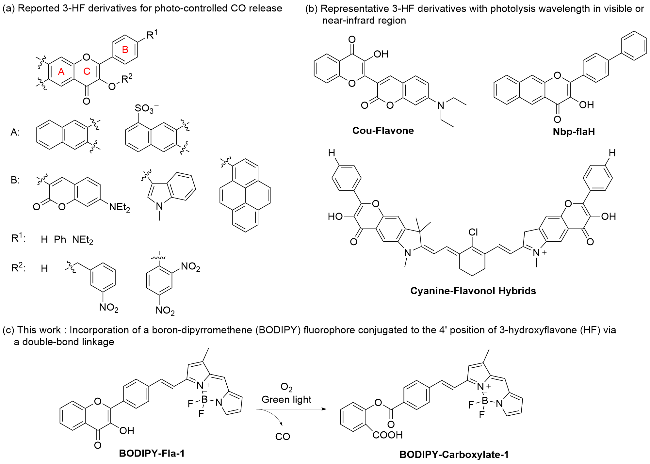
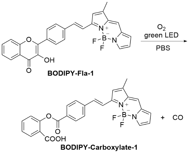
图1 (a)已报道的基于3-羟基黄酮醇结构衍生的光控CO给体; (b)光解波长在可见区或近红外区的3-羟基黄酮醇衍生物的代表性结构; (c)本工作: 3-羟基黄酮醇(HF)通过在B环4'位以双键共轭的连接方式与氟硼二吡咯(BODIPY)荧光团连接Figure 1 (a) Reported 3-HF derivatives for photo-controlled CO donors; (b) Representative structures of 3-HF derivatives with photolysis wavelength at visible or near-infrared region; (c) This work: incorporation of a boron-dipyrromethene (BODIPY) fluorophore conjugated to 4' position of 3-hydroxyflavone (HF) via a double-bond linkage |
1 结果与讨论
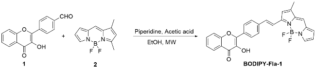
1.1 探针BODIPY-Fla-1的合成
1.2 BODIPY-Fla-1光解反应紫外-可见吸收光谱的变化
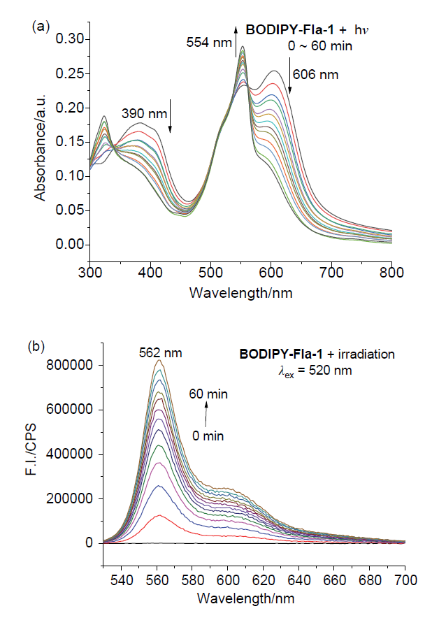
图2 (a) BODIPY-Fla-1 (10 μmol/L)随光解时间变化的紫外吸收光谱; (b) BODIPY-Fla-1 (10 μmol/L)随光解时间变化的荧光发射光谱图(λex=520 nm)Figure 2 (a) Time-dependent UV absorption spectra of BODIPY-Fla-1 (10 μmol/L) upon photolysis; (b) Time-dependent fluorescence emission spectrum of BODIPY-Fla-1 (10 μmol/L, λex=520 nm) upon photolysis All measurements were performed in 10 mmol/L pH=7.4 PBS buffer. Photolysis reactions were performed under a 15 W green LED light. |
1.3 BODIPY-Fla-1光解反应荧光光谱变化
1.4 BODIPY-Fla-1的光控释放CO的确认
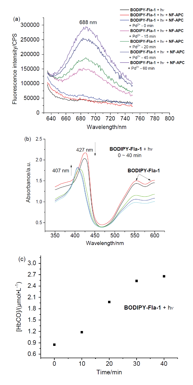
图3 (a) BODIPY-Fla-1 (10 μmol/L)绿光照射15 min后加入探针NF-APC (10 μmol/L)与Na2PdCl4 (60 μmol/L)后荧光发射谱图随时间的变化(λex=620 nm); (b)探针BODIPY-Fla-1 (50 μmol/L)与血红蛋白(4.2 μmol/L)共孵育条件下的紫外吸收光谱随绿光照射时间的变化(每10 min测一次); (c)相应生成的碳氧血红蛋白浓度计算值随光照时间的变化Figure 3 (a) Time-dependent fluorescence emission spectra of 10 μmol/L BODIPY-Fla-1 preirradiated with green LED light for 15 min incubated with NF-APC (10 μmol/L) and Na2PdCl4 (60 μmol/L) (λex=620 nm); (b) Time-dependent absorption spectra of the probe BODIPY-Fla-1 (50 μmol/L) coincubated with Hb (4.2 μmol/L) upon irradiated with green LED light; (c) Corresponding time-dependent calculated COHb concentration upon photoirradiation All measurements were performed in 10 mmol/L pH=7.4 PBS buffer, the light source is green LED, 15 W |
1.5 BODIPY-Fla-1光解生成的荧光产物的结构确证研究
1.6 BODIPY-Fla-1光解前后溶液聚集粒径变化研究
1.7 BODIPY-Fla-1绿光可控光解和荧光监测性能的循环测试验证
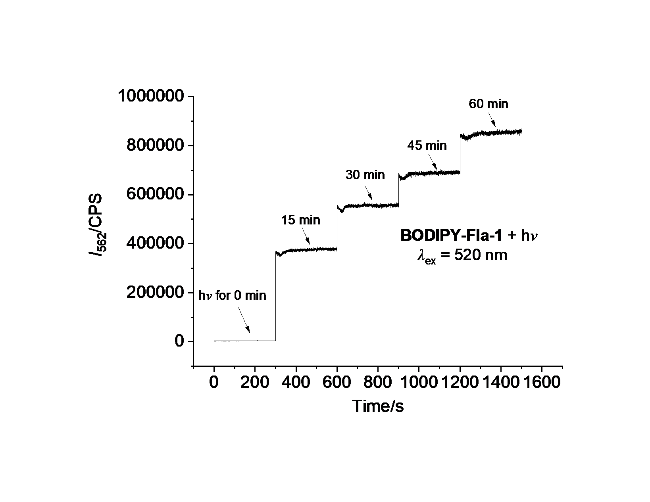
图5 探针BODIPY-Fla-1 (10 μmol/L)在PBS缓冲液(10 mmol/L, pH=7.4)中每隔15 min周期性照射(15 W, 绿色LED灯)后在562 nm处(λex=520 nm)的荧光强度随时间变化Figure 5 Time-dependent of fluorescent intensity changes of probe BODIPY-Fla-1 (10 μmol/L) at 562 nm (λex=520 nm) in PBS buffer (10 mmol/L, pH=7.4) after the periodic irradiation with green LED (15 W) in every 15 min |
1.8 BODIPY-Fla-1的光控CO释放的荧光跟踪成像和生物功能验证
1.8.1 细胞毒性实验
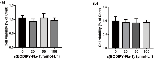
图6 (a)不同浓度的BODIPY-Fla-1作用于HeLa细胞8 h后的细胞存活率; (b)不同浓度的BODIPY-Fla-1作用于RAW264.7细胞8 h后的细胞存活率Figure 6 (a) Percentage of viable HeLa cells after treated with different concentrations of BODIPY-Fla-1 for 8 h; (b) Percentage of viable RAW264.7 cells after treated with different concentrations of BODIPY-Fla-1 for 8 h |
1.8.2 活细胞中CO光控释放和荧光成像实验
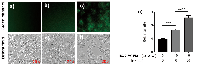
图7 探针BODIPY-Fla-1 (10 μmol/L)在RAW 264.7细胞中的CO释放荧光成像图: (a)细胞在正常培养基中孵育; (b)探针BODIPY-Fla-1 (10 µmol/L)孵育10 min后的细胞荧光图像; (c)探针BODIPY-Fla-1 (10 µmol/L)孵育10 min后再光照30 min后的细胞荧光图像; (d~f)相应的明场图像; (g)不同细胞组中荧光图像的相对荧光强度(于λex=340~380 nm, λem>425 nm处采集荧光图像, 光解所用光源为15 W绿色LED灯, 误差棒表示三个重复的标准偏差, ****P≤0.0001, ***P≤0.001)Figure 7 Fluorescence imaging of CO-release with probe BODIPY-Fla-1 in living RAW 264.7 cells. (a) Cells were incubated in normal media; (b) Fluorescence image of cells incubated in 10 μmol/L probe BODIPY-Fla-1 for 10 min; (c) Fluorescence image of cells incubated in 10 μmol/L probe BODIPY-Fla-1 for 10 min followed by 30 min of irradiation; (d~f) Corresponding bright-field images; (g) Relative fluorescence intensities of fluorescence images in different cell groups (Fluorescence images were captured with λex=340~380 nm and λem>425 nm; irradiation light source: green LED,15 W. The error bar represents the standard deviation of three repetitions, ****P≤0.0001, ***P≤0.001) |
1.8.3 活细胞中的CO光控释放对氧化应激的影响
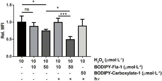


图8 不同浓度的BODIPY-Fla-1在绿光照射或黑暗条件下对A549细胞的抗氧化应激作用Figure 8 Anti-oxidative stress effect of different concentrations of BODIPY-Fla-1 on A549 cells in dark or under green light irradiation Error bars represent standard deviations of three replicate images in the same group, ***P≤0.001, *P≤0.1, ns: no significant difference. |