


1 Introduction
The genus Ainsliaea, belonging to the family Asteraceae, is primarily distributed in Southeast Asia. To date, 70 species of this genus have been identified worldwide with 44 species found in China.[1] Phytochemical studies have revealed that sesquiterpenoids are characteristic chemical constituents of Ainsliaea species. Numerous chemical compounds, such as triterpenoids, sesquiterpenoids, flavonoids, and phenylpropanoids, have been isolated from Ainsliaea species.[2,3] Among these, sesquiterpenoids are considered the main active components, exhibiting significant antitumor and anti-inflammatory activities.[4-6] Guaianolide sesquiterpenoids, as important secondary metabolites of natural products, have attracted considerable interest from researchers in natural product and medicinal chemistry due to their fascinating structures and diverse biological activities.[7-10]
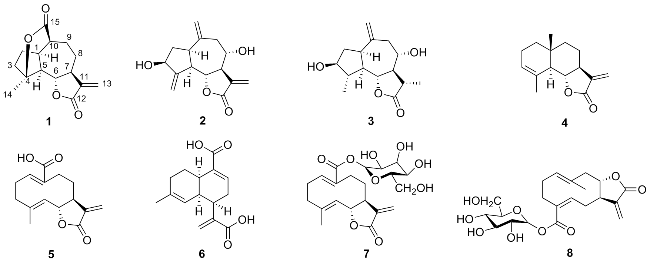
Eight compounds were isolated from Ainsliaea glabra. Among them, one is a previously undescribed guaianolide sesquiterpenoid (1), and seven are known compounds 2~8 (Figure 1). The known compounds were identified as eleganin (2),[11] isolipidiol (3),[12] α-cyclocostunolide (4),[13] taraxinic acid (5),[14] ainsliaea acid B (6),[15] taraxinic acid β-D-glucopyranosyl ester (7),[16] and germacra-7aH-1(10) E,4-Z,11(13)-trien-12,8a-olide-15-oic acid (15→1)-b-D- glu-cop-yranosyl ester (8).[17]
2 Results and discussion
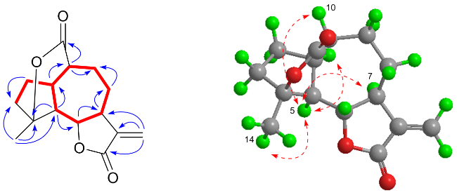
Ainsglaolide A (1) was obtained as white amorphous powder, and gave an elemental formula C15H18O4 from the (+)-HRESIMS ion at m/z 263.1274 [M+H]+, requiring 7 unsaturations. The IR spectrum revealed the existence of carbonyl (1770, 1725 cm-1). The interpretation of the 1D NMR date (Table 1) indicated distinctive signals for one terminal vinyl group (δC 139.0, 120.6), and two carbonyl groups (δC 173.0, 170.0). The rest of resonances were determined by the 13C NMR and distortionless enhancement by polarization transfer (DEPT) spectra as being one methyl, four methylenes, five methines (including one oxygenated), and one quaternary carbon. Based on the 1H-1H COSY correlations (Figure 2), one structural fragment of H-5/H-6/H-7/H2-8/H2-9/H-10/H-1/H2-2/H2-3 could be derived. Subsequently, the five-membered ring and the seven-membered ring were fused together at C-1 and C-5 by the HMBC correlations (Figure 2) from H3-14 (δH 1.49) to C-3 (δC 37.7), C-4 (δC 90.0) and C-5 (δC 50.8). Compound 1 was a guaianolide sesquiterpene, as evidenced by HMBC correlations from H2-13 (δH 6.21, 5.50) to C-7 (δC 44.8), C-11 (δC 139.0) and C-12 (δC 170.0), and the chemical shift of C-6 (δC 81.0). In the previous structural elucidation, compound 1 was found to possess two carbonyl groups, one double bond, and three cyclic systems. Based on degree of unsaturation analysis, it was deduced that the compound must contain an additional ring system. The characteristic chemical shift of C-4 at δC 90 indicates an oxidized quaternary carbon. The HMBC correlations observed between H-10 (δH 2.67) and C-4 (δC 90.0)/C-15 (δC 173.0) establish the formation of a six-membered lactone ring through ether linkage between C-4 and C-15. Notably, this configuration represents the sole chemically viable position for con-structing such a lactone ring. The structural assignment was further corroborated by excellent agreement between experimental mass spectrometric data and computational simulations. Consequently, the planar structure of 1 could be determined.
Table 1 1H NMR (400 MHz) and 13C NMR (100 MHz) data of compound 1 in CDCl3 |
| Position | δH, mult. (J/HZ) | δC |
|---|---|---|
| 1 | 2.62, t (5.7) | 40.8 |
| 2 | 2.10, overlap; 1.63, overlap | 30.7 |
| 3 | 1.97~1.89, m; 2.24, overlap | 37.7 |
| 4 | 90.0 | |
| 5 | 2.28, t (5.7) | 50.8 |
| 6 | 4.08, ddd (10.4, 5.7, 1.6) | 81.0 |
| 7 | 2.94, td (10.4, 1.6) | 44.8 |
| 8 | 2.16, overlap; 1.46~1.35, m | 26.8 |
| 9 | 2.46~2.38, m; 1.69, overlap | 31.8 |
| 10 | 2.67, br s | 47.9 |
| 11 | 139.0 | |
| 12 | 170.0 | |
| 13 | 6.21, dd (3.1, 1.6); 5.50, dd (3.1, 1.6) | 120.6 |
| 14 | 1.49, s | 21.8 |
| 15 | 173.0 |
 ), HMBC (
 ) and NOESY (
 ) correlations of compound 1
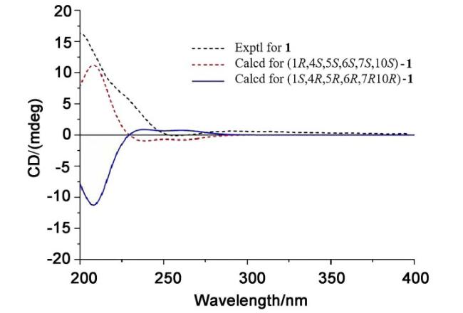
The NOE cross-peaks (Figure 2) of H-5/H3-14/H-1/H- 10/H-7 indicated these protons to be α-oriented and the six-membered lactone ring is β-oriented. Additionally, by contrasting theoretical and experimental electrostatic circular dichroism (ECD) curves (Figure 3), its absolute configuration (1R,4S,5S,6S,7S,10S) was verified. As a result, the relative configuration of 1 was established as shown (Figure 1).
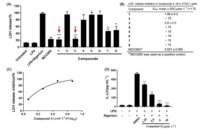
To assess the anti-inflammatory potential of the comp-ounds, we employed J774A.1 cells and employed lipopolysaccharide (LPS) and Nigericin as agonists to activate the NLRP3 inflammasome. MCC950, also recognized as CRID3, a potent cytokine release inhibitor and NLRP3 inflammasome antagonist, served as our positive control. Notably, compound 1 demonstrated a substantial reduction in lactate dehydrogenase (LDH) release in a dose-dependent manner (Figure 4A). Specifically, the IC50 values for com-pounds 1 and 3 were determined to be (1.86±0.2) and (2.90±0.3) μmol/L, respectively (Figure 4B). Additionally, compound 1 exhibited dose-dependent inhibition of LDH release, further confirming its inhibitory effect (Figure 4C). Further, compound 1 inhibited IL-1β level in a dose-de- pendent manner, ranging from 1.25 μmol/L to 10 μmol/L (Figure 4D).
Figure 4 Screening of anti-inflammatory compounds from Alisliaea glabra at LDH and elisa level(A) Primary evaluation of the impact of compounds 1 through 8 on lactate dehydrogenase (LDH) release was conducted (n=3), MCC950 served as the positive control in this assay. Compounds with lower LDH release were identified with a red arrow (B, C). Calculate IC50 for compounds with good anti-inflammatory activity. The LDH release inhibition rate of compound 1 (1.25, 2.5, 5, 10 μmol/L) was evaluated in J774A.1 cells of NLRP3 inflammasome-activated. (D) IL-1β level (ELISA). The data of are represented by mean±SD. The LPS+Nigericin group served as A control (A), *P<0.05, **P<0.01, ***P<0.001. n.s.=not significant (P>0.05). |
3 Conclusions
Eight sesquiterpenes were isolated from Ainsliaea glabra. Among them, a new guaianolide sesquiterpenoid, named ainsglaolide A (1), and seven known sesquiterpenoids 2~8 were identified. Compound 1 was a novel guaianolide sesquiterpenoid featuring a rare six-membered lactone ring formed between C-4 and C-15. Subsequent anti-inflammatory activity assessments of the isolated compounds revealed that compound 1 exhibited a potent inhibitory effect on NLRP3 inflammasome activation with an IC50 value of 1.86 μmol/L. Our results demonstrated that compound 1 dose-dependently suppressed LPS and nigericin-induced NLRP3 inflammasome activation in J774A.1 cells. This finding underscores compound 1 as a promising candidate for the treatment of diseases associated with hyperinflammatory responses. This compound provides a new solution for the treatment of diseases induced by excessive inflammatory response.
4 Experimental section
4.1 Instruments and reagents
UV spectra were run with a Hitachi UV-visible spectrophotometer, model number U-4100. Nicolet iS10 FT IR spectrometer was used to record IR spectra (KBr pellets). NMR spectra at 400 MHz were measured using a Bruker AscendTM spectrometer with tetramethylsilane as the internal standard. An Agilent1100 high performance liquid chromatography-time-of-flight mass spectrometer (HPLC-TOF-MS) was used to collect the HRESIMS data. Silica gel (80~100 mesh and 200~300 mesh; Qingdao Marine Chemical, Ltd.) and macroporous resin (D-101; Tianjin Haoju Science and Technology Ltd.) were used for column chromatography (CC). Thin-layer chromatography (TLC) was performed on silica gel plates (Qingdao Marine Chemical Co, Ltd.). An Agilent 1260 liquid chromatograph with an Agilent Zorbax SB-C18 column (250 mm×9.4 mm, i.d., 5 μmol/L) was used for semi-preparative HPLC.
Ainsliaea Glabra was collected from Mount Emei, Sichuan Province, People’s Republic of China in September 2018. The plant was identified by Chen Yu, Kunming Institute of Botany, Chinese Academy of Sciences. A voucher specimen (XWL 20180710) was deposited at the Key Laboratory of Medicinal Chemistry for Natural Resource, Ministry of Education, Yunnan University.
4.2 Experimental method
4.2.1 Extraction and isolation
The air-dried Ainsliaea glabra (11 kg) was extracted three a solo times with ethanol at 25 ℃. The combined ethanol extracts were vacuum-concentrated to obtain extracts, which were suspended in H2O and extracted with ethyl acetate to afford an EtOAc soluble extract. The EtOAc extracts (201 g) was divided into four parts (Fr.1~Fr.4) by using D-101 macroporous resin column eluted with MeOH/H2O (V∶V=50∶50, 70∶30, 90∶10, 100∶0). Fr.2 (28 g) was purified on a silica gel (80~100 mesh) with a petroleum ether (PE)/ethyl acetate (EtOAc) gradient elution system (V∶V=10∶1, 6∶1, 3∶1, 1∶1, EtOAc and MeOH) to obtained six fractions: Fr.2.1~Fr.2.6.
Fr.2.2 was eluted by preparative HPLC (MeCN/H2O, V∶V=80∶20) to obtain four fractions: Fr.2.2.1~Fr.2.2.4. Fr.2.2.1 was eluted by preparative thin layer chromatography (PE/EtOAc, V∶V=12∶3) to obtain compounds 1 (68.5 mg) and 4 (12.5 mg). Fr.2.3 was separated by column chromatography with silica gel, gradient elution of PE/ acetone system (V∶V=7∶1, 5∶1, 3∶1, 1∶1) to obtain four fractions: Fr.2.3.1~Fr.2.3.4. Further, Fr.2.3.2 was eluted by preparative thin layer chromatography (PE/ EtOAc, V∶V=5∶1) to obtain 2 (23.8 mg). To obtain compound 3 (44.4 mg), Fr.2.3.3 was separated by preparative TLC (PE/EtOAc, V∶V=3∶1). Fr.2.4 was separated by column chromatography with silica gel, gradient elution of PE/acetone system (V∶V=10∶1, 7∶1, 5∶1, 3∶1, 1∶1) to obtain five fractions: Fr.2.4.1~Fr.2.4.5. Further, Fr.2.4.3 was eluted by preparative thin layer chromatography (PE/ EtOAc, V∶V=7∶3) to obtain 6 (8.3 mg). Fr.2.4.1 was purified by semi-preparative HPLC (MeCN/H2O, V∶V= 70∶30) to obtain compound 5 (5.4 mg, tR=22.5 min). Fr.2.6 was subjected to middle chromatogram isolated gel (MCI) with MeOH/H2O gradient elution system (V∶V= 50∶50, 60∶40, 70∶30, 80∶20, and MeOH) to obtain five fractions: Fr.2.6.1~Fr.2.6.5. Compound 7 (12.9 mg) was obtained by separating Fr.2.6.1 on a Sephadex LH-20 gel column and eluting it with methanol. Fr.2.6.3 was purified by preparative HPLC (MeCN/H2O, V∶V=85∶15), and compound 8 (12.5 mg, tR=18.2 min) was obtained.
Ainsglaolide A (1): White amorphous powder; $[\alpha ]_{\text{D}}^{\text{25}}$ +32.6 (c 0.125), CH3OH; UV (MeOH) λmax [log ε/(L•mol-1•cm-1)]: 208 (3.95) nm; 1H NMR and 13C NMR data see Table 1; IR (KBr) νmax: 2924, 1770, 1725, 1251, 1162, 1001, 989 cm−1. HRMS (ESI) calcd for C15H19O4 [M+H]+ 263.1274, found 263.1278.
4.2.2 Quantum chemistry calculations
Calculations of 1 was made using Gaussian 09 and Gauss View 5.0 for theoretical calculations in quantum chemistry.[18] The program Spartan'14 initially carried out conformational searches. By DFT computations, the major conformers were optimized at the B3LYP/6-31G (d,p) level of theory in the gas phase. The theoretical ECD of compound 1 was conducted in CH3OH and polarizable continuum model (PCM) model using time-dependent density functional theory (TDDFT) at the B3LYP/6-31G (d,p) level. SpecDis software was used to calculate the ECD trend calculated.[19] By balancing the Boltzmann distribution rate of each geometric conformation, the ECD curve was produced.
4.2.3 Cell culture and stimulation
The J774A.1 macrophage cell line was acquired from the esteemed Kunming Institute of Zoology. These cells were sustained in a proliferative milieu comprising Dulbecco՚s Modified Eagle՚s Medium (DMEM), enriched with 10% heat-inactivated fetal bovine serum (FBS) to provide essential nutrients, and 1% penicillin-streptomycin antibiotic solution to ensure sterile conditions. Propagation occurred within T-75 polystyrene flasks, with meticulous environmental control maintained at 5% CO2 concentration and a temperature of 37 ℃ to mimic physiological conditions. Cell subculturing was initiated upon achieving 80%~90% confluency to maintain optimal cell density and health.
For targeted experimental procedures, J774A.1 cells were plated onto 24-well plates at a precise density of 2×10⁶ cells per well, allowing for cellular adherence overnight to ensure robust attachment. Subsequently, the culture medium was meticulously aspirated, and the cells were exposed to lipopolysaccharide (LPS) at a defined concentration of 200 ng/mL for a period of 3 h to induce an inflammatory response. Following LPS treatment, the medium was exchanged with Ultra-Fectin solution containing 10 μmol/L of compound 1 for an additional 30 min, aiming to modulate specific cellular pathways. To activate the NLRP3 inflammasome, a crucial component of innate immunity, the cells were further stimulated with nigericin at a concentration of 10 μmol/L for 1 h, precisely orchestrating the temporal sequence of stimulatory events to elucidate the underlying biological mechanisms.
4.2.4 Lactate dehydrogenase (LDH) assay
After activation of inflammasome treated by Alisliaea Glabra Hemsl, cell culture superserum was analyzed using LDH assay kit according to manufacturer՚s instructions.
4.2.5 Statistical significance analysis
The experimental data were expressed as mean±SD deviation, and were statistically analyzed by GraphPad Prism 7.0 software. Unidirectional analysis of variance (ANOVA) was used, followed by a Tukey Post Hoc test (T test) to analyze the statistical significance between multiple sets of data. P-values<0.05 indicates statistical significance.
Supporting Information The NMR and HRESIMS spectra of new compounds 1. The Supporting Information is available free of charge via the Internet at http://sioc- journal.cn.
(Lu, Y.)