


1 Introduction
The ocean covers 71% of the Earthʼs surface area and is home to approximately 87% of all life forms on Earth.[1] Marine microorganisms have advantages such as ease of large-scale production and genetic manipulation compared to traditional marine organisms like corals and sponges. Natural products derived from marine microorganisms have become valuable sources for drug lead discovery owing to their novel structures and extensive biological activities.[2-3] Dihydroisocoumarin compounds are an important class of natural products, commonly derived from marine microorganisms and plants.[4] As a member of the coumarin family, dihydroisocoumarins are characterized by a structure in which the carbonyl group is part of a six-membered pyran ring, which is fused directly to a benzene ring. Dihydrocoumarins have a wide range of biological activities, including anti-tumor, anti-inflammatory, antifouling, antiviral, anti-diabetic and α-glycosidase activities.[5-9] The phaeosphaerins A~E isolated from the fermentation broth of Phaeosphaeropsis sp. WP-26 from the South China Sea by Wang et al.[10] showed certain neuroprotective effects against H2O2 induced SH-SY5Y cell damage. Four novel dihydroisocoumarin compounds isolated by Yao et al. from ethanol extracts of pigeon pea leaves demonstrated cytotoxicity against various human tumor cells, with IC50 from 0.8 μmol/L to 1.4 μmol/L.[11] Two novel dihydroisocoumarin compounds isolated from the mangrove derived fungus Exserohilum rostratum NS- KS-3 by Bian et al.[12] exhibited α-glucosidase inhibitory activity. Nine novel dihydroisocoumarin dimers with unique linkage patterns isolated by Wang et al.[13] from the endophytic fungus Spegazzinia sp. MDW-573, some of which exhibited pro-angiogenic activity.
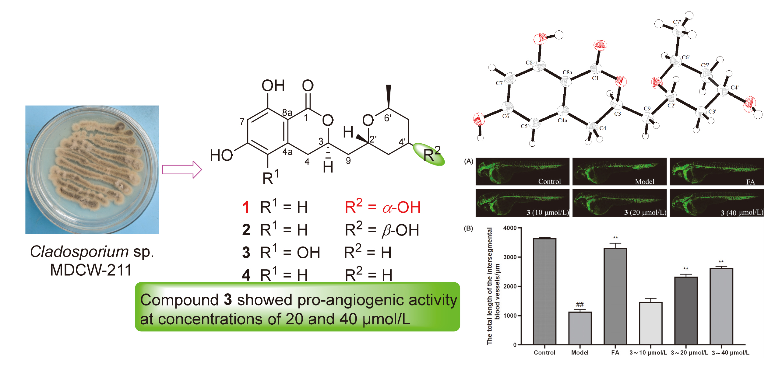
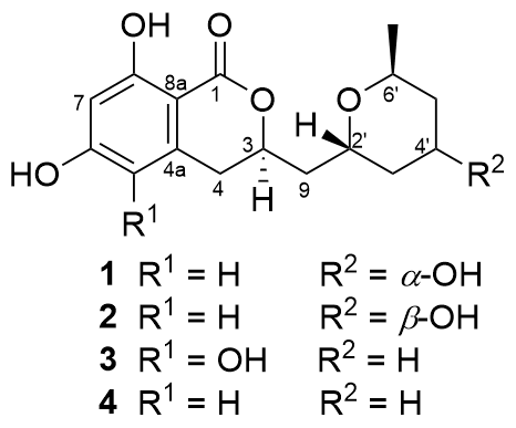
Our team has long been committed to the discovery of drug lead compounds derived from marine microorganisms. In this study, a fungal strain, Cladosporium sp. MDW-211, was isolated from crab samples collected on Weizhou Island, Guangxi. From its rice fermentation, one new dihydroisocoumarin, 4'S-hydroxyasperentin (1) and three known dihydroisocoumarins (2~4) were isolated and identified as illustrated in Figure 1. The absolute configuration of compound 1 was determined using single- crystal X-ray diffraction. The pro-angiogenic activities of compounds 1~4 were evaluated using a zebrafish model, and the results revealed that compound 3 exhibited significant proangiogenic activity.
2 Results and discussion
4'S-hydroxyasperentin (1) was obtained as yellow brown crystals. Its molecular formula C16H20O6 was determined by HRESIMS [M+Na]+ at m/z 331.1152 (calcd 331.1158). 13C NMR and HSQC spectra indicate the presence of 6 benzene ring carbons (δC 166.2, 165.5, 143.5, 107.9, 102.2, 101.6), one ester carbonyl group (δC 171.4), four methylene groups (δC/H 42.1/1.95, 41.8/1.95, 1.22, 40.0/1.75, 1.57, 34.4/2.87), four CH groups (δC/H 77.5/ 4.68, 69.1/4.22, 65.7/4.03, 64.5/4.03), and one methyl group (δC/H 18.8/1.24) in the molecule (Table 1).
Table 1 1H NMR (400 MHz) and 13C NMR (100 MHz) data of compound 1 in CD3OD |
| No. | δC | δH (J/Hz) |
|---|---|---|
| 1 | 171.4, C | |
| 3 | 77.5, CH | 4.63~4.73 (m, 1H) |
| 4 | 34.4, CH2 | 2.79~2.93 (m, 2H) |
| 4a | 143.5, C | |
| 5 | 107.9, CH | 6.20 (d, 2.5, 1H) |
| 6 | 166.2, C | |
| 7 | 102.2, CH | 6.20 (d, 2.5, 1H) |
| 8 | 165.5, C | |
| 8a | 101.6, C | |
| 9 | 42.1, CH2 | 1.83~1.99 (m, 2H) |
| 2' | 65.7, CH | 3.98~4.07 (overlap, 1H) |
| 3' | 41.8, CH2 | 1.83~1.99 (m, 1H) |
| 1.20~1.23 (overlap, 1H) | ||
| 4' | 64.5, CH | 3.98~4.07 (overlap, 1H) |
| 5' | 40.0, CH2 | 1.72~1.79 (m, 1H) |
| 1.52~1.62 (m, 1H) | ||
| 6' | 69.1, CH | 4.17~4.27 ((m, 1H) |
| 7' | 18.8, CH3 | 1.24 (d, 6.9, 3H) |
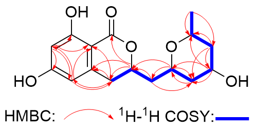
In the HMBC spectrum, H-7' showed correlation to C-5', proving the position of the methyl group. Besides, the following HMBC correlations were also observed: from H-3 to C-1 and C-4a, from H-4 to C-5 and C-8a, from H-5 to C-4, C-7 and C-8a, from H-7 to C-5, C-8 and C-8a, from H-9 to C-3', from H-2' to C-3 and C-3', from H-4' to C-2' and C-6', and from H-5' to C-3' and C-4'. The 1H-1H COSY spectrum demonstrated correlations from H-7' to H-6', from H-6' to H-5', from H-5' to H-4', from H-4' to H-3', from H-3' to H-2', from H-2'/H-9, from H-9 to H-3, and from H-3 to H-4 (Figure 2). Thus, the planar structure of compound 1 was determined to be the same as that of 4'-hydropiperentin with a different orientation of hydroxyl group at 4'-position. The NMR data of 1 are very similar to those of 4'-hydropiperentin,[14] with the differences of che- mical shifts mainly in the tetrahydropyran ring due to a different stereochemistry at 4'-position.
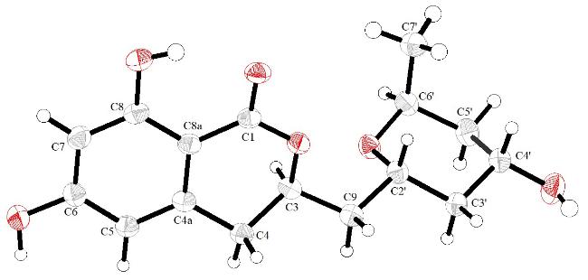
The absolute configuration of compound 1 was determined to be 3R,2'R,4'S,6'S by X-ray single crystal diffraction (Figure 3). Accordingly, the compound was named 4′S-hydroxyasperentin.
The pro-angiogenic effects of compounds 1~4 on the growth of intersegmental vessels in zebrafish were evaluated, as shown in Figure 4. In the blank control group, the intersegmental vessels of zebrafish grew normally, while in the model group, the vessel growth was significantly inhibited, indicating the successful establishment of the model. Compared with the model group, zebrafish treated with compound 3 exhibited a significant increase in intersegmental vessel length at concentrations of 20 and 40 μmol/L (P<0.01), indicating that compound 3 promotes angiogenesis at these concentrations.
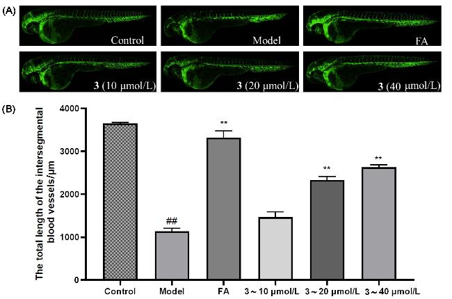


Figure 4 Influence of compounds on intersegmental vessels in zebrafish(A) Typical images of intersegmental vessels in zebrafish; (B) Quantitative analysis of intersegmental vessel length in zebrafish. Data represented as mean±SEM. Compared with the blank control group, ##: extremely significant difference (P<0.01); compared with the model group, **: extremely significant difference (P<0.01). Model, 0.2 µg/mL PTK787; FA, 40 μmol/L ferulic acid. |
3 Conclusions
This study investigates the marine-derived fungus Cladosporium sp. MDW-211, isolated from crab samples collected on Weizhou Island, Guangxi. One new dihydroisocoumarin (compound 1) and three known dihydroisocoumarins (compounds 2~4) were isolated from its rice fermentation. The absolute configuration of compound 1 was determined by single-crystal X-ray diffraction. Biological evaluation using a zebrafish model revealed that compound 3 exhibited significant pro-angiogenic activity at concentrations of 20 and 40 μmol/L.
4 Experimental section
4.1 General experimental procedures
IR spectra were obtained on a MAGNA-1R550 spectrophotometer in KBr discs. UV spectra were recorded on an Agilent Cary 60. Optical rotations were measured on a WZZ-2S/2SS digital polarimeter. NMR spectra were recorded on a Bruker Avance 400 spectrometer (Bruker, Fallanden, Switzerland), HR-ESI-MS spectra were obtained using a Mariner API-TOF. Semi-preparative high performance liquid chromatography (HPLC) was performed on a Waters 1525 system using a semi-prepara- tive C18 (Cosmosil, 10 ID×250 mm) column coupled with a Varian 330 detector.
4.2 Fungal materials
Cladosporium sp. MDW-211 was isolated from a crab sample collected at the Weizhou Island (N21°1′51′′, E109°7′55′) and identified by the 18S rDNA gene se-quence (GenBank accession No. PV849071).[15]
4.3 Fermentation, extraction and isolation
Cladosporium sp. MDW-211 was cultured at 28 ℃ for 4 d. Spores were cultivated in 1000 mL×3 Erlenmeyer flasks (120 mL seawater, 80 g rice). The flasks were cultured under static conditions for 30 d at room temperature. The culture was extracted with EtOAc to yield 581 mg. The extracts was further purified by semipreparative HPLC on an octadecylsilyl (ODS) column, eluting with 50% MeCN/H2O (10%~50%, 0.1% TFA, 10 mL/min) to yield compounds 1 (tR=13.6 min, 30.2 mg), 2 (tR=10.6 min, 6.8 mg), 3 (tR=18.4 min, 7.8 mg) and 4 (tR=34.4 min, 60.8 mg).
4'S-Hydroxyasperentin (1): Yellow brown crystal, m.p. 163.2~164.1 ℃; $[\alpha]_{\mathrm{D}}^{25}$-180.0 (c 0.3, MeOH); UV (MeOH) λmax [log ε/(L•mol-1•cm-1)]: 215 (1.97), 230 (1.10), 270 (1.34), 300 (0.57) nm; 1H NMR (400 MHz, CD3OD) and 13C NMR (100 MHz, CD3OD) see Table 1; IR (KBr) νmax: 3409, 2942, 1653, 1454, 1030 cm-1. Crystal data of 1: C16H20O6, Mr=308.334, orthorhombic, space group P212121 with a=0.86821(7) nm, b=1.22837(9) nm, c=1.38523(11) nm, α=β=γ=90°, V=1.4773(2) nm³, Z=4, Dcalcd=1.386 g/cm3, μ(Cu Kα)=0.887 mm-1; crystal size 0.20 mm×0.16 mm×0.08 mm, T=185.0 K. A total of 14938 reflections were measured, and 2914 [R(int)=0.0596, R(sigma)=0.0377] of them were used in the calculations. The final R1 was 0.0312 [I>2σ(I)], and the wR2 was 0.0850 (all data). Flack parameter 0.06(14).
4'-Hydroxyasperentin (2): White amorphous powder. 1H NMR (400 MHz, CD3OD) δ: 6.23 (d, J=2.3 Hz, 1H, H-7), 6.21 (d, J=2.3 Hz, 1H, H-5), 4.58~4.68 (m, 1H, H-3), 4.35~4.42 (m, 1H, H-2'), 3.90~4.00 (m, 1H, H-4'), 3.73~3.82 (m, 1H, H-6'), 2.85~3.00 (overlap, 2H, H-4), 2.24~2.34 (m, 1H, H-9a), 1.90~1.96 (m, 1H, H-5'a), 1.74~1.86 (m, 2H, H-9b, 3'a), 1.58~1.68 (m, 1H, H-3'b), 1.28~1.36 (m, 1H, H-5'b), 1.17 (d, J=6.2 Hz, 3H, H-7'); 13C NMR (100 MHz, CD3OD) δ: 171.3 (C-1), 166.3 (C-6), 165.6 (C-8), 143.5 (C-4a), 107.9 (C-7), 102.2 (CH, C-5), 101.6 (C-8a), 77.9 (CH, C-3), 70.0 (CH, C-2'), 66.3 (CH, C-6'), 64.7 (CH, C-4'), 43.6 (CH2, C-5'), 39.1 (CH2, C-3'), 37.3 (CH2, C-9), 34.2 (CH₂, C-4), 22.1 (CH3, C-7'). LR-ESI-MS calcd for C16H20O6Na [M+Na]+ 331.1, found 331.6.
Cladomarine (3): White amorphous powder. 1H NMR (400 MHz, CD3OD) δ: 6.27 (s, 1H, H-7), 4.57~4.65 (m, 1H, H-3), 4.10~4.18 (m, 1H, H-2'), 3.87~3.96 (m, 1H, H-6'), 3.17 (dd, J=16.8, 3.4 Hz, 1H, H-4a), 2.66 (dd, J=16.8, 11.2 Hz, 1H, H-4b), 2.15 (ddd, J=14.7, 11.0, 3.7 Hz, 1H, H-9a), 1.79 (ddd, J=14.7, 9.1, 3.2 Hz, 1H, H-9b), 1.71~1.75 (m, 1H, H-3'a), 1.62~1.71 (m, 3H, H-4'a, 4'b, 5'b), 1.28~1.36 (m, 2H, H-3'b, 5'a), 1.18 (d, J=6.2 Hz, 3H, H-7'); 13C NMR (100 MHz, CD3OD) δ: 171.9 (C-1), 159.0 (C-8), 155.5 (C-6), 135.8 (C-5), 126.1 (C-4a), 101.9 (CH, C-7), 100.2 (C-8a), 77.7 (CH, C-3), 68.4 (CH, C-2', 6'), 39.4 (CH, C-9), 32.7 (CH2, C-5'), 31.4 (CH2, C-3'), 28.5 (CH2, C-4), 20.0 (CH3, C-7'), 19.3 (CH2, C-4'). LR-ESI-MS calcd for C16H21O6 [M+H]+ 309.1, found 309.6.
Asperentin (4): White amorphous powder. 1H NMR (400 MHz, CD3OD) δ: 6.22 (d, J=2.3 Hz, 1H, H-5), 6.20 (d, J=2.3 Hz, 1H, H-7), 4.60~4.70 (m, 1H, H-3), 4.10~4.18 (m, 1H, H-2'), 3.88~3.96 (m, 1H, H-6'), 2.81~2.97 (m, 2H, H-4), 2.12 (ddd, J=14.3, 10.4, 3.4 Hz, 1H, H-9a), 1.63~1.81 (overlap, 5H, H-9b, 3', 4'), 1.79 (ddd, J=14.7, 9.1, 3.2 Hz, 1H, H-9b), 1.71~1.75 (m, 1H, H-3'a), 1.62~1.71 (m, 3H, H-4'a, 4'b, 5'b), 1.27~1.45 (m, 2H, H-5'), 1.18 (d, J=6.4 Hz, 3H, H-7'); 13C NMR (100 MHz, CD3OD) δ: 171.4 (C-1), 166.2 (C-6), 165.6 (C-8), 143.5 (C-4a), 107.9 (C-5), 102.2 (CH, C-7), 101.6 (C-8a), 77.8 (CH, C-3), 68.4 (CH, C-6'), 68.3 (CH, C-2'), 39.1 (CH2, C-9), 34.3 (CH2, C-4), 32.7 (CH2, C-5'), 31.4 (CH2, C-3'), 20.0 (CH3, C-7'), 19.3 (CH2, C-3'). LR-ESI-MS calcd for C16H21O5 [M+H]+ 293.1, found 293.4.
4.4 Pro-angiogenic activity assay
The experiment was completed in the Engineering Research Center of Zebrafish Models for Human Diseases and Drug Screening of Shandong Province.[16] The experiment procedures were conducted according to the standard ethical guidelines that were approved by the Ethics Committee of the Biology Institute of Shandong Academy of Science (SWS20240204). Based on the transgenic Tg (fli-1: EGFP) zebrafish model, the proangiogenic activity of the isolated compounds was tested. Firstly, zebrafish eggs developed at (28±0.5) ℃ for 20~24 h were collected, and 1 mg/mL Pronase E was added to remove the egg membrane to obtain zebrafish embryos. 2 mL of fish culture water, 0.2 µg/mL PTK787, test samples (10, 20, 40 μmol/L), and 10 zebrafish embryos were added to a 24 well plate in sequence as the experimental group. The system with only fish culture water and zebrafish embryos added was used as the normal control group, the system with fish culture water, PTK787, and zebrafish embryos added was used as the model group, and the system with fish culture water, 40 μmol/L ferulic acid, PTK787, and zebrafish embryos added was used as the positive drug group. Each group had three parallel experiments. After culturing the experimental system at (28±0.5) ℃ for 24 h, the growth of intersegmental vessels in zebrafish was observed using an OLYMPUS fluorescence microscope, and the proangiogenic activity of each sample was determined by Image-Pro Plus 5.1 and GraphPad Prism 10.
Supporting Information HR-ESI-MS, 1D NMR and 2D NMR, IR, UV spectra, and X-ray crystallographic information file (CIF) of compound 1. The Supporting Information is available free of charge via the Internet at http://sioc-journal.cn/.
(Lu, Y.)