
荧光可调控的碳量子点的电化学制备及性质研究
收稿日期: 2013-10-09
网络出版日期: 2013-12-25
基金资助
项目受国家自然科学基金(Nos. 21073018,21233003)资助.
Electrochemical Preparation of Color-Tunable Fluorescent Carbon Quantum Dots
Received date: 2013-10-09
Online published: 2013-12-25
Supported by
Project supported by the National Natural Science Foundation of China (Nos. 21073018, 21233003).
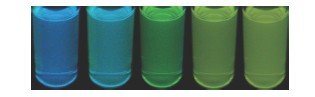
采用循环伏安法(Cyclic Voltammetry,CV)在碱性条件下电解石墨棒,得到水溶性的荧光碳量子点. 通过透射电子显微镜(TEM)、拉曼光谱(Raman spectrum)、原子力显微镜(AFM)对所制备的碳量子点进行形貌及结构表征,发现该碳量子点由1~4层石墨烯片层堆积形成,粒径在19 nm左右,厚度在1 nm左右. 通过荧光光谱(PL)、紫外可见吸收光谱(UV-vis)、傅里叶变换红外光谱(FTIR)、X射线光电子能谱(XPS)对所制备的碳量子点进行性质测定,发现该碳量子点在400和525 nm处有两个荧光发射峰,且通过控制扫描周数可以调节两个发射峰的相对强度,从而调控碳量子点的荧光颜色:随着扫描周数的增加,400 nm处发射峰的相对强度逐渐减小,而525 nm处发射峰的相对强度逐渐增大,两个荧光发射峰分别与碳量子点的π-π共轭体系和含氧官能团的n-π共轭体系有关.
李腾飞 , 李昳玮 , 肖璐 , 余洪涛 , 范楼珍 . 荧光可调控的碳量子点的电化学制备及性质研究[J]. 化学学报, 2014 , 72(2) : 227 -232 . DOI: 10.6023/A13101036
Carbon quantum dots have attracted much attention in the fields of bioimaging, biolabeling and drug delivery. Theoretical and experimental studies have shown that carbon quantum dots are expected to show unique optical properties due to their quantum confinement and edge effect. In this report, water-soluble and color-tunable fluorescent carbon dots were prepared by cyclic voltammetry (CV) in alkaline condition. The structure of the carbon dots was confirmed by means of transmission electron microscope (TEM), Raman spectrum and atomic force microscope (AFM). The finding shows that the carbon quantum dots have a uniform diameter around 19 nm, and are mainly consist of 1 to 4 layers of graphene with a mass of oxygen-containing functional groups. Their features and properties were characterized by photoluminescence spectra (PL), UV-visible spectroscopy (UV-vis), fourier transform infrared spectroscopy (FTIR), and X-ray photoelectron spectroscopy (XPS). The results indicated that the carbon quantum dots have two fluorescent emission peaks at 400 nm and 525 nm. The former peak was associated with the π-π conjugated system of carbon dots, which showed an excitation-wavelength dependent feature, while the latter peak was associated with n-π conjugated system of oxygen-containing functional groups, which remained unshifted when excited by different excitation wavelengths, suggesting a novel kind of fluorescent feature and mechanism different from those of previously reported carbon quantum dots depending on excitation wavelengths. The fluorescent color of carbon quantum dots could be controlled by the cycles of CV. As the increase of CV cycles, the relative content of oxygen-containing functional groups increased, leading to the decrease of the relative intensity of fluorescent peaks at 400 nm and the increase of the relative intensity of fluorescent peak at 525 nm; and the color of the fluorescent changes from blue to yellow. The color-tunable fluorescent carbon quantum dots showed a high water solubility and good photostability, which indicates that the carbon quantum dots might be used as a bioimaging marker in cell imaging.
Key words: electrochemistry; fluorescence; carbon quantum dots
[1] Terasaki, M. Methods Cell Biol. 1989, 29, 125.
[2] Jaiswal, J. K.; Goldman, E. R.; Mattoussi, H.; Simon, S. M. Nature Methods 2004, 1, 73.
[3] Xie, W. J.; Fu, Y. Y.; Ma, H.; Zhang, M.; Fan, L. Z. Acta Chim. Sinica 2012, 70, 2169. (谢文菁, 傅英懿, 马红, 张沫, 范楼珍, 化学学报, 2012, 70, 2169.)
[4] Zhu, J.; Liao, L.; Zhu, L. N.; Kong, J. L.; Liu, B. H. Acta Chim. Sinica 2012, 71, 69. (朱杰, 廖蕾, 朱丽娜, 孔继烈, 刘宝红, 化学学报, 2012, 71, 69.)
[5] Faklaris, O.; Joshi, V.; Irinopoulou, T.; Tauc, P.; Sennour, M.; Girard, H.; Gesset, C. l.; Arnault, J.-C.; Thorel, A.; Boudou, J.-P. ACS nano 2009, 3, 3955.
[6] Riggs, J. E.; Guo, Z.; Carroll, D. L.; Sun, Y.-P. J. Am. Chem. Soc. 2000, 122, 5879.
[7] Yifeng, E.; Bai, L.; Fan, L.; Han, M.; Zhang, X.; Yang, S. J. Mater. Chem. 2011, 21, 819.
[8] Hu, S.-L.; Niu, K.-Y.; Sun, J.; Yang, J.; Zhao, N.-Q.; Du, X.-W. J. Mater. Chem. 2009, 19, 484.
[9] Ray, S.; Saha, A.; Jana, N. R.; Sarkar, R. J. Phys. Chem. C 2009, 113, 18546.
[10] Zhou, J.; Booker, C.; Li, R.; Zhou, X.; Sham, T.-K.; Sun, X.; Ding, Z. J. Am. Chem. Soc. 2007, 129, 744.
[11] Li, Y.; Hu, Y.; Zhao, Y.; Shi, G.; Deng, L.; Hou, Y.; Qu, L. Adv. Mater. 2011, 23, 776.
[12] Chien, C. T.; Li, S. S.; Lai, W. J.; Yeh, Y. C.; Chen, H. A.; Chen, I.; Chen, L. C.; Chen, K. H.; Nemoto, T.; Isoda, S. Angew. Chem., Int. Ed. 2012, 51, 6662.
[13] Kudin, K. N.; Ozbas, B.; Schniepp, H. C.; Prud'Homme, R. K.; Aksay, I. A.; Car, R. Nano Lett. 2008, 8, 36.
[14] Zhao, Q.-L.; Zhang, Z.-L.; Huang, B.-H.; Peng, J.; Zhang, M.; Pang, D.-W. Chem. Commun. 2008, 5116.
[15] Liu, N.; Luo, F.; Wu, H.; Liu, Y.; Zhang, C.; Chen, J. Adv. Funct. Mater. 2008, 18, 1518.Lu, J.; Yang, J.-X.; Wang, J.; Lim, A.; Wang, S.; Loh, K. P. ACS nano 2009, 3, 2367.
/
| 〈 |
|
〉 |
