Acta Chimica Sinica ›› 2021, Vol. 79 ›› Issue (1): 87-92.DOI: 10.6023/A20080399 Previous Articles Next Articles
Article
任江波a, 王蕾b, 郭锐a, 唐永和a, 周红梅a, 林伟英a,c,*( )
)
投稿日期:2020-08-31
发布日期:2020-10-17
通讯作者:
林伟英
作者简介:基金资助:
Jiangbo Rena, Lei Wangb, Rui Guoa, Yonghe Tanga, Hongmei Zhoua, Weiying Lina,c,*()
Received:2020-08-31
Published:2020-10-17
Contact:
Weiying Lin
Supported by:Share
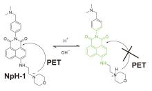
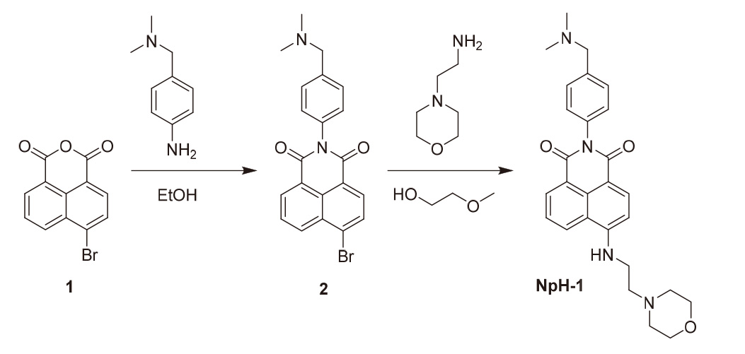
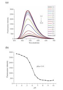
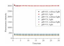
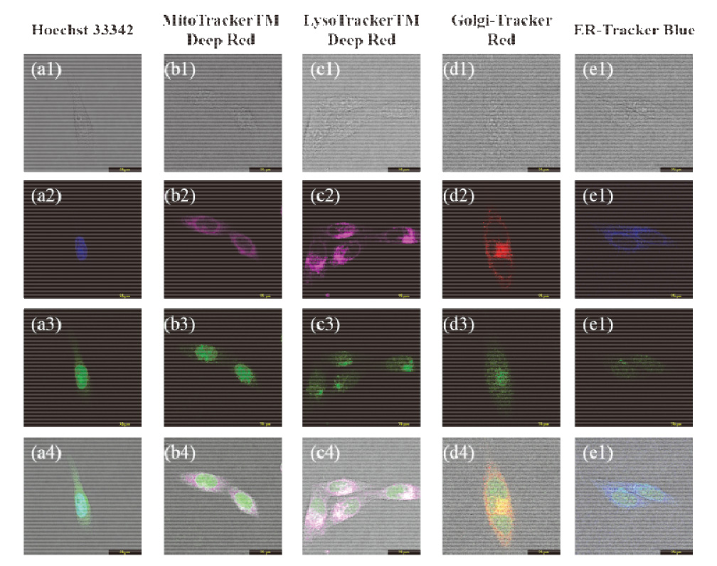
Jiangbo Ren, Lei Wang, Rui Guo, Yonghe Tang, Hongmei Zhou, Weiying Lin. A Naphthalimide-Based Fluorescent Probe for Detecting Intracellular pH and Its Biological Imaging Application[J]. Acta Chimica Sinica, 2021, 79(1): 87-92.


| [1] |
Xu. Xu. K.; Klibanov A. M. J. Am. Chem. Soc. 1996, 118 (1), 9815.
|
| [2] |
Tian H.; Gan J.; Chen K.; He J.; Song Q.L.; Hou X. Y. J. Mater. Chem . 2002, 12 ( 5),1262.
|
| [3] |
Burleigh S. C.; van de Laar T.; Stroop C. J.; van Grunsven W. M.; O'Donoghue N.; Rudd P. M.; Davey G. P. BMC Biotechnol. 2011, 11(1), 95.
|
| [4] |
Zhou X.; Su F.; Lu H.; Senechal-Willis P.; Tian Y.; Johnson R. H.; Meldrum D. R. Biomaterials 2012, 33(1), 171.
|
| [5] |
Luzio J. P.; Pryor P. R.; Bright N. A. Nat. Rev. Mol. Cell Bio. 2007, 8 ( 8),622.
|
| [6] |
Reinheckel T.; Deussing J.; Roth W.; Peters C. Biol. Chem. 2001, 382 ( 5),735.
|
| [7] |
Watts C. Biochim. Biophys. Acta, Proteins Proteomics 2012, 1824(1), 14.
|
| [8] |
Zhao H. Traffic 2012, 13(10), 1307.
|
| [9] |
Mohamed M. M.; Sloane B. F. Nat. Rev. Cancer 2006, 6(10), 764.
|
| [10] |
Groth-Pedersen L.; Jaattela M. Cancer Lett. 2013, 332(2), 265.
|
| [11] |
Cirman T.; Oresic K.; Mazovec G. D.; Turk V.; Reed J. C.; Myers R. M.; Salvesen G. S.; Turk B. J. Biol. Chem. 2004, 279 ( 5),3578.
|
| [12] |
Hesse S. J.A.; Ruijter G. J.G.; Dijkema C.; Visser J. J. Biotechnol. 2000, 77(1), 5.
|
| [13] |
Schartner E. P.; Henderson M. R.; Purdey M.; Dhatrak D.; Monro T. M.; Gill P. G.; Callen D. F. Cancer Res. 2016, 76 ( 23),6795.
|
| [14] |
Newport J. W.; Forbes D. J. Annu. Rev. Biochem. 1987, 56(1), 535.
|
| [15] |
Shen W. H.; Balajee A. S.; Wang J.; Wu H.; Eng C.; Pandolfi P. P.; Yin Y. Cell 2007, 128(1), 157.
|
| [16] |
Zhou C. M.S. Thesis, Liaoning University, Shen-yang , 2013.
|
|
(周超, 硕士论文, 辽宁大学, 沈阳,2013). (in Chinese)
|
|
| [17] |
Singha S.; Kim D.; Seo H.; Cho S.W.; Ahn K. H. Chem. Soc. Rev. 2015, 44 ( 13),4367.
|
| [18] |
Li X.; Gao X.; Shi W.; Ma H. Chem. Rev. 2014, 114(1), 590.
|
| [19] |
Lv H.; Yang X.F.; Zhong Y.; Guo Y.; Li Z.; Li H. Anal. Chem. 2014, 86 ( 3),1800.
|
| [20] |
Carter K. P.; Young A. M.; Palmer A. E. Chem. Rev. 2014, 114 ( 8),4564.
|
| [21] |
Li L.; Li P.; Fang J.; Li Q.; Xiao H.; Zhou H.; Tang B. Anal. Chem. 2015, 87(12), 6057.
|
| [22] |
Sun R.; Liu W.; Xu Y.; Lu J.; Ge J.; Ihara M. Chem. Commun. 2013, 49(91), 10709.
|
| [23] |
Zhang Y.; Fang H.; Zhang X.; Wang S.; Xing G. ChemistrySelect 2016, 1(1), 1.
|
| [24] |
Wang, X.L.; Li, X.J.; Sun, R.; Xu, Y.J.; Ge J. F. Analyst. 2016, 141(10), 2962.
|
| [25] |
Song G. J.; Bai S.Y.; Luo J.; Cao X. Q.; Zhao B. X. J. Fluoresc . 2016, 26 ( 6),2079.
|
| [26] |
Zhang S.; Chen T.; Lee H.; Bi J.; Ghosh A.; Fang M.; Qian Z.; Xie F.; Ainsley J.; Christov C.; Luo F.; Zhao F.; Liu H. ACS Sensors 2017, 2(7), 924.
|
| [27] |
Zhang, Y; Wang, Z.-L; Tao,Y; Xu-X; Fang-H; Wang,S.-F. Chin. J. Org. Chem. 2018, 38(10), 2693 . (in Chinese)
|
|
张燕, 王忠龙, 陶钰, 徐徐, 方华, 王石发, 有机化学, 2018, 38(10), 2693.
|
|
| [28] |
Xiang, D.-C; Liu, H; Meng, Q.-H; Lan, M.-B; Wei, G. Acta Chim. Sinica 2013, 71(10), 1435 . (in Chinese)
|
|
向德成, 刘恒, 孟庆华, 蓝闽波, 卫钢, 化学学报, 2013, 71(10), 1435.
|
|
| [29] |
Wu, P.-Q; Liang, C.-H. Int. J. Med. Radiol. 2016, 39(04), 410 . (in Chinese)
|
|
吴佩琪, 梁长虹. 国际医学放射学杂志, 2016, 39(04), 410.
|
|
| [30] |
Guan, X.-L; Li, Z.-F; Wang, L; Liu, M.-N; Wang, K.-L; Yang, X.-Q; Li, Y.-L; Hu, L.-L; Zhao, X.-L; Lai, S.-J; Lei, Z.-Q. Acta Chim. Sinica 2019, 77(12), 1268 . (in Chinese)
|
|
关晓琳, 李志飞, 王林, 刘美娜, 王凯龙, 杨学琴, 李亚丽, 胡丽丽, 赵小龙, 来守军, 雷自强, 化学学报, 2019, 77(12), 1268.
|
|
| [31] |
Ma, Y; Chen, K.-X; Guo, Z.-L; Liu, S.-J; Zhao, Q; Huang, W.-Y. Acta Chim. Sinica 2020, 78(01), 23 . (in Chinese)
|
|
马云, 陈可欣, 郭则灵, 刘淑娟, 赵强, 黄维扬, 化学学报, 2020, 78(01), 23.
|
|
| [32] |
Li, Y; Zhan, H; Li, M. New Chem. Mater. 2019, 47(08), 135 . (in Chinese)
|
|
李阳, 张辉, 李明, 化工新型材料, 2019, 47(08), 135.
|
|
| [33] |
Liu Y.; Zhou J.; Wang L.; Hu X.; Liu X.; Liu M.; Cao Z.; Shangguan D.; Tan W. J. Am. Chem. Soc. 2016, 138 ( 38),12368.
|
| [34] |
Cao, X.-J; Chen, L.-N; Zhang, X; Liu, J.-T; Chen, M.-Y; Wu, Q.-R; Miao, J.-Y; Zhao, B.-X. Anal. Chim. Acta 2016, 920, 86.
|
| [1] | Jianqiang Chen, Gangguo Zhu, Jie Wu. Recent Advances in Nickel-Catalyzed Ring Opening Cross-Coupling of Aziridines [J]. Acta Chimica Sinica, 2024, 82(2): 190-212. |
| [2] | Cheng-Qiang Wang, Chao Feng. Applications of Nucleophilic Fluorine Sources in the Selective Fluorofunctionalization of Unsaturated Carbon-Carbon Bonds [J]. Acta Chimica Sinica, 2024, 82(2): 160-170. |
| [3] | Jinglin Yi, Mao Chen. Photo-Induced Copolymerization of Chlorotrifluoroethylene and Methyl Isopropenyl Ether★ [J]. Acta Chimica Sinica, 2024, 82(2): 126-131. |
| [4] | Zhiqiang Wang, Jinzhan Su. Investigation of the Kinetic Properties and Photoelectrochemical Water Splitting of Cu3V2O8/ZnO Photoanode Modified by Cobalt Phosphate [J]. Acta Chimica Sinica, 2024, 82(1): 26-35. |
| [5] | Chenyu Liao, Shanwei Guo, Meiwei Huang, Yong Guo, Qing-Yun Chen, Chao Liu, Yunwen Zhang. Synthesis and Properties of Fluoroether Phosphocholine [J]. Acta Chimica Sinica, 2024, 82(1): 46-52. |
| [6] | Yuqing Shi, Mingzhu Chu, Bo Han, Haojie Ma, Ran Li, Xueyan Hou, Yuqi Zhang, Ji-Jiang Wang. Smart Two-dimensional Photonic Crystal Hydrogel for Accurate Detection of Hg2+ [J]. Acta Chimica Sinica, 2024, 82(1): 9-15. |
| [7] | Ruxin Zeng, Peng R. Chen. RNA-Binding Proteome Analysis and Functional Explorations★ [J]. Acta Chimica Sinica, 2024, 82(1): 53-61. |
| [8] | Ye Tian, Duanhui Si, Shuiying Gao, Rong Cao. Ultra-Long Organic Room Temperature Phosphorescence of Phthalic Acid Derivative Modified Polymer★ [J]. Acta Chimica Sinica, 2023, 81(9): 1129-1134. |
| [9] | Yuhan Wu, Dongdong Zhang, Hongyu Yin, Zhengnan Chen, Wen Zhao, Yuhua Chi. Density Functional Theory Study of Janus In2S2X Photocatalytic Reduction of CO2 under “Double Carbon” Target [J]. Acta Chimica Sinica, 2023, 81(9): 1148-1156. |
| [10] | Fengjie Ge, Kaizhi Zhang, Qingpeng Cao, Hui Xu, Tao Zhou, Wenhao Zhang, Xinxin Ban, Xiaobo Zhang, Na Li, Peng Zhu. Design, Synthesis and Electroluminescence Performance of Flexible Fluorenyl Block Delayed Fluorescence Dimers [J]. Acta Chimica Sinica, 2023, 81(9): 1157-1166. |
| [11] | Jiao Kong, Lin Du, Xiangyang Li, Jidong Zhu, Ya-Qiu Long. Small Molecule Degraders Targeting the SHP2E76A Mutant Effectively Inhibiting the Proliferation of Wild-type and Mutant SHP2 Dependent Tumor Cells [J]. Acta Chimica Sinica, 2023, 81(9): 1120-1128. |
| [12] | Yuchun Han, Yilin Wang. Retrospect and Prospect of Long-lasting Antibacterial Materials★ [J]. Acta Chimica Sinica, 2023, 81(9): 1196-1201. |
| [13] | Ruxin Tian, Miao Yang, Guo Chen, Jiangshan Liu, Mengmei Yuan, Hong Yuan, Shuxin Ouyang, Tierui Zhang. Ru/Quartz Filter Paper: A Recyclable Photothermocatalytic Film for CO2 Methanation★ [J]. Acta Chimica Sinica, 2023, 81(8): 869-873. |
| [14] | Mei Hong, Jinqiang Gao, Tong Li, Shihe Yang. In-situ Etching Strategy for Manipulation of Hierarchical Zeolite and Its Application★ [J]. Acta Chimica Sinica, 2023, 81(8): 937-948. |
| [15] | Yongxue Li, Yu Liu. Supramolecular Secondary Assembly Based on Amphiphilic Calix[4]arenes and Its Biological Applications★ [J]. Acta Chimica Sinica, 2023, 81(8): 928-936. |
| Viewed | ||||||
|
Full text |
|
|||||
|
Abstract |
|
|||||
