化学学报 ›› 2023, Vol. 81 ›› Issue (8): 990-1001.DOI: 10.6023/A23040166 上一篇 下一篇
所属专题: 庆祝《化学学报》创刊90周年合辑
综述
杨宇洁a, 巩宇锈a, 顾天航a,*( ), 张伟贤a,b,*()
投稿日期:2023-04-26
发布日期:2023-09-14
作者简介: |
杨宇洁, 同济大学环境科学与工程学院2021级硕士生, 研究方向为纳米零价铁环境应用. |
 |
巩宇锈, 同济大学环境科学与工程学院2017级博士, 研究方向为纳米零价铁修复重金属污染土壤. |
 |
顾天航, 同济大学环境科学与工程学院2018级博士生, 研究方向为纳米零价铁富集水中稀有元素, 已发表SCI论文10余篇. |
 |
张伟贤, 教授、博士生导师, 国家特聘专家, 2011年起任污染控制与资源化研究国家重点实验室主任. 1984年毕业于同济大学, 1996年获美国约翰•霍普金斯大学(The Johns Hopkins University)环境工程博士学位, 曾任美国里海大学(Lehigh University)教授. 主持过国家自然科学基金海外及港澳学者合作研究基金及多项国家自然科学基金项目. 长期致力于环境中重金属及持久性有机污染物的基础与应用研究, 是环境纳米技术的先驱之一, 纳米零价铁技术的创始研究者. 在纳米零价铁合成、表征、污染物反应机理、应用于地下水修复及废水处理方面发表了系列经典论文. |
基金资助:
Yujie Yanga, Yuxiu Gonga, Tianhang Gua(), Wei-xian Zhanga,b()
Received:2023-04-26
Published:2023-09-14
Contact:
*E-mail: tianhanggu@tongji.edu.cn; zhangwx@tongji.edu.cn
About author:Supported by:文章分享

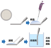
电子显微镜是表征微观粒子结构的必要手段, 但预处理过程会对含水样品结构造成破坏, 导致结果失真. 冷冻电子显微镜(Cryo-EM)技术通过将样品在低温下快速冷冻, 使水合样品处于无定形玻璃态冰层内, 在保障高真空度要求的同时减少电子辐射对样品的破坏, 目前已成为高分辨地观察含水样品原始状态下结构最先进的手段, 在生物、材料等领域应用广泛. 本综述总结了冷冻电镜在电子元件、成像技术与分辨率等方面的进展, 综述了样品制备方法及三维重构技术. 鉴于绝大多数环境样品为含水样品, 冷冻电镜技术在大气、水体、土壤等环境介质中的应用扩宽和深化了对环境中微纳米粒子本身形态结构和粒子间相互作用关系的理解. 我们期待冷冻电镜为环境科学研究带来突破性贡献.
杨宇洁, 巩宇锈, 顾天航, 张伟贤. 冷冻电子显微镜技术进展及环境研究应用★[J]. 化学学报, 2023, 81(8): 990-1001.
Yujie Yang, Yuxiu Gong, Tianhang Gu, Wei-xian Zhang. Progress and Environmental Research Applications of Cryo-Electron Microscopy★[J]. Acta Chimica Sinica, 2023, 81(8): 990-1001.
| 冷冻电镜种类 | 原理 | 代表性研究 | 参考文献 |
|---|---|---|---|
| 冷冻透射电子显微镜 | 将平行电子束投射到样品上, 电子与样品中的 原子碰撞散射, 从而形成投影图像 | 金、银纳米颗粒 | [ |
| 噬菌体的蛋白质外壳 | [ | ||
| 蒙脱石-二氧化硅悬浊液 | [ | ||
| 气溶胶颗粒 | [ | ||
| MS2噬菌体 | [ | ||
| 冷冻扫描电子显微镜 | 利用高能电子束扫描样品, 在其表面形成二次电子、 背散电子等, 从而转换产生图像 | 石油工业中的乳液和悬浮液 | [ |
| A. Borkumensis细菌生物膜 | [ | ||
| 乳胶/陶瓷纳米颗粒涂层 | [ | ||
| 海藻酸盐水凝胶 | [ | ||
| 活性污泥 | [ | ||
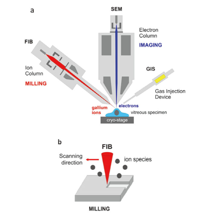
| 冷冻聚焦离子束扫描 电子显微镜 | 利用聚焦离子束扫描样品得到图像 | 枯草芽孢杆菌孢子 | [ |
| 新型冠状病毒 | [ | ||
| 黏土的孔隙结构 | [ | ||
| 冷冻扫描透射电子显 微镜 | 利用聚焦后的电子束斑逐点扫描样品, 在样品下方收 集散射或透射电子, 从而形成明、暗场像 | 铁蛋白 | [ |
| 锂金属电池中固液界面和枝晶 的结构 | [ |
| 冷冻电镜种类 | 原理 | 代表性研究 | 参考文献 |
|---|---|---|---|
| 冷冻透射电子显微镜 | 将平行电子束投射到样品上, 电子与样品中的 原子碰撞散射, 从而形成投影图像 | 金、银纳米颗粒 | [ |
| 噬菌体的蛋白质外壳 | [ | ||
| 蒙脱石-二氧化硅悬浊液 | [ | ||
| 气溶胶颗粒 | [ | ||
| MS2噬菌体 | [ | ||
| 冷冻扫描电子显微镜 | 利用高能电子束扫描样品, 在其表面形成二次电子、 背散电子等, 从而转换产生图像 | 石油工业中的乳液和悬浮液 | [ |
| A. Borkumensis细菌生物膜 | [ | ||
| 乳胶/陶瓷纳米颗粒涂层 | [ | ||
| 海藻酸盐水凝胶 | [ | ||
| 活性污泥 | [ | ||
| 冷冻聚焦离子束扫描 电子显微镜 | 利用聚焦离子束扫描样品得到图像 | 枯草芽孢杆菌孢子 | [ |
| 新型冠状病毒 | [ | ||
| 黏土的孔隙结构 | [ | ||
| 冷冻扫描透射电子显 微镜 | 利用聚焦后的电子束斑逐点扫描样品, 在样品下方收 集散射或透射电子, 从而形成明、暗场像 | 铁蛋白 | [ |
| 锂金属电池中固液界面和枝晶 的结构 | [ |


| [1] |
Ball P. Chem. Rev. 2008, 108, 74.
doi: 10.1021/cr068037a |
| [2] |
Ball P. Proc. Natl. Acad. Sci. U. S. A. 2017, 114, 13327.
doi: 10.1073/pnas.1703781114 |
| [3] |
Yao H. P.; Song Y. T.; Chen Y.; Wu N. P.; Xu J. L.; Sun C. J.; Zhang J. X.; Weng T. H.; Zhang Z. Y.; Wu Z. G.; Cheng L. F.; Shi D. R.; Lu X. Y.; Lei J. L.; Crispin M.; Shi Y. G.; Li L. J.; Li S. Cell 2020, 183, 730.
doi: 10.1016/j.cell.2020.09.018 |
| [4] |
Adrian M.; Dubochet J.; Lepault J.; Mcdowall A. W. Nature 1984, 308, 32.
doi: 10.1038/308032a0 |
| [5] |
Mendonca L.; Howe A.; Gilchrist J. B.; Sheng Y. W.; Sun D. P.; Knight M. L.; Zanetti-Domingues L. C.; Bateman B.; Krebs A. S.; Chen L.; Radecke J.; Li V. V. D.; Ni T.; Kounatidis I.; Koronfel M. A.; Szynkiewicz M.; Harkiolaki M.; Martin-Fernandez M. L.; James W.; Zhang P. J. Nat. Commun. 2021, 12, 4629.
doi: 10.1038/s41467-021-24887-y |
| [6] |
Fernandez-Leiro R.; Scheres S. H. Nature 2016, 537, 339.
doi: 10.1038/nature19948 |
| [7] |
Fitzpatrick A. W. P.; Falcon B.; He S.; Murzin A. G.; Murshudov G.; Garringer H. J.; Crowther R. A.; Ghetti B.; Goedert M.; Scheres S. H. W. Nature 2017, 547, 185.
doi: 10.1038/nature23002 |
| [8] |
Shi Y.; Murzin A. G.; Falcon B.; Epstein A.; Machin J.; Tempest P.; Newell K. L.; Vidal R.; Garringer H. J.; Sahara N.; Higuchi M.; Ghetti B.; Jang M. K.; Scheres S. H. W.; Goedert M. Acta Neuropathol. 2021, 141, 697.
doi: 10.1007/s00401-021-02294-3 |
| [9] |
Falcon B.; Zhang W. J.; Murzin A. G.; Murshudov G.; Garringer H. J.; Vidal R.; Crowther R. A.; Ghetti B.; Scheres S. H. W.; Goedert M. Nature 2018, 561, 137.
doi: 10.1038/s41586-018-0454-y |
| [10] |
Falcon B.; Zivanov J.; Zhang W. J.; Murzin A. G.; Garringer H. J.; Vidal R.; Crowther R. A.; Newell K. L.; Ghetti B.; Goedert M.; Scheres S. H. W. Nature 2019, 568, 420.
doi: 10.1038/s41586-019-1026-5 |
| [11] |
Zhang W. J.; Tarutani A.; Newell K. L.; Murzin A. G.; Matsubara T.; Falcon B.; Vidal R.; Garringer H. J.; Shi Y.; Ikeuchi T.; Murayama S.; Ghetti B.; Hasegawa M.; Goedert M.; Scheres S. H. W. Nature 2020, 580, 283.
doi: 10.1038/s41586-020-2043-0 |
| [12] |
Zhu K. F.; Yuan C.; Du Y. M.; Sun K. L.; Zhang X. K.; Vogel H.; Jia X. D.; Gao Y. Z.; Zhang Q. F.; Wang D. P.; Zhang H. W. Mil. Med. Res. 2023, 10.
|
| [13] |
Cameroni E.; Bowen J. E.; Rosen L. E.; Saliba C.; Zepeda S. K.; Culap K.; Pinto D.; VanBlargan L. A.; De Marco A.; di Iulio J.; Zatta F.; Kaiser H.; Noack J.; Farhat N.; Czudnochowski N.; Havenar-Daughton C.; Sprouse K. R.; Dillen J. R.; Powell A. E.; Chen A.; Maher C.; Yin L.; Sun D.; Soriaga L.; Bassi J.; Silacci-Fregni C.; Gustafsson C.; Franko N. M.; Logue J.; Iqbal N. T.; Mazzitelli I.; Geffner J.; Grifantini R.; Chu H.; Gori A.; Riva A.; Giannini O.; Ceschi A.; Ferrari P.; Cippa P. E.; Franzetti-Pellanda A.; Garzoni C.; Halfmann P. J.; Kawaoka Y.; Hebner C.; Purcell L. A.; Piccoli L.; Pizzuto M. S.; Walls A. C.; Diamond M. S.; Telenti A.; Virgin H. W.; Lanzavecchia A.; Snell G.; Veesler D.; Corti D. Nature 2022, 602, 664.
doi: 10.1038/s41586-021-04386-2 |
| [14] |
Li Y. Z.; Huang W.; Li Y. B.; Pei A.; Boyle D. T.; Cui Y. Joule 2018, 2, 2167.
doi: 10.1016/j.joule.2018.08.004 |
| [15] |
Cheng D. Y.; Wynn T. A.; Wang X. F.; Wang S.; Zhang M. H.; Shimizu R.; Bai S.; Nguyen H.; Fang C. C.; Kim M. C.; Li W. K.; Lu B. Y.; Kim S. J.; Meng Y. S. Joule 2020, 4, 2484.
doi: 10.1016/j.joule.2020.08.013 |
| [16] |
Liu Y. J.; Ju Z. J.; Zhang B. L.; Wang Y.; Nai J. W.; Liu T. F.; Tao X. Y. Acc. Chem. Res. 2021, 54, 2088.
doi: 10.1021/acs.accounts.1c00120 |
| [17] |
Li Y. Z.; Wang K. C.; Zhou W. J.; Li Y. B.; Vila R.; Huang W.; Wang H. X.; Chen G. X.; Wu G. H.; Tsao Y. C.; Wang H. S.; Sinclair R.; Chiu W.; Cui Y. Matter 2019, 1, 428.
doi: 10.1016/j.matt.2019.06.001 |
| [18] |
Wang F. B.; Gnewou O.; Solemanifar A.; Conticello V. P.; Egelman E. H. Chem. Rev. 2022, 122, 14055.
doi: 10.1021/acs.chemrev.1c00753 |
| [19] |
Schrinner M.; Polzer F.; Mei Y.; Lu Y.; Haupt B.; Ballauff M.; Goldel A.; Drechsler M.; Preussner J.; Glatzel U. Macromol. Chem. Phys. 2007, 208, 1542.
doi: 10.1002/(ISSN)1521-3935 |
| [20] |
Lu Y.; Mei Y.; Schrinner M.; Ballauff M.; Moller M. W. J. Phys. Chem. C 2007, 111, 7676.
doi: 10.1021/jp070973m |
| [21] |
Bayfield O. W.; Klimuk E.; Winkler D. C.; Hesketh E. L.; Chechik M.; Cheng N. Q.; Dykeman E. C.; Minakhin L.; Ranson N. A.; Severinov K.; Steven A. C.; Antson A. A. Proc. Natl. Acad. Sci. U. S. A. 2019, 116, 3556.
doi: 10.1073/pnas.1813204116 |
| [22] |
Hilhorst J.; Meester V.; Groeneveld E.; Dhont J. K. G.; Lekkerkerker H. N. W. J. Phys. Chem. B 2014, 118, 11816.
doi: 10.1021/jp504217m |
| [23] |
Li W. J.; Teng X. M.; Chen X. Y.; Liu L.; Xu L.; Zhang J.; Wang Y. Y.; Zhang Y.; Shi Z. B. Environ. Sci. Technol. 2021, 55, 16639.
|
| [24] |
Dai X. H.; Li Z. H.; Lai M.; Shu S.; Du Y. S.; Zhou Z. H.; Sun R. Nature 2017, 541, 112.
doi: 10.1038/nature20589 |
| [25] |
Mikula R. J.; Munoz V. A. Colloids Surf. A 2000, 174, 23.
doi: 10.1016/S0927-7757(00)00518-5 |
| [26] |
Omarova M.; Swientoniewski L. T.; Tsengam I. K. M.; Blake D. A.; John V.; McCormick A.; Bothun G. D.; Raghavan S. R.; Bose A. ACS Sustainable Chem. Eng. 2019, 7, 14490.
doi: 10.1021/acssuschemeng.9b01923 |
| [27] |
Luo H.; Scriven L. E.; Francis L. F. J. Colloid Interface Sci. 2007, 316, 500.
doi: 10.1016/j.jcis.2007.07.047 |
| [28] |
Aston R.; Sewell K.; Klein T.; Lawrie G.; Grondahl L. Eur. Polym. J. 2016, 82, 1.
doi: 10.1016/j.eurpolymj.2016.06.025 |
| [29] |
Liu N.; Gong Y. X.; Peng X. X.; Li S. L.; Zhang W. X. J. Hazard. Mater. 2022, 432, 128683.
doi: 10.1016/j.jhazmat.2022.128683 |
| [30] |
Krysiak-Baltyn K.; Cavalida R.; Thwaites B.; Reeve P. J.; Scales P. J.; Van den Akker B.; Ong L.; Martin G. J. O.; Stickland A. D.; Gras S. L. J. Water Process Eng. 2019, 29, 100785.
doi: 10.1016/j.jwpe.2019.100785 |
| [31] |
Schertel A.; Snaidero N.; Han H. M.; Ruhwedel T.; Laue M.; Grabenbauer M.; Mobius W. J. Struct. Biol. 2013, 184, 355.
doi: 10.1016/j.jsb.2013.09.024 |
| [32] |
Lubelli B.; de Winter D. A. M.; Post J. A.; van Hees R. P. J.; Drury M. R. Appl. Clay Sci. 2013, 80, 358.
|
| [33] |
Houben L.; Weissman H.; Wolf S. G.; Rybtchinski B. Nature 2020, 579, 540.
doi: 10.1038/s41586-020-2104-4 |
| [34] |
Zachman M. J.; Tu Z. Y.; Choudhury S.; Archer L. A.; Kourkoutis L. F. Nature 2018, 560, 345.
doi: 10.1038/s41586-018-0397-3 |
| [35] |
Xu B. J.; Liu L. Protein Sci. 2020, 29, 872.
doi: 10.1002/pro.v29.4 |
| [36] |
De Rosier D. J.; Klug A. Nature 1968, 217, 130.
doi: 10.1038/217130a0 |
| [37] |
Taylor K. A.; Glaeser R. M. Science 1974, 186, 1036.
doi: 10.1126/science.186.4168.1036 |
| [38] |
Dubochet J.; Adrian M.; Chang J. J.; Homo J. C.; Lepault J.; Mcdowall A. W.; Schultz P. Q. Rev. Biophys. 1988, 21, 129.
doi: 10.1017/S0033583500004297 |
| [39] |
Dubochet J. Methods Cell Biol. 2007, 79, 7.
|
| [40] |
Taylor K. A.; Glaeser R. M. J. Ultrastruct. Res. 1976, 55, 448.
doi: 10.1016/S0022-5320(76)80099-8 |
| [41] |
Limmer D. T.; Chandler D. Proc. Natl. Acad. Sci. U. S. A. 2014, 111, 9413.
doi: 10.1073/pnas.1407277111 |
| [42] |
Dubochet J.; Mcdowall A. W. J. Microsc. 1981, 124, 3.
|
| [43] |
Dubochet J.; Mcdowall A. W.; Lepault J. Biol. Cell 1982, 45, 456.
|
| [44] |
Frank J.; Shimkin B.; Dowse H. Ultramicroscopy 1981, 6, 343.
doi: 10.1016/S0304-3991(81)80236-7 |
| [45] |
Vanheel M.; Frank J. Ultramicroscopy 1981, 6, 187.
|
| [46] |
Vanheel M.; Keegstra W. Ultramicroscopy 1981, 7, 113.
doi: 10.1016/0304-3991(81)90001-2 |
| [47] |
Henderson R.; Baldwin J. M.; Ceska T. A.; Zemlin F.; Beckmann E.; Downing K. H. J. Mol. Biol. 1990, 213, 899.
doi: 10.1016/S0022-2836(05)80271-2 |
| [48] |
Liao M. F.; Cao E. H.; Julius D.; Cheng Y. F. Nature 2013, 504, 107.
doi: 10.1038/nature12822 |
| [49] |
Merk A.; Bartesaghi A.; Banerjee S.; Falconieri V.; Rao P.; Davis M. I.; Pragani R.; Boxer M. B.; Earl L. A.; Milne J. L. S.; Subramaniam S. Cell 2016, 165, 1698.
doi: 10.1016/j.cell.2016.05.040 |
| [50] |
Yip K. M.; Fischer N.; Paknia E.; Chari A.; Stark H. Nature 2020, 587, 157.
doi: 10.1038/s41586-020-2833-4 |
| [51] |
Sente A.; Nakane T.; Kotecha A.; Aricescu A. R.; Scheres S. H. W. Acta Crystallogr. A 2020, 76, 220.
|
| [52] |
Cui H.; Hodgdon T. K.; Kaler E. W.; Abezgauz L.; Danino D.; Lubovsky M.; Talmon Y.; Pochan D. J. Soft Matter 2007, 3, 945.
doi: 10.1039/b704194b |
| [53] |
Rigort A.; Plitzko J. M. Arch. Biochem. Biophys. 2015, 581, 122.
doi: 10.1016/j.abb.2015.02.009 |
| [54] |
Volkert C. A.; Minor A. M. MRS Bull. 2007, 32, 389.
doi: 10.1557/mrs2007.62 |
| [55] |
Shen P. S. Anal. Bioanal. Chem. 2018, 410, 2053.
doi: 10.1007/s00216-018-0899-8 |
| [56] |
Rykaczewski K.; Landin T.; Walker M. L.; Scott J. H. J.; Varanasi K. K. ACS Nano 2012, 6, 9326.
doi: 10.1021/nn304250e |
| [57] |
Stewart P. L. Wiley Interdiscip. Rev.: Nanomed. Nanobiotechnol. 2017, 9, 1417.
|
| [58] |
Hayles M. F.; De Winter D. A. M. J. Microsc. 2021, 281, 138.
doi: 10.1111/jmi.v281.2 |
| [59] |
Gong Y. X.; Gu T. H.; Ling L.; Qiu R. L.; Zhang W. X. J. Hazard. Mater. 2022, 436, 129192.
doi: 10.1016/j.jhazmat.2022.129192 |
| [60] |
Thompson R. F.; Walker M.; Siebert C. A.; Muench S. P.; Ranson N. A. Methods 2016, 100, 3.
doi: 10.1016/j.ymeth.2016.02.017 |
| [61] |
Milne J. L. S.; Borgnia M. J.; Bartesaghi A.; Tran E. E. H.; Earl L. A.; Schauder D. M.; Lengyel J.; Pierson J.; Patwardhan A.; Subramaniam S. FEBS J. 2013, 280, 28.
doi: 10.1111/febs.12078 |
| [62] |
Zhong S.; Pochan D. J. Polym. Rev. 2010, 50, 287.
doi: 10.1080/15583724.2010.493254 |
| [63] |
Newcomb C. J.; Moyer T. J.; Lee S. S.; Stupp S. I. Curr. Opin. Colloid Interface Sci. 2012, 17, 350.
doi: 10.1016/j.cocis.2012.09.004 |
| [64] |
Danino D.; Gupta R.; Satyavolu J.; Talmon Y. J. Colloid Interface Sci. 2002, 249, 180.
doi: 10.1006/jcis.2002.8229 |
| [65] |
Unwin P. N. T.; Henderson R. J. Mol. Biol. 1975, 94, 425.
doi: 10.1016/0022-2836(75)90212-0 |
| [66] |
Sander B.; Golas M. M.; Stark H. J. Struct. Biol. 2003, 143, 219.
doi: 10.1016/j.jsb.2003.08.001 |
| [67] |
Joyeux L.; Penczek P. A. Ultramicroscopy 2002, 92, 33.
doi: 10.1016/S0304-3991(01)00154-1 |
| [68] |
Nogales E. Nat. Methods 2016, 13, 24.
|
| [69] |
Sigworth F. J. J. Struct. Biol. 1998, 122, 328.
doi: 10.1006/jsbi.1998.4014 |
| [70] |
Scheres S. H. W.; Gao H. X.; Valle M.; Herman G. T.; Eggermont P. P. B.; Frank J.; Carazo J. M. Nat. Methods 2007, 4, 27.
doi: 10.1126/science.4.79.27.b |
| [71] |
Liao M.; Cao E.; Julius D.; Cheng Y. Curr. Opin. Struct. Biol. 2014, 27, 1.
doi: 10.1016/j.sbi.2014.02.005 |
| [72] |
Cheng Y. F. Cell 2015, 161, 450.
doi: 10.1016/j.cell.2015.03.049 |
| [73] |
Pyle E.; Zanetti G. Biochem. J. 2021, 478, 1827.
doi: 10.1042/BCJ20200715 |
| [74] |
Wagner J.; Schaffer M.; Fernandez-Busnadiego R. FEBS Lett. 2017, 591, 2520.
doi: 10.1002/feb2.2017.591.issue-17 |
| [75] |
Comolli L. R.; Downing K. H. J. Struct. Biol. 2005, 152, 149.
doi: 10.1016/j.jsb.2005.08.004 |
| [76] |
Bammes B. E.; Jakana J.; Schmid M. F.; Chiu W. J. Struct. Biol. 2010, 169, 331.
doi: 10.1016/j.jsb.2009.11.001 |
| [77] |
Mcewen B. F.; Downing K. H.; Glaeser R. M. Ultramicroscopy 1995, 60, 357.
doi: 10.1016/0304-3991(95)00082-8 |
| [78] |
McMullan G.; Faruqi A. R.; Henderson R. Methods Enzymol. 2016, 579, 1.
|
| [79] |
McMullan G.; Chen S.; Henderson R.; Faruqi A. R. Ultramicroscopy 2009, 109, 1126.
doi: 10.1016/j.ultramic.2009.04.002 |
| [80] |
Lucic V.; Forster F.; Baumeister W. Annu. Rev. Biochem. 2005, 74, 833.
doi: 10.1146/biochem.2005.74.issue-1 |
| [81] |
Kramer M.; Rolf C.; Luebke A.; Afchine A.; Spelten N.; Costa A.; Meyer J.; Zoger M.; Smith J.; Herman R. L.; Buchholz B.; Ebert V.; Baumgardner D.; Borrmann S.; Klingebiel M.; Avallone L. Atmos. Chem. Phys. 2016, 16, 3463.
doi: 10.5194/acp-16-3463-2016 |
| [82] |
Magee N.; Boaggio K.; Staskiewicz S.; Lynn A.; Zhao X. Y.; Tusay N.; Schuh T.; Bandamede M.; Bancroft L.; Connelly D.; Hurler K.; Miner B.; Khoudary E. Atmos. Chem. Phys. 2021, 21, 7171.
doi: 10.5194/acp-21-7171-2021 |
| [83] |
Fiegel J.; Clarke R.; Edwards D. A. Drug Discov. Today 2006, 11, 51.
doi: 10.1016/S1359-6446(05)03687-1 |
| [84] |
Yao M. S. J. Aerosol Sci. 2018, 119, 91.
doi: 10.1016/j.jaerosci.2018.01.009 |
| [85] |
Tellier R.; Li Y. G.; Cowling B. J.; Tang J. W. BMC Infect. Dis. 2019, 19, 101.
doi: 10.1186/s12879-019-3707-y |
| [86] |
Moschovis P. P.; Yonker L. M.; Shah J.; Singh D.; Demokritou P.; Kinane T. B. Pediatr. Pulmonol. 2021, 56, 6.
|
| [87] |
Tellier R. J. R. Soc., Interface 2009, 6, 783.
|
| [88] |
Cowling B. J.; Ip D. K. M.; Fang V. J.; Suntarattiwong P.; Olsen S. J.; Levy J.; Uyeki T. M.; Leung G. M.; Peiris J. S. M.; Chotpitayasunondh T.; Nishiura H.; Simmerman J. M. Nat. Commun. 2013, 4, 1935.
doi: 10.1038/ncomms2922 |
| [89] |
Brankston G.; Gitterman L.; Hirji Z.; Lemieux C.; Gardam M. Lancet Infect. Dis. 2007, 7, 758.
doi: 10.1016/S1473-3099(07)70268-2 |
| [90] |
Walls A. C.; Park Y. J.; Tortorici M. A.; Wall A.; McGuire A. T.; Veesler D. Cell 2020, 183, 1735.
doi: 10.1016/j.cell.2020.11.032 |
| [91] |
Zhu Z. Y.; Liang A. X.; Haotian R. L.; Tang S. S.; Liu M.; Xie B. T.; Luo A. Q. Acta Chim. Sinica 2023, 81, 253. (in Chinese)
doi: 10.6023/A22120483 |
|
( 朱子煜, 梁阿新, 浩天瑞霖, 唐珊珊, 刘淼, 解炳腾, 罗爱芹, 化学学报, 2023, 81, 253.)
|
|
| [92] |
Wrapp D.; Wang N. S.; Corbett K. S.; Goldsmith J. A.; Hsieh C. L.; Abiona O.; Graham B. S.; McLellan J. S. Science 2020, 367, 1260.
doi: 10.1126/science.abb2507 |
| [93] |
Xu C.; Wang Y. X.; Liu C. X.; Zhang C.; Han W. Y.; Hong X. Y.; Wang Y. F.; Hong Q.; Wang S. T.; Zhao Q. Y.; Wang Y. L.; Yang Y.; Chen K. J.; Zheng W.; Kong L. L.; Wang F. F.; Zuo Q. Y.; Huang Z.; Cong Y. Sci. Adv. 2021, 7, 5575.
|
| [94] |
Ito S.; Gorb S. N. J. R. Soc., Interface 2019, 16, 0269.
|
| [95] |
Tsou C. H.; Cheng P. C.; Tseng C. M.; Yen H. J.; Fu Y. L.; You T. R.; Walden D. B. Plant Reprod. 2015, 28, 47.
doi: 10.1007/s00497-015-0257-3 |
| [96] |
Karolyi F.; Gorb S. N.; Krenn H. W. Arthropod-Plant Interact. 2009, 3, 1.
doi: 10.1007/s11829-008-9052-5 |
| [97] |
Trotsenko Y. A.; Murrell J. C. Adv. Appl. Microbiol. 2008, 63, 183.
|
| [98] |
Semrau J. D.; DiSpirito A. A.; Yoon S. FEMS Microbiol. Rev. 2010, 34, 496.
doi: 10.1111/j.1574-6976.2010.00212.x |
| [99] |
Wang V. C. C.; Maji S.; Chen P. R. Y.; Lee H. K.; Yu S. S. F.; Chan S. I. Chem. Rev. 2017, 117, 8574.
doi: 10.1021/acs.chemrev.6b00624 |
| [100] |
Chang W. H.; Lin H. H.; Tsai I. K.; Huang S. H.; Chung S. C.; Tu I. P.; Yu S. S. F.; Chan S. I. J. Am. Chem. Soc. 2021, 143, 9922.
doi: 10.1021/jacs.1c04082 |
| [101] |
Yang S. M.; Liu A. R.; Liu J.; Liu Z. L.; Zhang W. X. Acta Chim. Sinica 2022, 80, 1536. (in Chinese)
doi: 10.6023/A22080345 |
|
( 杨思明, 刘爱荣, 刘静, 刘钊丽, 张伟贤, 化学学报, 2022, 80, 1536.)
|
|
| [102] |
Hochella M. F.; Mogk D. W.; Ranville J.; Allen I. C.; Luther G. W.; Marr L. C.; McGrail B. P.; Murayama M.; Qafoku N. P.; Rosso K. M.; Sahai N.; Schroeder P. A.; Vikesland P.; Westerhoff P.; Yang Y. Science 2019, 363, 1414.
|
| [103] |
Tadesse L. F.; Ho C. S.; Chen D. H.; Arami H.; Banaei N.; Gambhir S. S.; Jeffrey S. S.; Saleh A. A. E.; Dionne J. Nano Lett. 2020, 20, 7655.
doi: 10.1021/acs.nanolett.0c03189 |
| [104] |
Yin L. P.; Zhang M.; Zhang Y. L.; Qiao F. L. Acta Oceanol. Sin. 2018, 37, 69.
|
| [105] |
Wise C. F.; Wise J. T. F.; Wise S. S.; Thompson W. D.; Wise J. P.; Wise J. P. Aquat. Toxicol. 2014, 152, 335.
doi: 10.1016/j.aquatox.2014.04.020 |
| [106] |
DeLeo D. M.; Ruiz-Ramos D. V.; Baums I. B.; Cordes E. E. Deep-Sea Res. 2016, 129, 137.
|
| [107] |
Alexander F. J.; King C. K.; Reichelt-Brushett A. J.; Harrison P. L. Environ. Toxicol. Chem. 2017, 36, 1563.
doi: 10.1002/etc.3679 |
| [108] |
Gong H. Y.; Bao M. T.; Pi G. L.; Li Y. M.; Wang A. Q.; Wang Z. N. ACS Sustainable Chem. Eng. 2016, 4, 169.
doi: 10.1021/acssuschemeng.5b00935 |
| [109] |
Tchounwou P. B.; Yedjou C. G.; Patlolla A. K.; Sutton, D. J. Exp. Suppl. 2012, 101, 133.
|
| [110] |
Ahmad F.; Ashraf N.; Zhou R. B.; Chen J. J.; Liu Y. L.; Zeng X. B.; Zhao F. Z.; Yin D. C. J. Hazard. Mater. 2019, 380, 120906.
doi: 10.1016/j.jhazmat.2019.120906 |
| [111] |
Negre M.; Leone P.; Trichet J.; Defarge C.; Boero V.; Gennari M. Geoderma 2004, 121, 1.
doi: 10.1016/j.geoderma.2003.09.011 |
| [1] | 邓权政, 毛文婷, 韩璐. 介观尺度多孔材料的电子显微学结构解析[J]. 化学学报, 2022, 80(8): 1203-1216. |
| 阅读次数 | ||||||
|
全文 |
|
|||||
|
摘要 |
|
|||||
