



1 Introduction
Cucurbiturils (CB[n]s) are an important class of supramolecular hosts,[1-3] which are valued for their high affinity and selectivity for guests in aqueous solution.[4] Over the years, many research groups have devoted themselves to improving the understanding of the mechanism of CB[n]s formation and host-guest complexation by CB[n]s.[5-6] In this context, glycoluril, the rigid subunit of CB[n]s, has been decorated to function as molecular clips for the assembly of tennis-shaped supramolecular capsules[7-8] and precursors for the preparation of a variety of extended acyclic cucurbit[n]urils,[9-16] both of which have found wide applications in host-guest binding.[17] Moreover, o- and p- xylylene-flanked diphenylglycolurils have been demonstrated to possess a unique concave shape, which has been widely used as building block for design of advanced clip hosts,[18] which can encapsulate aromatic guests with association constants reaching to the 106 L•mol-1 level,[19] while water-soluble pyridinium-derived clip molecules have been revealed to form nano-sized aggregates in water.[20] Given their high rigidity and hydrophobicity, we envision that the unique clip shape of this family of glycoluril derivatives may be used to design new amphiphilic monomers that self-assemble into highly stable nanoparticles for developing encapsulating-based biofunctions. However, such a potential has not been explored.
Intracellular delivery of exogenous DNA plays a paramount role in the development of gene therapy and editing.[21-23] Owing to their clinical applications and great potentials, short DNA strands have been important targets of delivery strategies. The past two decades have witnessed important advance for the development of delivery strategies, which include antibody conjugates,[24] molecular scale targeted ligand-oligonucleotide conjugates,[25] polymer-based nanoparticles,[26] macromolecular carriers such as dendrimers or dynamic polymers,[27-28] oligomeric peptides[29] and supramolecular polymers.[30-31] In spite of these advances, the intrinsic structural complexity and challenges for establishing supervision-accepting mechanism and quality control largely limit their practical applications. In principle, single-molecule carriers have the advantages of simple and stable structures, predictable activity, highest-level quality control and diverse dosing protocols.[32] However, the effort making use of this strategy has largely focused on cell-penetrating peptides[29,33-34] and amphiphilic macrocycles,[35] such as cyclodextrin,[36-37] calixarene[38-39] or pillararene[40-41] derivatives, which, however, are typically structurally complicated and thereby difficult to prepare and purify. Thus, the development of structurally simple organic molecules that can aggregate into stable nanoparticles for intracellular DNA delivery is highly valuable. Here we report that tetracationic glycoluril amphiphiles can self-assemble into highly stable nanoparticles. We demonstrate that these single-molecule aggregates can encapsulate both single- and double-stranded DNA (ss- & ds-DNA) to achieve efficient delivery of the included DNA into normal and cancer cells.
2 Results and discussion
A new class of glycoluril-based amphiphilic small molecule compounds M1~M4 were designed and prepared (Scheme 1). Two methyl-substituted glycoluril were equipped with amphiphilic aromatic arms at two sides, with either imidazolium, for M1 and M3, or pyridinium, for M2 and M4, salts decorated at the side chains of the aromatic groups. For the synthesis of M1~M4, firstly, compound 1 was produced by dimethyl glycoluril and paraformaldehyde in the presence of HCl according to literature report.[42] Compound 1 was then reacted with dibromide 2, which was synthesized from hydroquinone for 2a or commercially available 2b, to give tetrabromide 3 in 64%~66% yields. Tetrabromide 3 was then treated with 1-methyl-1H-imidazole or pyridine to afford compounds M1~M4 in 87%~93% yields.
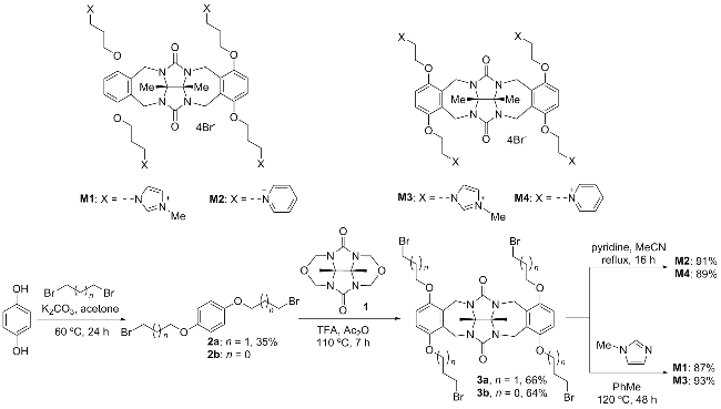
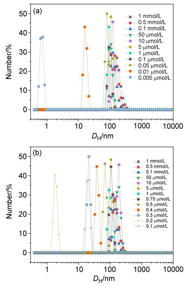
Because of the unique structures of glycoluril derivatives, M1~M4 have the C-shaped character and be able to behave as molecular clips. These amphiphilic molecular clips (AMCs) possess four cations which exhibit good water solubility. Among them, the solubility of M4 in phosphate buffer solution (PBS) buffer was as high as 97 mmol/L, while the solubility of M3, M2 and M1 was 86, 41 and 38 mmol/L, respectively. Firstly, dynamic light scattering (DLS) experiments were carried out on solutions of M1~M4 in water. As shown in Figure 1a, for imidazolium salt M1, it had a hydrodynamic diameter (DH) of >100 nm at the low concentration of 0.05 μmol/L. The DH decreased to 15.7 nm when the concentration of M1 was decreased to 0.01 μmol/L. As to pyridinium salt M2 (Figure 1b), it had a DH greater than 100 nm in the concentration range from 0.75 μmol/L to 1 mmol/L, and when the concentration gradually decreased from 0.5 μmol/L to 0.1 μmol/L, the DH decreased from 68.1 nm to 1.74 nm. Compared to that of M2, M1 has larger DH at low concentration, which indicates that the self-assembly of M1 is more stable and the stable self-aggregation can be maintained at the wide concentration range from 0.05 μmol/L to 1 mmol/L. M3 and M4 possess shorter alkyl chains than those of M1 and M2. To reach a 100 nm of DH, the concentration of M3 should be higher than 1 μmol/L and that of M4 be higher than 50 μmol/L. The aggregations of M3 and M4 were less stable at low concentrations, especially for M4. The results indicate that the self-assembly of M1~M3 is more stable and can form nanoparticles with a certain size, which lays the foundation for the delivery of sDNA. An amphiphilic glycoluril dicarboxylate has been revealed to aggregate into particles of mesoscopic size which is driven hydrophobically,[20] while acyclic cucurbituril-based supramolecular amphiphiles have been reported to form nanoparticles in water through the aggregation of the hydrophobic glycoluril-based backbone.[43-44] We thus tentatively proposed that compounds M1~M4 underwent similar glycoluril aggregation to give rise to nanoparticles with average, concentration-dependent sizes (Figure 2). The aggregation was expected to be irregular, but the size of the resulting nanoparticles was concentration-depen- dent.
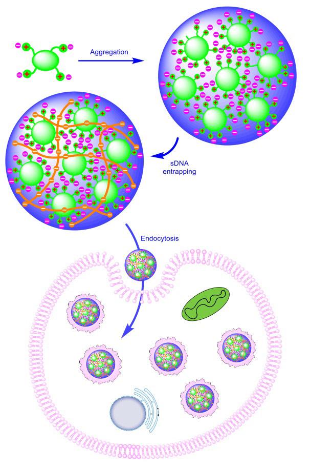
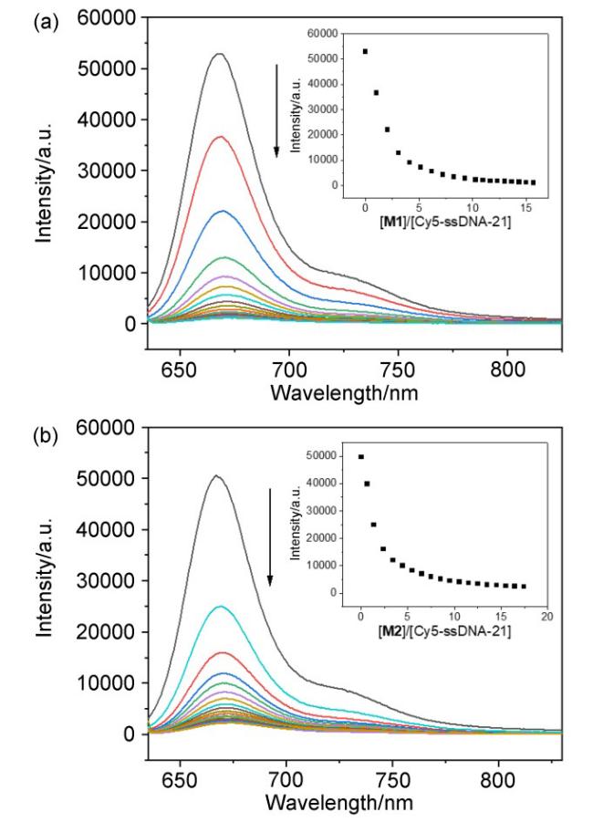
Fluorescent titration experiments were performed in water to study the interactions between each of the four AMC aggregates and Cy5-ssDNA-21/Cy5-dsDNA-21, which were fluorescently labelled 21 nucleotide single- stranded and double-stranded DNA, respectively. Cy5- ssDNA-21 and Cy5-dsDNA-21 have been previously used to evaluate in vitro DNA delivery activity of porous framework-type or ionic supramolecular polymer carriers.[28,30-31] For all fluorescent titration experiments, the concentration of ssDNA was kept consistent (0.25 μmol/ L). In particular, dropwise addition of M1 or M2 to the solution of Cy5-ssDNA-21 (Figures 3a and 3b) or Cy5- dsDNA-21 led to significant quenching of the fluorescence of the Cy5 probe attached to one end of the DNA sequences, and dropwise addition of M3 or M4 to Cy5-ssDNA-21 or Cy5-dsDNA-21 did not, which suggested that the interactions of M3 or M4 with Cy5-ssDNA-21 or Cy5-dsDNA- 21 were substantially weaker than those of M1 and M2. In addition, for Cy5-ssDNA-21, the quenching of Cy5 probe showed a significant inflection point when about 6-fold equivalents of M1 or 5-fold equivalents of M2 were added, respectively, while for Cy5-dsDNA-21, the quenching of Cy5 probe showed a significant inflection point when about 20-fold equivalents of M1 or 4-fold equivalents of M2 were added. In contrast, the addition of excess of M3 or M4 did not cause a significant quenching of the fluorescence of sDNA. The above significant quenching of fluorescence suggested that ss- and ds-DNAs were encapsulated inside the AMC aggregates formed by M1 or M2, respectively, through multivalent intermolecular ion-pair- ing electrostatic interactions (Figure 2), whereas M3 and M4 did not strongly interact with the two sDNAs. For M1 and M2, DNA adhering to the surface of their aggregates would only cause the quenching of the fluorescence of the molecules of the surface layer. Thus, complete quenching of the fluorescence excluded this possibility.
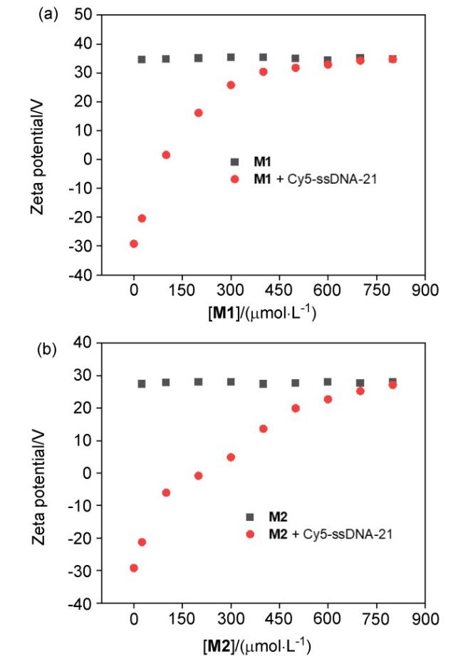
We further assessed the stability of the AMC aggregates of the two compounds through DLS experiments and whether short DNA could be encapsulated into the interior of their aggregates by measuring the zeta potential (Figures 4). Cy5-ssDNA-21 and Cy5-dsDNA-21 (10 μmol/L) exhibited a potential of -29.3 and -43.5 mV, respectively, while M1 and M2 (25~800 μmol/L) exhibited ca. 35 and 28 mV zeta potential, respectively, indicating that the AMC aggregates were positively charged. Dropwise addition of a certain concentration of the aggregates to the solution of Cy5-ssDNA-21 or Cy5-dsDNA-21 caused continuous shifting of their potentials toward positive charge. When the dropwise addition of the aggregates reached a critical concentration, the potential values would tend to coincide with those of the pure solution of the aggregates. These observations again supported the mechanism that the aggregates encapsulated sDNA into their interior after a critical concentration, and also excluded that the mechanism that the DNA chains were adsorbed on the surface of the nanoaggregates. DLS experiments were also carried for M2 and its mixture with dsDNA after the solutions were left to stand for 24 h, which gave rise to results comparable with those obtained without standing, indicating that both nanoparticles were stable. Moreover, the nanoparticles also had narrow polydispersity index.
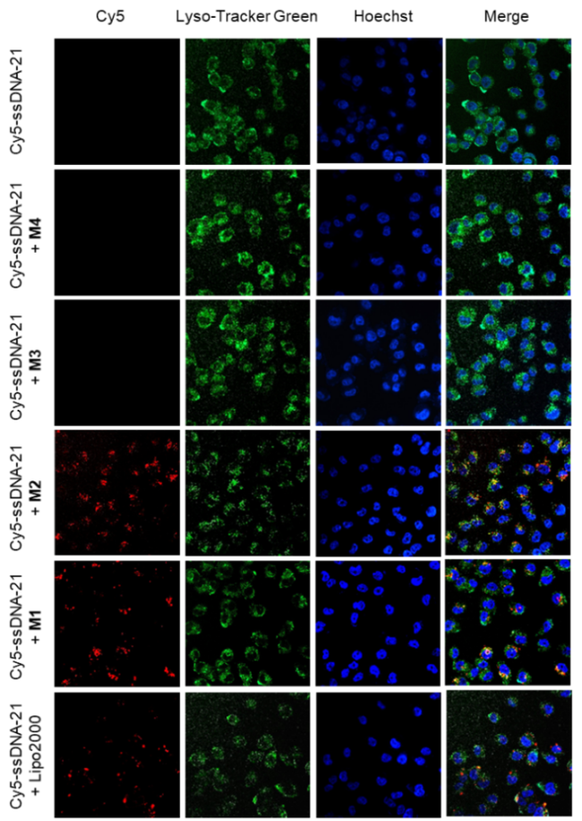
Confocal laser scanning microscopy (CLSM) was used to assess the ability of the AMC aggregates to deliver Cy5-ssDNA-21 to MCF-7/ADR cells (Figure 5). The nuclei and lysosomes of the cells were stained with Hoechst dye and Lyso-Tracker Green dye, respectively. The cells were incubated with a mixture of one of the AMC aggregates (20 μg/mL) and Cy5-ssDNA-21 (2.5 μg/mL) for 2 h, and then the intensity of Cy5 fluorescence was observed in the images. It can be seen that the images showed the characteristic fluorescence of Cy5 probe under the condition of the presence of compound M1 or M2. In contrast, the sample in the presence of M3 or M4 did not exhibit the fluorescence, indicating that they did not have the ability to deliver sDNA. In the absence of the AMC assembly, for the same dose of free Cy5-ssDNA-21, the image did not show the characteristic fluorescence of Cy5 probe. Meanwhile, a mixture of commercial reagent Lipo2000 (20 μg/mL) and Cy5-ssDNA-21 (2.5 μg/mL) was incubated under the same conditions for 2 h. A weak fluorescence was observed, but no uniform delivery was achieved. The above phenomena clearly indicate that M1 and M2 have the ability to deliver sDNA into the MCF-7/ADR cells.
We continued to use confocal laser scanning microscopy to assess the ability of the four AMC aggregates to deliver short double-stranded DNA into MCF-7/ADR cells. A concentration of 5 μg/mL Cy5-dsDNA-21 was mixed with one of the four AMC aggregates (40 μg/mL) and incubated with MCF-7/ADR cells for 2 h. Similar to the results of the Cy5-ssDNA-21 delivery experiments, the two groups with the addition of M1 or M2 delivery agents showed stronger characteristic fluorescence of Cy5 probe, while the two groups with the addition of M3 or M4 still did not show the above fluorescence, which further supported that M1 and M2 also had better delivery ability for short double- stranded DNA. When MCF-7/ADR cells were incubated for 2 h with a mixture of the commercial reagent Lipo2000 at a concentration of 40 μg/mL and Cy5-dsDNA-21 at 5 μg/mL, the characteristic fluorescence of Cy5 could be observed, but the fluorescence intensity was weaker than that of the M2 group. The above experimental results indicated that M1 and M2 also had better delivery ability for short double-stranded DNA.
The CLSM imaging experiments have demonstrated that compounds M1 and M2 have the ability to deliver sDNA into MCF-7/ADR cells. Therefore, we performed additional flow cytometry experiments to quantitatively assess the magnitude of their delivery capacity. First, we used flow cytometry to study the capacity of four AMC aggregates to delivery Cy5-ssDNA-21. The concentration of the aggregate and Lipo2000 was kept at 20 μg/mL, and 2.5 μg/mL Cy5-ssDNA-21 was used. Cells were incubated using a mixture of their solutions, and the percentage of DNA internalization was determined after 2 h. A percentage of less than 5% was showed for the internalization of Cy5-ss- DNA-21 with none or the addition of M3 or M4, again demonstrating the absence of the ability of M3 and M4 to deliver sDNA. For MCF-7/ADR cells, it exhibited an 82.9% cellular internalization of Cy5-ssDNA-21 when using M2 as the delivery agent, which was higher than the percentage of cellular internalization for Lipo2000, which was 63.1% at the same dose. And the ssDNA delivery efficiency for M1 was similar to that of Lipo2000, which was 59.3%.
We then proceeded to use flow cytometry to investigate the ability of the four AMC aggregates to deliver short double-stranded DNA into cells (Figure 6a). Cy5-ds- DNA-21 was used at a dose of 5 μg/mL, while at the same time the concentrations of the four aggregates and Lipo2000 were changed to 40 μg/mL, and the incubation time was all 2 h. It can be observed that the percentage of internalization of the DNA by the cells was increased in all cases. For MCF-7/ADR cells, the percentage of cellular internalization was 86.3% and 94% when M1 and M2 were used as delivery agents, respectively, which were higher than that of the commercial reagent, Lipo2000 (65.3%). When the cells were incubated with M3 or M4 as delivery agents, the percentage of cell internalization was still below 5% after 2 h, indicating that they have little or no delivery capacity for short double-stranded DNA. H9C2 normal cell line and B16 cancer cell line were further used to assess the ability of M1~M4 to deliver both types of DNA into cells. Either M1 or M2 exhibited high DNA delivery efficiency for H9C2 cell. Although it was slightly lower than that of Lipo2000 (93.4%), the percentage of internalization of both ssDNA and dsDNA by M1 and M2 was higher than 86%. For B16 cells, the aggregate M2 was the best DNA delivery agent than other aggregates as well as Lipo2000. The percentage of internalization by M2 was 70.1% for ssDNA and 53.4% for dsDNA. This indicated that M1 and M2 could serve as nanoparticle carriers for short double-stranded DNA cellular delivery through the well-established endocytosis mechanism (Figure 2), as revealed for different kinds of nano carriers.[28,45-46] The lowered delivery efficiency of M3 and M4 may be attributed to their shorter ethylidene linkers that reduced their amphiphilicity and the stability of the resulting nanoparticles. Short linkage may also weaken their encapsulation for DNA since it should increase the hindrance of the hydrophobic glycouril unit for highly hydrophilic, multianionic DNA chains.
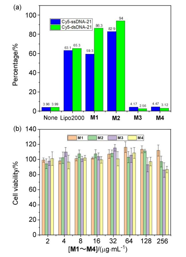


Figure 6 (a) Delivery (internalization) of Cy5-ssDNA-21 (2.5 μg/mL) and Cy5-dsDNA-21 (5 μg/mL) into MCF-7/ADR cell line by M1~M4 (20 μg/mL for ssDNA and 40 μg/mL for dsDNA) and Lipo2000 (20 μg/mL for ssDNA and 40 μg/mL for dsDNA) after incubation in complete 1640 medium for 2 h, and (b) cell viability values (%) of MCF-7/ADR cell line evaluated by CCK-8 proliferation tests versus the incubation concentration of M1~M4 (the cells (≈2×10⁴ per well) were incubated with M1~M4 at 37 ℃ for 24 h. Error bars represent the s.d. of uncertainty for each point) |
We have demonstrated the sDNA delivery ability of AMC aggregates to normal or cancer cells, and we subsequently used the Cell Counting Kit-8 assay to assess the vitro cytotoxicity of four aggregates, using MCF-7/ADR, H9C2, and B16 cells. For all cells involved in the assay, cell viability remained above 80% even at a concentration of 256 μg/mL the aggregates. If a 5% hemolysis rate was acceptable, the hemolysis rates of the four aggregates on human and Sprague-Dawley (SD) rat erythrocytes at 256 μg/mL were still well below the above standard. These further demonstrated the vitro safety of the four aggregates.
3 Conclusions
In summary, we have designed and prepared amphiphilic molecular clips for intracellular delivery of short DNA based on the dimethyl glycoluril backbone. The new amphiphilic molecules can self-assemble to form highly stable nanoparticles of defined sizes, depending on the concentrations. The nanoparticles can be driven by multivalent ion-pairing electrostatic interactions to include short DNA, and their nanoscale sizes endows them with the ability to function as carriers to realize intracellular delivery of the included DNA into cancer cells. The results show that single rigid molecules can rival polymeric systems by aggregation into stable nano-scaled particles to attain delivery function. In the future, the rigid backbone of the amphiphiles will be optimized to obtain molecules that not only display enhanced DNA delivery, but also can be applied for drug delivery.
4 Experimental section
4.1 General methods and materials
All reagents and solvents were commercially available and used as received without further purification. 1H NMR and 13C NMR spectra were recorded on Bruker AVANCE III HD 400 MHz and 500 MHz (125 MHz) instruments, respectively. Dynamic light scattering experiments were conducted on a Malvern Zetasizer Nano ZS90 using a monochromatic coherent He-Ne laser (633 nm) as the light source and a detector that detected the scattered light at an angle of 90°. Fluorescent measurements were performed on a VARIAN CARY Eclipse Fluorescence Spectrophotometer. Fluorescent microscopic experiments were performed on a VisiScope of Visitron Systems GmbH. Flow cytometric experiments were conducted with a Gallios 3L 10C flow cytometry system (Beckman Coulter, USA). Cell viability was measured by a Microplate Reader (BioTek Epoch 2). Fetal bovine serum (FBS), 1640 Medium, DMEM Medium were purchased from Thermo Fisher Scientific. Cell Counting Kit-8 (CCK-8) was purchased from Beyotime Biotechnology. Cy5-ssDNA-21 and Cy5- dsDNA-21 were commercials purchased from Shanghai HuaGen Biotech Co., Ltd. Lipo2000 was purchased from Thermo Fisher Scientific Co. Dimethyl glycoluril,[9] 1,[31] 2a,[31] 2b,[31] 3a,[31] 3b,[42] M1,[31] and M3[31,42] were prepared according to reported procedures.
4.2 Synthesis of compound M2
A solution of compound 3a (130 mg, 0.14 mmol) in acetonitrile (2.5 mL) was treated with pyridine (125 μL, 1.4 mmol). The mixture was refluxed for 36 h. The reaction mixture was cooled to 25 ℃ and the solvent was evaporated under vacuum. The residue was dissolved in H2O (5 mL), filtered through celite, and concentrated in vacuo to afford the title compound as a brown solid (198 mg, 91%). 1H NMR (400 MHz, D2O) δ: 8.76 (d, J=5.8 Hz, 8H), 8.49 (t, J=7.8 Hz, 4H), 7.90~7.84 (m, 8H), 6.63 (s, 4H), 5.12 (d, J=16.4 Hz, 4H), 4.84 (d, J=6.8 Hz, 8H), 4.11 (d, J=16.4 Hz, 4H), 4.00~3.95 (m, 4H), 3.71~3.66 (m, 4H), 2.48 (dd, J=11.9, 5.7 Hz, 8H), 1.87 (s, 6H); 13C NMR (126 MHz, D2O) δ: 157.79, 149.43, 145.68, 144.38, 128.21, 127.82, 112.67, 78.87, 64.98, 58.98, 34.92, 29.75, 15.46; HRMS calcd for C54H62N8O6Br4 1159.2316 [M-Br]+, found 1159.2314.
4.3 Synthesis of compound M4
A solution of compound 3b (121 mg, 0.14 mmol) in acetonitrile (2.5 mL) was treated with pyridine (125 μL, 1.4 mmol). The mixture was refluxed for 36 h. The reaction mixture was cooled to 25 ℃ and the solvent was evaporated under vacuum. The residue was dissolved in H2O (5 mL), filtered through celite, and concentrated in vacuo to afford the title compound as a white solid (167 mg, 89%). 1H NMR (400 MHz, D2O) δ: 8.89 (d, J=5.9 Hz, 8H), 8.51 (t, J=7.7 Hz, 4H), 7.95 (t, J=7.1 Hz, 8H), 6.59 (s, 4H), 5.06 (s, 8H), 5.02 (s, 4H), 4.59~4.52 (m, 4H), 4.47~4.40 (m, 4H), 4.09 (d, J=16.3 Hz, 4H), 1.84 (s, 6H); 13C NMR (126 MHz, D2O) δ: 157.63, 149.16, 146.01, 144.71, 128.27, 128.04, 113.29, 78.77, 67.25, 34.86, 15.56; HRMS calcd for C50H54N8O6Br4 1103.1688 [M-Br]+, found 1103.1718.
Supporting Information Supplemental results for dynamic light scattering (DLS) experiments, fluorescence spectra, zeta potentials, confocal laser scanning microscopy (CLSM) experiments, flow cytometric experiments, cell viability values (%), hemolytic activity and NMR spectra of compounds. The Supporting Information is available free of charge via the Internet at http://sioc-journal.cn.
(Zhao, C.)