

1 Introduction
X-ray scintillators, capable of converting high-energy X-ray into visible low-energy light, are widely used in the fields of industrial non-destructive testing,[1-3] medical imaging,[4-8] etc. The most researched scintillators are inorganic compounds, such as CsI:Tl, bismuth germanate (BGO), and CsPbBr3, which demonstrate strong X-ray absorption capability and excellent X-ray scintillation properties, but generally suffer from the disadvantages of ultrahigh preparation temperature, high toxicity, and inferior environmental stability.[9-10] Pure organic scintillators, on the other hand, are of significant advantages such as low preparation temperature, ease of modification, and the ability to fabricate large-area flexible scintillation screens.[11-16] Despite these benefits, pure organic scintillators, typically composed of elements with small atomic numbers like carbon and hydrogen, have weak X-ray absorption, leading to low radioluminescence (RL) efficiency and posing a significant challenge to their widespread application. Combined the advantages of inorganic and organic scintillators, organic-inorganic hybrid scintillators simultaneously exhibit accomplished X-ray scintillation performance and simple preparation process, possessing more practical application prospect.[17-19]
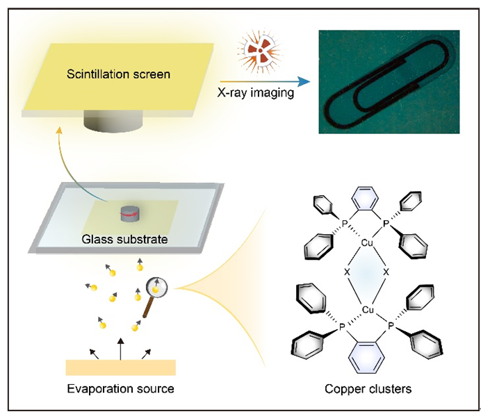
Recently, organic-inorganic hybrid Cu(I) clusters,[20-27] exhibiting the superior RL efficiencies in the solid state, have been a research focus in the scintillator field. Cu(I) clusters contain a rigid [CuX]n (X=Cl, Br, I) core (Figure 1a), which plays an important role for high photoluminescence quantum yield (PLQY) due to the inhibited structural relaxation and strong X-ray absorption owing to high atomic number copper and halogen atoms. Under X-ray excitation, the RL of Cu(I) clusters are mainly derived from metal/halogen-to-ligand [(M+X)LCT] excited state.[20,28] Due to the heavy atom effect, both 1(M+X)LCT and 3(M+X)LCT excitons can emit RL, enabling 100% exciton utilization. Benefited from these inherent advantages, the single crystals of Cu(I) clusters show the comparable scintillation performance with the commercial inorganic counterparts. However, it is extremely challenging for them to cultivate large area single crystals for X-ray imaging due to the weak intermolecular interactions and complex molecular structures. Hence, they are usually doped into polymer matrix to prepare scintillator films, whereas the polymer matrix has low X-ray absorption ability and is hardly radioluminescent, resulting in deteriorated scintillation efficiency. In addition, the uneven dispersion of scintillators within the polymer matrix can also lead to poor X-ray imaging resolution. Hence, there is an urgent need for innovative film fabrication techniques for Cu(I) cluster scintillators.
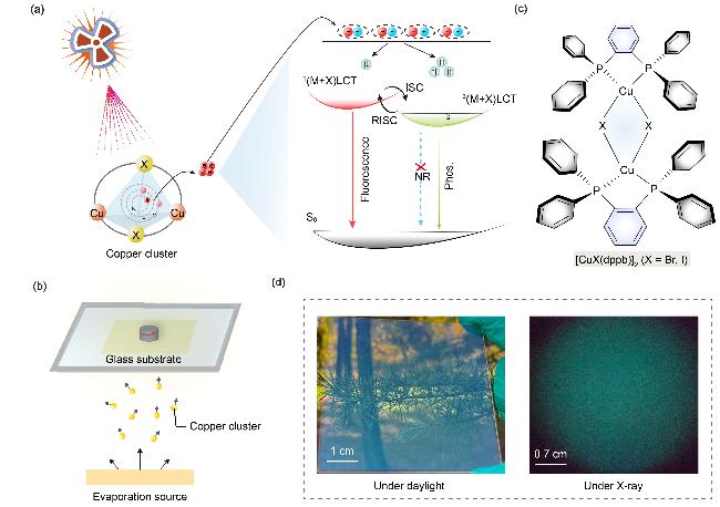
Figure 1 (a) Schematic diagram of the RL mechanism of Cu(I) clusters (Phos: phosphorescence, NR: non-radiative transition); (b) Schematic diagram of preparation of scintillation films by vacuum evaporation deposition method; (c) Chemical structures of [CuX(dppb)]2 complexes; (d) Evaporation-processed films of [CuX(dppb)]2 complexes under daylight (left) and X-ray (right) |
Vacuum evaporation deposition that can prepare high-quality films over large area has been extensively applied in organic light-emitting diodes,[29-31] perovskite photovoltaics,[32-34] and organic field-effect transistors.[35-37] We assumed that the pure Cu(I) cluster films prepared by vacuum evaporation deposition would be homogeneous without sacrificing the scintillation performance (Figure 1b). To proof our hypothesis, two dinuclear Cu(I) clusters [CuX(dppb)]2 (X=Br, I) with admirable thermal stability were prepared (Figure 1c). The decomposition temperatures of the two clusters are higher than 440 ℃, making them suitable for vacuum evaporation deposition. Due to the strong X-ray absorption and high exciton utilization, the clusters display excellent X-ray scintillation performance with the high light yields of up to 19356.7 photons/MeV. The scintillation screen based on the Cu(I) clusters prepared by vacuum evaporation deposition is smooth and uniform with a small surface roughness value of 1.04 nm (Figure 1d), which is exploitable in X-ray imaging for various objects. The current work suggests great potential of vacuum evaporation deposition in fabricating scintillation screens for X-ray radiography.
2 Results and discussion
The dinuclear Cu(I)-halide clusters, [CuBr(dppb)]2 and [CuI(dppb)]2, could be synthesized in a large scale through a simple room-temperature solution reaction of 1,2-bis(diphenylphosphino)benzene (dppb) with CuX (X=Br or I) and purified by vacuum sublimation. The chemical structures of the products were confirmed by proton (1H) and carbon (13C) nuclear magnetic resonance (NMR) and crystallographic analyses. The thermal stability of [CuX- (dppb)]2 was subsequently determined by thermogravimetric analysis (TGA). [CuX(dppb)]2 clusters exhibit a high decomposition temperature of up to 440 ℃, indicating that the material has good thermal stability which is conducive to fabricate evaporation-processed scintillator film.
The basic photophysical properties of the two clusters were investigated through ultraviolet-visible (UV-Vis) absorption spectroscopy and photoluminescence (PL) spectroscopy. Both the pure [CuBr(dppb)]2 and [CuI- (dppb)]2 films exhibit two absorption peaks at ca. 290 and ca. 330 nm (Figure 2a). The ca. 290 nm absorption is attributed to π-π* transition of dppb ligand, while ca. 330 nm absorption should be assigned to CuX-involved electronic transition. The steady-state PL (SSPL) spectra of [CuBr-(dppb)]2 and [CuI(dppb)]2 crystals are similar with the bluish green emission bands centered at 505 nm, which shows the lifetimes of ca. 4 µs at room temperature (Figure 2b). Notably, the SSPL spectrum of [CuBr(dppb)]2 crystal is red-shifted than that of [CuI(dppb)]2 crystals. The reason is that Br- has stronger ligand field strength than I-, which would increase the energy separation of d-orbitals, resulting in a smaller metal-to-ligand charge transfer (MLCT) energy. In addition, both [CuBr(dppb)]2 and [CuI(dppb)]2 crystals exhibit the satisfactorily high photoluminescence quantum yields (PLQYs) of 49.5% and 56.9% respectively (Figure 2b), indicating that non-radia- tive transition is inhibited, which might be due to the tight molecular packing in crystalline state. With the temperature increasing from 78 to 288 K, the lifetimes of the [CuI(dppb)]2 crystals constantly decrease (Figure 2c), illustrating that the bluish green emission is mainly derived from phosphorescence. The phosphorescence-dominant emission is further confirmed by temperature-dependent SSPL spectra, which show slightly decreased emission intensity with increasing temperature from 80 to 180 K (Figure 2d). The emission has virtually no change above 180 K, which also manifests the rigid structure of [CuI- (dppb)]2 crystals.
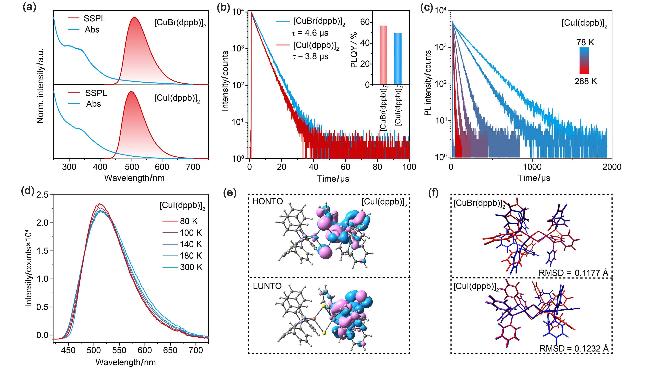
Figure 2 (a) Normalized UV-Vis absorption spectra and steady-state emission spectra (excitation wavelength: 365 nm, temperature: 298 K) in pure films and in crystalline state for [CuX(dppb)]2 clusters; (b) Lifetime curves of [CuBr(dppb)]2 and [CuI(dppb)]2 crystals monitored at 510 and 500 nm, respectively (inset shows the PLQYs of the complex crystals); (c) Plot of the emission decay lifetime of [CuI(dppb)]2 versus temperature monitored at 505 nm; (d) Temperature dependence of SSPL spectra for [CuI(dppb)]2 crystals from 80 to 300 K; (e) Calculated HONTO and LUNTO distributions of [CuI(dppb)]2 at triplet (T1) state; (f) Diagram of geometry deviations between ground state (S0) and singlet (S1) states for the [CuBr(dppb)]2 (top) and [CuI(dppb)]2 (bottom) complexes (the structures at S0 and S1 states are depicted in deep-blue and red, respectively) |
Theoretical calculations were performed to get an insight into the photophysical properties of [CuX(dppb)]2 clusters. The frontier molecular orbits of the two clusters are similar, with the highest occupied molecular orbital (HOMO) and lowest unoccupied molecular orbital (LUMO) located on Cu2X2 core and dppb ligand, respectively, indicating their charge transfer characteristic. The separated HOMO and LUMO distributions render the small single-triplet energy gaps (ΔEST), which are 0.15 and 0.13 eV for [CuBr(dppb)]2 and [CuI(dppb)]2, respectively. The small ΔEST indicate that the clusters may have thermally activated delayed fluorescence (TADF) properties.[38] The natural transition orbital (NTO) calculations were also performed to analyze the electron transition components of [CuX(dppb)]2 at triplet (T1) states. The highest occupied NTOs (HONTOs) are primarily distributed on Cu2X2 core and dppb ligand (Figure 2e), while the lowest unoccupied NTO (LUNTOs) are dominated by dppb ligand; the overlaps between HONTOs and LUNTOs for [CuBr(dppb)]2 and [CuI(dppb)]2 are 38.68% and 35.14%, respectively, guaranteeing the high PLQYs.[39] Additionally, the molecular cores of the two copper clusters show minimal conformational variation between the singlet (S1) and ground (S0) states with a low root mean square displacement (RMSD) values of approximately 0.12 Å (Figure 2f). This indicates that they have a rigid core structure, which can suppress the non-radiative transition and improve the luminous efficiency.
The excellent crystal luminescence and high atomic number of halogen atoms (Br, I) forebode the promising potential of [CuX(dppb)]2 clusters as X-ray scintillators. Therefore, a systematic study of the scintillation performance of [CuX(dppb)]2 under X-ray excitation was conducted. Since the X-ray absorption coefficient (α) is proportional to the fourth power of the atomic number,[40] [CuI(dppb)]2 (Zmax=53, Kα=33.17 keV) containing iodine atoms exhibits the stronger X-ray absorption in comparison with [CuBr(dppb)]2 (Zmax=35, Kα=13.47 keV) (Figure 3a). Subsequently, the attenuation of [CuX- (dppb)]2 as a function of thickness was calculated at an X-ray photon energy of 68 keV. It was found that a 0.4-mm-thick [CuI(dppb)]2 can attenuate 99% of X-ray photon energy, while [CuBr(dppb)]2 requires a thickness of 0.68 nm (Figure 3b). Figure 3c shows the RL spectra of 50 mg [CuX(dppb)]2 crystals compared to commercial scintillators CsI:Tl, Ce:YAP, and Ce:YAG powders under the same X-ray dose rate irradiation. [CuX(dppb)]2 crystals manifest the excellent X-ray scintillation with RL intensity comparable to these commercial scintillators (Figure 3d). Although the PLQY of [CuI(dppb)]2 crystal is lower than [CuBr(dppb)]2 crystal, it has higher RL intensity probably due to its stronger X-ray absorption of I atom. Benefited from the strong X-ray absorption and high exciton utilization, [CuI(dppb)]2 crystal exhibits a high light yield of up to 19356.7 photons/MeV. The RL spectra of [CuX(dppb)]2 at different X-ray dose rates were recorded. As shown in Figure 3e and Figure S14 (see Supporting Information), the RL intensity of [CuX(dppb)]2 increases monotonically and exhibits a good linear response in the range of 0.688 μGy/s to 278 μGy/s. The limit of detection (LOD) values at a signal-to-noise ratio of 3 were evaluated to be 186 and 158 nGy/s for [CuBr- (dppb)]2 and [CuI(dppb)]2, respectively (Figure 3f), which are significantly lower than the limit for medical X-ray diagnostics (5.5 μGy/s). Moreover, the [CuI- (dppb)]2 crystals reveal excellent radiation stability with RL intensity keeping nearly unchanged for 130 on-off excitation circles (Figure 3g). The radiation intensities maintain over 99% even after continuous irradiation at a dose rate of 278 μGy/s for 1800 s.
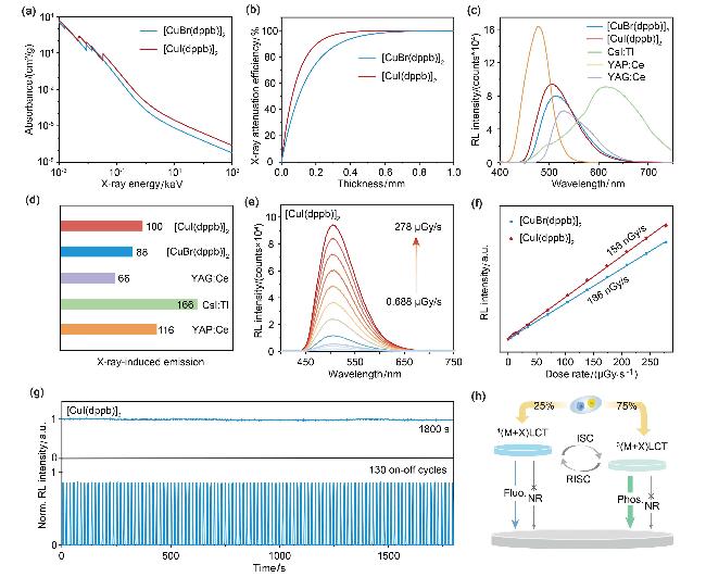
Figure 3 (a) Simulated X-ray absorption spectra and (b) X-ray attenuation efficiency as a function of material thickness of [CuX(dppb)]2 clusters; (c) RL spectra of [CuX(dppb)]2 crystals compared to commercial CsI:Tl, YAP:Ce, YAG:Ce scintillators under X-ray irradiation at a dose rate of 278 μGy/s; (d) Comparison of the integrated areas of RL spectra for the different types of scintillators; (e) RL spectra of [CuI(dppb)]2 crystal at different dose rates; (f) Linear behavior of the RL intensity for [CuI(dppb)]2 crystal as a function of X-ray irradiation dose rate; (g) X-ray photostability for [CuI(dppb)]2 crystal under continuous irradiation (top) and repeated on-off cycles (bottom) at 278 μGy/s; (h) RL mechanism for [CuX(dppb)]2 under X-ray excitation |
The RL and SSPL (Figure 2a) spectra of [CuX(dppb)]2 crystals closely align with the emission peak at ca. 500 nm, indicating that the emissions might arise from the same radiative channels under UV and X-ray irradiation. Thus, a rational mechanism for the RL of [CuX(dppb)]2 crystals can be proposed. Upon X-ray excitation, the heavy atoms (Br and I atoms) in [CuX(dppb)]2 interact with X-ray photons through the photoelectric effect and Compton scattering,[41] releasing high-energy electrons. These hot electrons further interact with atoms, producing a large number of secondary electrons. After losing excess energy, the electrons are eventually captured by the ligands, forming hole-electron pairs. The recombination of hole-electron pairs produces 25% singlet excitons and 75% triplet excitons according to the spin rules (Figure 3h). Due to the small ΔEST between the 1(M+X)LCT and 3(M+X)LCT states, the S1 and T1 excitons of [CuX(dppb)]2 are interconvertible through intersystem crossing (ISC) and reverse intersystem crossing (RISC). The RL emission mainly comes from phosphorescence generated by 3(M+X)LCT excitons, and partly comes from fluorescence derived from 1(M+X)LCT excitons.
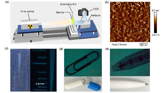
Benefiting from the excellent X-ray scintillation performance and thermal stability, [CuI(dppb)]2 was used to fabricate scintillation screen through vacuum evaporation deposition for X-ray imaging (Figure 4a). Atomic force microscopy (AFM) images indicate that the vacuum- evaporated [CuI(dppb)]2 film has a smooth and uniform surface with a small surface roughness value of 1.04 nm (Figure 4b). Line pair card imaging further demonstrates its high spatial resolution, where the line pairs corresponding to >8 lp/mm is identifiable (Figure 4c). Then the film was further applied to X-ray imaging for various objects (Figures 4c and 4d). Under X-ray irradiation, the paper clip inside a capsule and the internal structure of an electronic chip could be clearly revealed, highlighting the potential applications of such scintillator films in non-destructive testing and medical imaging.
Figure 4 (a) Schematic diagram of the optical apparatus for X-ray imaging; (b) AFM image of [CuI(dppb)]2 scintillator film; (c) Brightfield photograph (left) and X-ray imaging (right) of the standard resolution test template; Brightfield photographs (bottom) and X-ray imaging photos (top) of (d) a capsule fitted with a paper clip and (e) a ballpoint pen |
3 Conclusions
In summary, we have proposed an effective strategy to prepare X-ray imaging screen through vacuum evaporation deposition. Two coper clusters [CuBr(dppb)]2 and [CuI- (dppb)]2 appropriate for evaporation process were synthesized, which exhibit meritorious luminous efficiencies due to the rigid molecular structures. The X-ray scintillation performance of the coper clusters is comparable to those commercial inorganic scintillators with a high light yield of up to 19356.7 photons/MeV and a detection limit as low as 158 nGy/s. The thin film of [CuI(dppb)]2 fabricated by vacuum evaporation deposition is smooth and uniform, which has been applied to X-ray imaging for various objects. Though the imaging spatial resolution of the thin film needs to be improved, the current work highlights the po-tential of vacuum evaporation deposition method in the development of high-quality imaging screen for advanced X-ray radiography.
4 Experimental section
4.1 Instruments and reagents
1H and 13C nuclear magnetic resonance spectra (NMR) were recorded on a Bruker Ultra Shield Plus 400 MHz instruments with DMSO-d6, CDCl3 or CD2Cl2 as the solvent and tetramethylsilane (TMS) as the internal standard. All chemicals and solvents, unless otherwise stated, were purchased from commercial suppliers and used as received. 1,2-Bis(diphenylphosphanyl)benzene was purcha- sed from Shanghai Macklin Biochemical Technology Co., Ltd. CsI:Tl, Ce:YAP, and Ce:YAG scintillators were purchased from Shanghai Epic Crystal Co., Ltd.
4.2 Synthesis and characterization
1,2-Bis(diphenylphosphanyl)benzene (446.5 mg, 1 mmol, 1 equiv.) was added to a suspension of copper(I) bromide or iodide (1 mmol, 1 equiv.) in 50 mL of dry acetonitrile, and the mixture was heated under reflux for 4 h. A large amount of light-green percipient emerged during the reaction. After cooling to room temperature, the percipients were filtered, washed by dichloromethane, and dried in vacuum drying oven. The crude product was further purified through train sublimation to give small crystalline grains.
[CuBr(dppb)]2: Light green crystals (156 mg, 34.9%). m.p. 383.5 ℃; 1H NMR (400 MHz, DMSO-d6) δ: 7.65~7.55 (m, 6H), 7.41~7.31 (m, 32H), 7.16~7.14 (t, J=8.0 Hz, 5H), 6.98 (d, J=6.9 Hz, 5H); 13C NMR (101 MHz, CDCl3) δ: 143.31, 143.01, 142.71, 133.98, 133.90, 133.85, 133.82, 132.83, 132.68, 132.52, 129.56, 128.95, 128.10, 128.05, 128.00. HRMS (MALDI) calcd for C61H52Br2- Cu2P4 [M+H]+ 1175.9665, found 1177.1802.
[CuI(dppb)]2: Light green crystals (167 mg, 37.4%). m.p. 390.4 ℃; 1H NMR (400 MHz, CD2Cl2) δ: 7.29~7.26 (m, 4H), 7.19~7.10 (m, 28H), 7.02~6.98 (m, 16H); 13C NMR (101 MHz, CD2Cl2) δ: 143.31, 143.02, 142.72, 134.27, 134.09, 134.02, 133.94, 133.00, 132.86, 132.71, 130.03, 129.19, 128.12, 128.07, 128.03. HRMS (MALDI) calcd for C61H52I2Cu2P4 [M+H]+ 1271.9388, found 1272.9470.
4.3 Thermal properties
Thermogravimetric analyses (TGA) were conducted on a DTG-60 Shimadzu thermal analyst system under a heating rate of 10 ℃/min and a nitrogen flow rate of 50 cm3/min. The differential scanning calorimetry (DSC) analyses were performed on a Shimadzu DSC-60A instrument under a heating rate of 10 ℃/min and a nitrogen flow rate of 20 cm3/min.
4.4 Basic photophysical characterization
UV-Vis and fluorescence spectra were measured using a Jasco V-750 spectrophotometer and an Edinburgh FLS- 1000 fluorescence spectrometer, respectively. The PLQY was determined with an Edinburgh FLS980 fluorescence spectrophotometer, which is fitted with an integrating sphere. Fluorescence decay measurements were performed using Variable Pulsed Lasers (VPL-375).
4.5 Theoretical calculation
Theoretical calculations were performed on Gaussian 09 program. B3LYP functional was used for structural optimization of both ground and excited states using 6-31G* basis set for carbon, hydrogen, and phosphorus atoms, and Lanl2dz pseudopotential basis set for Cu, I and Br atoms. Vibrational frequency calculations were subsequently carried out to confirm that all these optimized structures are corresponding to the minima on the potential energy surfaces. NTO analysis was performed based on TD-DFT results to offer a compact orbital representation for the electronic transition density matrix. RMSD was calculated using the VMD software.
4.6 X-ray luminescence properties
4.6.1 Scintillator film preparation
In a general procedure, the quartz sheet substrate (9 cm×9 cm) was washed with deionized water, acetone, and ethanol for 30 min in an ultrasonic water bath. The scintillator film with a thickness of 500 nm was deposited on a quartz sheet substrate by adding 100 mg of [CuI(dppb)]2 crystals to a crucible followed by evaporating them at a rate of 0.1 nm/s.
4.6.2 X-ray absorption curve simulation
Simulated X-ray absorption was performed on the National Institute of Standards and Technology (NIST) website. The molecular formula of the material was entered into the XCOM: Photon Cross Sections Database, in which the energy setting was set at a minimum of 0.001 MeV and a maximum of 100000 MeV. Then the data were exported and plotted in Origin software to obtain the simulated X-ray absorption spectrum.
4.6.3 Attenuation efficiency of the scintillator as a function of thickness
The X-ray absorption curves of the materials were obtained in the simulation software Photon Cross Sections Database (XCOM). The Y-axis data was selected for mathematical analysis to obtain the interpolation value, and 5000 points were taken. The X-value at the beginning was set to 1, and the X-value at the end was set to 68 to obtain a numerical interval, and the sum of the numerical interval was multiplied by the corresponding percentage to obtain the photon cross-section function c(E), and then the X-ray attenuation efficiency (AE) was calculated by the formula:
AE(%)=(1-e-c(E)ρd)×100%
where e is natural constant, E, ρ, and d represent the corresponding photon energy, scintillator density, and scintillator film thickness, respectively.
4.6.4 Calculation of X-ray light yield
The light yields of crystal samples were evaluated using a commercial scintillator YAG:Ce (light yield of 14,000 photons/MeV) as a reference by comparing the integrated area of RL. The RL curves were obtained using a spectrometer (FS5, Edinburgh) under identical measurement conditions (dose rate: 278 μGy/s). The samples used for light yield measurement are crystalline powders, which exhibit a polydisperse particle size distribution within the micrometer range.
4.6.5 Calculation of X-ray detection limits
The detection limit was calculated using the 3σ/k method. By measuring the dose-dependent RL spectra, a linear relationship between RL intensity and dose rate was obtained, with the slope k representing the sensitivity of the X-ray detector. σ represents the signal-to-noise ratio of the instrument.
4.6.6 X-ray image acquisition and processing
X-ray imaging was performed using an imaging device built with an X-ray tube (Tungsten Target, Moxtek) and a commercial camera (EOS R5, Canon). The ISO, aperture, and exposure time were set to 1000, f2.8, and 30 s for the samples under ambient light, and set to 8000, f2.8, and 30 s for the X-ray excitation samples, respectively. The X-ray tube voltage and current were set to 50 kV and 100 μA respectively. The raw X-ray images were further processed using Adobe Illustrator software.
Supporting Information 1H NMR and 13C NMR spectra of products [CuBr(dppb)]2 and [CuI(dppb)]2, X-ray single crystal data for products [CuBr(dppb)]2 and [CuI- (dppb)]2. The Supporting Information is available free of charge via the Internet at http://sioc-journal.cn.
(Lu, Y.)