

1 Introduction
The traditional Chinese medicine Notopterygium incisum consists of the dried rhizome and root of Notopterygium incisum Ting ex H. T. Chang or Notopterygium forbesii H. de Boiss. As an endemic medicinal plant resource in China, N. incisum is widely distributed across regions such as Tibet, Qinghai, Gansu, Yunnan, and Sichuan.[1] It thrives in distinctive growing environments characterized by high altitudes, cool temperatures, and shade tolerance. N. incisum is commonly utilized to treat various conditions wind-cold, headaches, paralysis, rheumatic disorders, and pain in the shoulders and back.[2]
N. incisum exhibits a complex chemical composition along with a diverse array of pharmacological activities including anti-inflammatory, antibacterial, antioxidant, antiviral, antipyretic, and analgesic properties.[3-7] To date, various chemical constituents such as volatile oils, terpenoids, coumarins, glycosides, organic acids, and phenolic compounds[8-12] have been isolated and identified from N. incisum. These components may partially account for the observed pharmacological activities of this species.
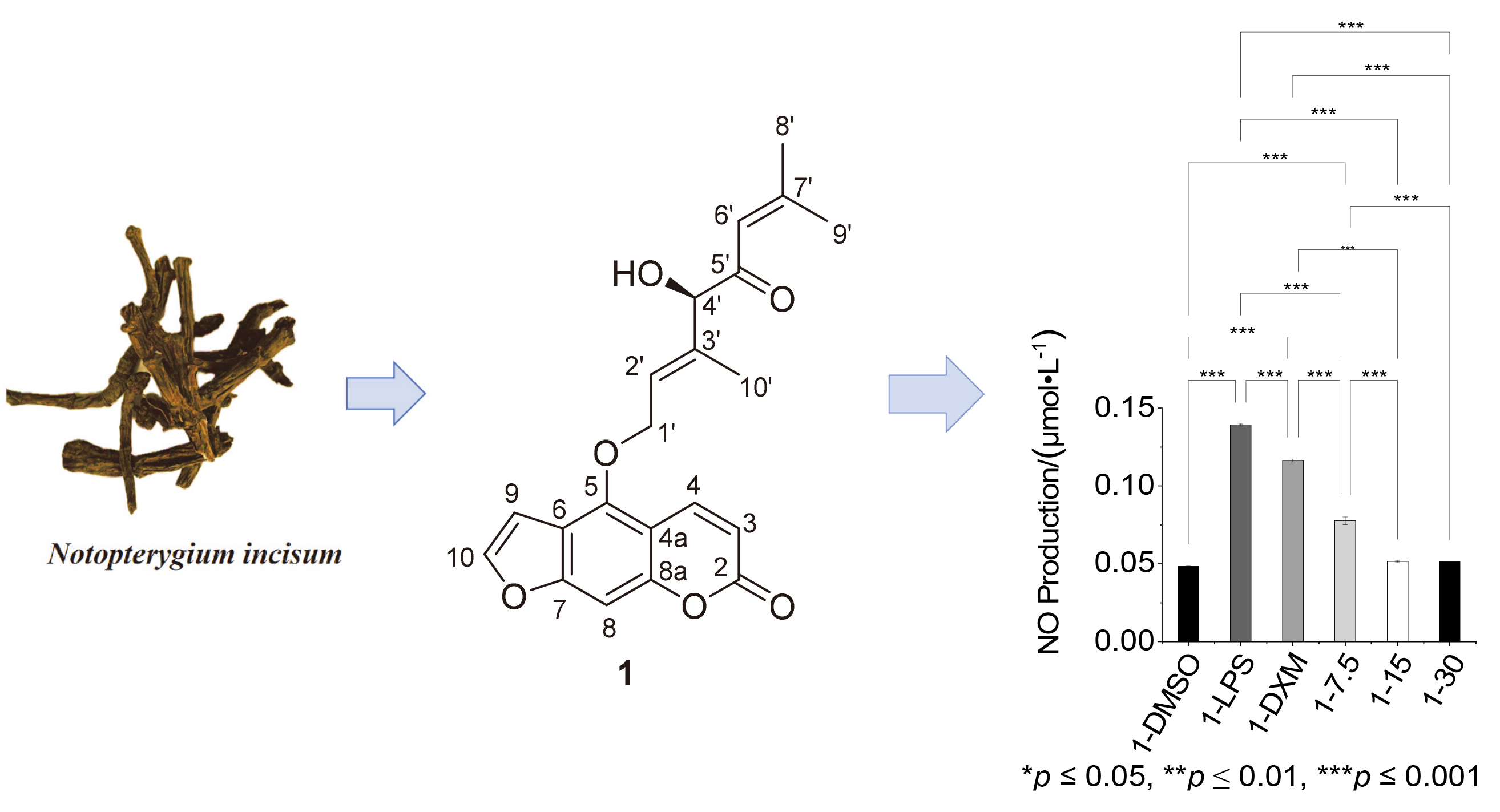
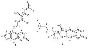
As part of our ongoing efforts to explore natural products with novel structures and anti-inflammatory activities derived from traditional Chinese medicines in southwestern China, the discovery of two new linear furocoumarins extracted from the root of N. incisum was reported (Figure 1). This paper details the structural elucidation of these compounds and evaluates their potential anti-inflammatory activities.
2 Results and discussion
Compound 1 was obtained as a yellow gum. The HRESIMS data revealed its molecular formula is C21H20O6. The IR bands indicated the presence of hydroxyl (3428 cm-1) and carbonyl (1732 cm-1) groups. The 1H NMR, 13C NMR and HSQC spectra of 1 indicated the presence of a set of typical signals of a linear furanocoumarin moiety [δH: 8.17 (d, J=9.8 Hz, 1H, H-4), 7.61 (d, J=2.3 Hz, 1H, H-10), 7.18 (s, 1H, H-8), 6.95 (d, J=2.3 Hz, 1H, H-9), 6.29 (d, J=9.8 Hz, 1H, H-3); δC: 161.2 (C-2), 158.3 (C-7), 152.8 (C-8a), 148.6 (C-5), 145.3 (C-10), 139.4 (C-4), 114.0 (C-6), 113.1 (C-3), 107.5 (C-4a), 105.0 (C-9), 94.6 (C-8)], along with a carbonyl group [δC: 197.7 (C-5')]; two trisubstituted double bonds [δH: 6.05~6.07 (m, 1H, H-7') and 6.00 (t, J=5.8 Hz, 1H, H-2'); δC 161.7 (C-7'), 139.3 (C-3'), 126.3 (C-2'), and 118.4 (C-6')]; an oxy-methylene [δH: 5.07 (d, J=6.7 Hz, 2H, H-1'); δC: 69.2 (C-1')]; three methyls [δH: 2.24 (s, 3H, H-9'), 1.91 (s, 3H, H-8'), and 1.55 (s, 3H, H-10'); δC: 28.3 (C-8'), 21.7 (C-9'), 11.8 (C-4')].
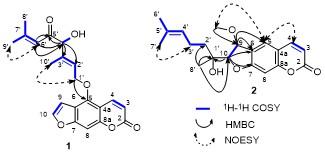
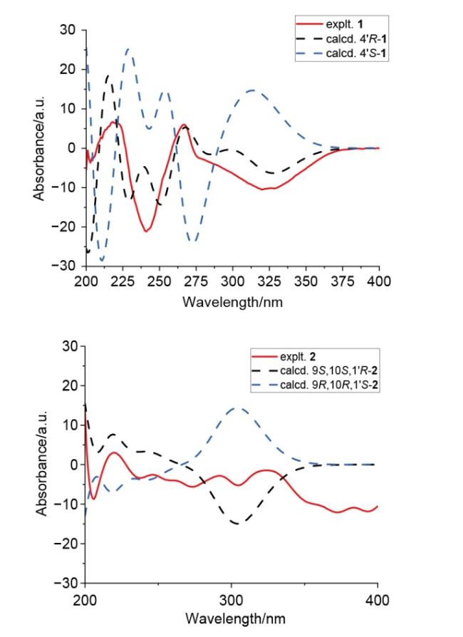
The 1H-1H COSY correlations (Figure 2) of H-1'/H-2'/ H-10', of H-4/4-OH, as well as H-6'/H-8'/H-9' combined with HMBC correlations between H-4' and C-2', C-3', C-5', C-6'; between H-6' and C-4', C-5' confirmed the structure of the branched chain. The HMBC correlations between H-1' and C-5 indicated the branched chain was located at C-5. The NOESY correlations (Figure 2) of H-1'/H-10' indicated the E-configuration of Δ2',3' thereby confirming planar structure of compound 1. The absolute configuration of 1 was deduced to be 4'R based on subsequent ECD calculations (Figure 3).[13]
Compound 2 was obtained as yellow gum. Its molecular formula was deduced to be C20H24O5 based on the HRESIMS ion peak at m/z 345.1698 [M+H]+ (calcd for C20H25O5+, 345.1697). The IR bands indicated the presence of hydroxyl (3428 cm-1) and carbonyl (1724 cm-1) groups. The 1D and 2D NMR spectra indicated compound 2 owns a coumarin moiety [δH: 7.64 (d, J=9.8 Hz, 1H, H-4), 7.48 (s, 1H, H-5), 6.81 (s, 1H, H-8), 6.25 (d, 1H, J=9.8 Hz, H-3); δC: 164.0 (C-7), 161.2 (C-2), 157.2 (C-8a), 143.8 (C-4), 125.3 (C-6), 124.6 (C-5), 113.2 (C-4a), 112.9 (C-3), 98.8 (C-8)]; along with a trisubstituted double bond [δH: 5.14 (t, J=7.2 Hz, 1H, H-4'); δC: 132.9 (C-5'), 123.9 (C-4')]; two oxy-methines [δH: 5.08 (d, J=9.8 Hz, 1H, H-9), 4.55 (d, J=4.7 Hz, 1H, H-10); δC: 94.5 (C-10), 80.1 (C-9)]; an oxy-methyl [δH: 3.44 (s, 3H, 9-OMe); δC: 55.7 (9-OMe)]; two methylenes [δH: 2.17 (q, J=8.1 Hz, 2H, H-3'), 1.57~1.58 (m, 2H, H-2'); δC: 37.9 (C-2'), 22.1 (C-3')]; and three methyls [δH: 1.70 (s, 3H, H-6'), 1.65 (s, 3H, H-7') and 1.20 (s, 3H, H-8'); δC: 25.9 (C-6'), 22.3 (C-8'), 17.9 (C-7')].
The 1H-1H COSY correlations (Figure 2) of H-9/H-10 along with the HMBC correlations (Figure 2) between H-9 and C-5, C-6; between H-10 and C-6, C-7 indicated the presence of a dihydrofuran ring, which contributes to the formation of a linear furanocoumarin moiety. The 1H-1H COSY correlations of H-3'/H-4' /H-5', H-5'/H-7', and H-5'/H-8' combined with the NOESY correlations (Figure 2) of H-3'/H-7' and H-4'/H-6' formed the structure of the branched chain. The HMBC correlations between 9-OMe and C-9 as well as the 1H-1H COSY correlations of 9- OMe/H-5 confirmed the location of the oxy-methyl group. Additionally, the HMBC correlations between H-8' and C-1', C-2', and C-10 ultimately confirmed the planar structure of 2. The coupling constants between H-9 and H-10, approximately 4.5 Hz, indicated that these protons are in different orientation relative to the furan ring.[12] The relative configuration of 2 was deduced to be 9S*,10S*, 1'R* based on DP4+ analysis. The absolute configuration of 2 was finally confirmed to be 9S,10S,1'R by subsequent ECD calculation.
Compounds 1 and 2 were evaluated for their anti-infla- mmatory potential by assessing their inhibition of nitric oxide (NO) expression in lipopolysaccharide (LPS)-stimu- lated RAW 264.7 cells. Compound 1 exhibited a stronger inhibitory effect, reducing NO levels at a concentration of 7.5 µmol/L, which was superior to that of the positive control, dexamethasone (DXM).
3 Conclusions
Two new linear furocoumarins were successfully isolated from N. incisum. Their structural elucidation was achieved by comprehensive spectroscopic analysis combined with ECD calculation. Notably, compound 1 exhibited significant anti-inflammatory potential by suppressing the expression of key pro-inflammatory cytokines (NO) in LPS-stimulated RAW264.7 macrophages at a concentration of 7.5 μmol/L. These findings not only expand the chemical diversity of furocoumarins derived from N. incisum but also highlight compound 1 as a promising candidate for further exploration of its anti-inflammatory mechanisms and therapeutic applications.
4 Experimental section
4.1 General experimental procedures
Optical rotations were measured using a Perkin-Elmer 241 Polarimeter (Perkin Elmer, Inc., Waltham, MA, USA). UV and CD spectra were recorded on a Chirascan circular dichroism spectrometer (Applied Photophysics Ltd., Leat- herhead, UK). The IR spectra were acquired using an Agilent Cary 600 FT-IR infrared spectrometer (Agilent Tech- nologies, Santa Clara, USA). Mass spectra were obtained from an ultra-performance liquid chromatography system coupled with a Q Exactive Quadrupole-Electro-static Field Orbital Trap high-resolution mass spectrometer (Thermo Fisher Scientific, Bremen, Germany). NMR spectra were measured with a Bruker Ascend 600 MHz spectrometer (Bruker, Karlsruhe, Germany), utilizing TMS as the internal standard. Fractionation was conducted using column chromatography with silica gel (200~300 mesh). The samples were fractionated by a dynamic axial compression column (Hanbon Sci. & Tech, Huaian, China). Purification was performed using a NP7000 preparative high-perfor- mance liquid chromatograph (Hanbon Sci. & Tech, Huai- an, China), equipped with an C18 5 μm semipreparative column (10 mm×250 mm). Methanol, acetone, and ethyl acetate were obtained as analytical grade, while dimethyl sulfoxide (chromatographic grade) was purchased from Chengdu Kelong Chemical Co., Ltd. Dichloromethane (analytically pure) and methanol (chromatography preparative grade) were sourced from Chengdu Jinshan Chemical Reagent Co., Ltd.
4.2 Material
The roots and rhizomes of N. incisum were obtained from Sichuan Neautus Pharmaceutical Co., Ltd. and authenticated by Guang-Zhi Wang, a professor of the School of Pharmacy, Chengdu University of Traditional Chinese Medicine. A specimen (20221225) has been deposited in the Laboratory of Traditional Chinese Medicine Chemistry, School of Pharmacy, Chengdu University of Traditional Chinese Medicine.
4.3 Extraction and isolation
30 kg of N. incisum roots and rhizomes were powdered and extracted using 95% ethanol, resulting in the recovery of 3.3 kg of dried extract. This extract was then dispersed in pure water and applied to an activated D101 macropo- rous resin column (80 mm×1200 mm). The mixture was allowed to adsorb and was subsequently washed overnight with a methanol/water gradient (0∶100, 30∶70, 60∶40, 95∶5) as the mobile phase. Fractionation was performed with five column volumes for each gradient elution. The portion eluted with 60% methanol (101.3 g) was placed in a silica gel column and eluted with a mixture of petroleum ether and acetone (V∶V=100∶0~50∶50), yielding eight fractions (Fr.1 to Fr.8). Fraction 3 (18.5 g) was further processed on a silica gel column using petroleum ether and ethyl acetate as mobile phases (V∶V=100∶0~50∶50), resulting in sixteen sub-fractions (Fr.3.1~Fr.3.16), Fr3.6 was separated using a Sephadex LH-20 column and eluted with dichloromethane and methanol (V∶V=50∶50) generating five sub-fractions (Fr.3.6.1~Fr.3.6.5). Fr.3.6.4 was further purified by semi-pHPLC using a C18 column (5 μm, 10 mm×250 mm) and afforded compound 1 (methanol-water: V∶V=60∶40, 3 mL/min, tR=51.0 min, 1.38 mg). Fraction 4 (22.3 g) was further processed on a silica gel column using petroleum ether and ethyl acetate as mobile phases (V∶V=100∶0~50∶50), resulting in twelve sub-fractions (Fr.4.1~Fr.4.12), Fr.4.7 was separated using a Sephadex LH-20 column and eluted with dichloromethane/methanol (V∶V=50∶50) generating five sub-fractions (Fr.4.7.1~Fr.4.7.5). Fr.4.7.4 was further purified by semi-pHPLC using a C18 column (5 μm, 10 mm×250 mm) and yielded compound 2 (methanol-water: V∶V=62∶38, 3 mL/min, tR=39.0 min, 1.12 mg).
Notoprenylate L (1): Pale yellow amorphous powder. $[\alpha]_{\mathrm{D}}^{20}$-100.0 (c 0. 046, MeOH); UV (MeOH) λmax: 221 (3.05), 246 (2.66), 265 (1.60), 310 (1.15) nm; CD (c 1.41 mmol/L, MeOH) λmax (Δε): 218 (6.72), 241 (-21.19), 267 (6.01), 320 (-10.48) nm; 1H NMR (600 MHz, CDCl3) δ: 8.17 (d, J=10.3 Hz, 1H), 7.61 (d, J=2.3 Hz, 1H), 7.18 (s, 1H), 6.95 (d, J=1.3 Hz, 1H), 6.29 (d, J=9.8 Hz, 1H), 6.07~6.05 (m, 1H), 6.00 (t, J=5.8 Hz, 1H), 5.07 (d, J=6.7 Hz, 3H), 4.52 (d, J=4.1 Hz, 2H), 4.23 (d, J=4.4 Hz, 1H), 2.24 (s, 3H), 1.91 (s, 3H), 1.55 (s, 3H); 13C NMR (151 MHz, CDCl3) δ: 197.65, 161.68, 161.24, 158.30, 152.82, 148.63, 145.27, 139.35, 139.31, 126.31, 118.40, 113.95, 113.06, 107.46, 104.98, 94.63, 82.51, 69.15, 28.29, 21.71, 11.81; IR (KBr) ν: 3428, 2925, 2852, 1732, 1629, 1456, 1390, 1204, 1132, 1076, 1029, 827, 750 cm-1; HRESIMS calcd for C21H23O5 [M+H]+ 369.1333, found 369.1328.
Nototerprinol K (2): Pale yellow amorphous powder; $[\alpha]_{\mathrm{D}}^{20}$+2.5 (c 0. 040, MeOH), UV (MeOH) λmax: 203 (1.85), 220 (1.18), 293 (0.35), 326 (0.39) nm; CD (c 1.41 mmol/L, MeOH) λmax (Δε): 204 (3.09), 226 (-0.70) nm; 1H NMR (600 MHz, CDCl3) δ: 7.64 (d, J=9.5 Hz, 1H), 7.48 (s, 1H), 6.81 (s, 1H), 6.25 (d, J=9.5 Hz, 1H), 5.14 (t, J=7.2 Hz, 1H), 5.08 (s, 1H), 4.55 (s, 0H), 3.49 (s, 1H), 3.44 (s, 2H), 2.17 (q, J=8.2 Hz, 2H), 1.70 (s, 3H), 1.65 (s, 3H), 1.20 (s, 3H); 13C NMR (151 MHz, CDCl3) δ: 163.99, 161.19, 157.17, 143.82, 132.87, 125.30, 124.55, 123.89, 113.17, 112.94, 98.80, 94.53, 80.09, 73.48, 55.74, 37.87, 25.88, 22.34, 22.05, 17.88; IR (KBr) ν: 3428, 2923, 2850, 1724, 1625, 1392, 1124 cm-1; HRESIMS calcd for C20H25O5 [M+H]+ 345.1697, found 345.1698.
4.4 CCK-8 assay
RAW264.7 cells were procured from the Cell Resource Center at the Shanghai Institutes for Biological Sciences, Chinese Academy of Sciences. The cells were cultured in dulbecco’s modified eagle medium (DMEM) supplemented with 10% fetal bovine serum (FBS) and 1% penicillin-streptomycin at 37 ℃ in a humidified atmosphere containing 5% CO2. RAW264.7 macrophages were seeded at a density of 5×10³ cells per well in a 96-well cell culture plate. After an overnight incubation, the old DMEM was discarded, and fresh DMEM containing various concentrations of compounds 1 or 2 was introduced, with dimethyl sulfoxide (DMSO) serving as a blank control. Following a 24-hour incubation period, 10 µL of CCK-8 reagent was added to each well and incubated for 1 h. Finally, the absorbance was measured using a microplate reader (Thermo Scientific, N16443, USA) at a wavelength of 450 nm.
4.5 Measurement of NO production
RAW264.7 macrophages were seeded at a density of 2×10⁵ cells per well in a 6-well plate and incubated overnight. The cells were subsequently treated with DMSO (Gibco), 30 µmol/L DXM (Dexamethasone, Beyotime), or compound 1 or 2 at concentrations of 7.5, 15, and 30 µmol/L for a duration of 2 h, followed by stimulation with 1 µg/mL LPS (Beyotime). After a 24-hour incubation period, NO production was assessed using the Nitric Oxide Assay Kit (Beyotime, S0021S, China).
Supporting Information HRESIMS, NMR, IR and UV of compounds 1 and 2, and details of chemical calculation. The Supporting Information is available free of charge via the Internet at http://sioc-journal.cn.
(Cheng, F.)