

1 Introduction
Streptomyces species are renowned as prolific producers of structurally diverse natural products with broad bioactivities, particularly in antibiotic production.[1] Whole- genome sequencing and bioinformatics analyses have demonstrated that these microorganisms possess large linear chromosomes harboring more than 20 secondary metabolite biosynthetic gene clusters (BGCs), with over 80% remaining cryptic or poorly characterized.[2] In the post- genomic era, microbial genome mining has emerged as a powerful alternative to conventional approaches for discovering novel secondary metabolites.[3] However, a major challenge in genome mining lies in the fact that most BGCs remain transcriptionally silent under standard laboratory cultivation conditions. This necessitates the development of innovative strategies to activate these silent gene clusters,[4] of which Streptomyces antibiotic regulatory proteins (SARPs) represent a class of pathway-specific transcriptional activators that play pivotal roles in regulating antibiotic biosynthesis in Streptomyces species.
Streptomyces aureus SP-371, the known producer of agricultural-bactericide aureonuclemycin, exhibits significant potential for secondary metabolite biosynthesis as evidenced by the identification of ca. 34 biosynthetic gene clusters (BGCs) through anti-SMASH genome analysis.[5-7] Previous research on this strain led to the discovery of eight aromatic polyketides featuring two types of frameworks, two pentacyclic isomers and six glycosylated tetracyc- lines[8] and one spiro-naphthoquinone[9] by overexpressing the regulatory SARP gene, along with aureonuclemycin. In our continuing efforts to explore the structural diversity of natural products from S. aureus SP-371, we chemically investigated its ethyl acetate extract from large-scale fermentation, leading to discover a new sesquiterpene-lactone 1 (Figure 1), neopentalenolactone D1, bearing a unique carboxamide functional group. Herein, we reported the isolation, structural elucidation, and preliminary evaluation of biological activities for compound 1.
2 Results and discussion
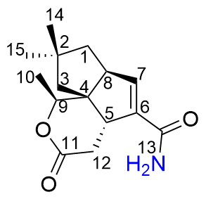
Compound 1 was obtained as a white amorphous powder. Its molecular ion peak was determined by HRESIMS at m/z 264.1594 [M+H]+ (calcd for C15H22NO3 264.1600), corresponding to a molecular formula C15H21NO3 with six degrees of unsaturation. The ¹H NMR spectrum displayed characteristic signals including two singlet methyl groups at δH 1.06 (s, 3H) and 1.07 (s, 3H), one doublet methyl group at δH 1.38 (d, J=6.4 Hz, 3H), and one olefinic proton at δH 6.50~6.52 (m, 1H). The ¹³C NMR spectrum showed 15 distinct carbon signals, featuring: one ester carbonyls (δC 176.05), one amide carbonyl (δC 169.59), two olefinic carbons (δC 140.15 and 143.94), and one oxygenated methine (δC 80.18) (Table 1). The 1H-1H correlation spectroscopy (COSY) spectrum indicated the presence of three isolated proton sequences of H2-1/H-8/H-7, H-9/H3-10 and H-5/ H2-12 (Figure 2). The above NMR data suggests that 1 is a sesquiterpene structurally analogous to neopentalenolactone D (previously identified as its methyl ester[10-11]) except for the major chemical shift differences observed at C-13 (δ +2.23), C-6 (δ +2.51), and C-7 (δ -5.71). The heteronuclear multiple bond correlations (HMBC) observed between H-9 to C-10 and C-11, and H-1 to C-7, and H-7 to C-13 provided further confirmation that 1 shares the same molecular skeleton with neopentalenolactone D. In combine with the molecular formula, the number of hydrogens directly bonded to carbon and the index of unsaturation, there is an NH2 group to be assigned. Based on the molecular scaffold established by 1D and 2D NMR, it is reasonable that the extra NH2 group was linked to C-13 carbonyl, which was confirmed by the chemical shift differences at C-13, C-6, C-7, thus the chemical structure of compound 1 was established as the amide derivative of neopentalenolactone D (Figure 2).
Table 1 1H NMR (400 MHz) and 13C NMR (100 MHz) spectroscopic data of 1 in CD3OD |
| No. | δC | δH |
|---|---|---|
| 1 | 45.24 (CH2) | 1.71 (dd, J=13.3, 10.2 Hz, H1α), 1.53~1.57 (m, H1β) |
| 2 | 42.36 (C) | |
| 3 | 48.98 (CH2) | 1.42~1.46 (m, H3β), 1.95~2.01 (m, H3α) |
| 4 | 58.82 (C) | |
| 5 | 54.07 (CH) | 3.31~3.33 (m, 1H) |
| 6 | 140.15 (C) | |
| 7 | 143.94 (CH) | 6.50~6.52 (m, 1H) |
| 8 | 56.96 (CH) | 3.09 (ddd, J=10.0, 5.6, 2.8 Hz, 1H) |
| 9 | 80.18 (CH) | 4.52 (q, J=6.4 Hz, 1H) |
| 10 | 15.57 (CH3) | 1.38 (d, J=6.4 Hz, 1H) |
| 11 | 176.05 (C) | |
| 12 | 34.01 (CH2) | 2.95 (dd, J=15.2, 6.9 Hz, H12β), 2.51 (dd, J=15.1, 10.7 Hz, H12α) |
| 13 | 169.59 (C) | |
| 14 | 31.59 (CH3) | 1.07 (s, 3H) |
| 15 | 29.48 (CH3) | 1.06 (s, 3H) |
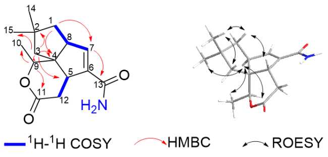
The relative configuration of compound 1 was unambiguously established through rotating frame overhauser effect correlations (ROESY) experiments. The nuclear overhauser effect spectroscopy (NOESY) of H3-14 with H-1β/H-5/H-3β, H-5 with H-3β/H-12β, indicated that H3-14, H1β, H-3β, H-5 and H-12β were in β-orientation, while the ROESY correlations of H3-15 with H-1α/H-3α, H-8 with H-9 indicated that H3-15, H1α, H-3α, H-8 and H-9 were in α-orientation (Figure 2). Then its relative configuration was deduced as shown in Figure 1, which is consistent with neopentalenolactone D. Here we named compound 1 as neopentalenolactone D1.
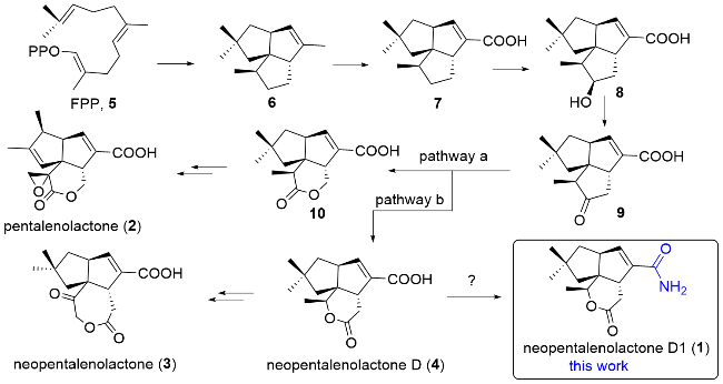
Pentalenolactone-type compounds are widely occurring sesquiterpenoid antibiotics, including pentalenolactone (2) that has been isolated from a variety of Streptomyces species[10,12-14] and neopentalenolactone (3).[10] Multiple pentalenolactone BGCs have been identified, including pen from S. exfoliatus, pnt from S. arenae, ptl from S. avermitilis and pll from Streptomyces sp. Northern Regional Research Laboratory (NRRL) S-4. The biochemical functions of open reading frames (ORFs) as well as the biosynthetic pathway have been well characterized,[15-18] of which pentalenolactone (2) and neopentalenolactone (3) are biosynthesized from the common precursor 9 through pathway a and pathway b, respectively (Scheme 1). A high number of biosynthetic intermediates and shunt metabolites in the conversion from farnesyl pyrophosphate (FPP, 5) to pentalenolactone have been isolated from several pentalenolactone producers.[15,19-21] Since neopentalenolactone D1 was a derivative of neopentalenolactone D, we hypothesized that there is a BGC responsible for neopentalenolactone biosynthesis. From the anti-SMASH analysis, we identified one terpene BGC, here designated as the psu cluster, which is predicted to synthesize neopentalenolactone D. The ca.12-kb psu biosynthetic gene cluster (BGC) contained 10 open reading frames (ORFs), showing an average sequence similarity >65% to those of the pnt cluster from S. arenaria and the ptl cluster from S. avermitilis. Neopentalenolactone D1 may be the shunt metabolite of neopentalenolactone D divergent from precursor 9 in pathway b. As the amidation of terminal carboxylic acid was an enzymatic reaction in organism, such as asparagine synthase in primary metabolism. It is possible that an amidotransferase enzyme existed beyond the cluster responsible for amidation because there are no gene encoding amidotransferase in psu gene cluster.
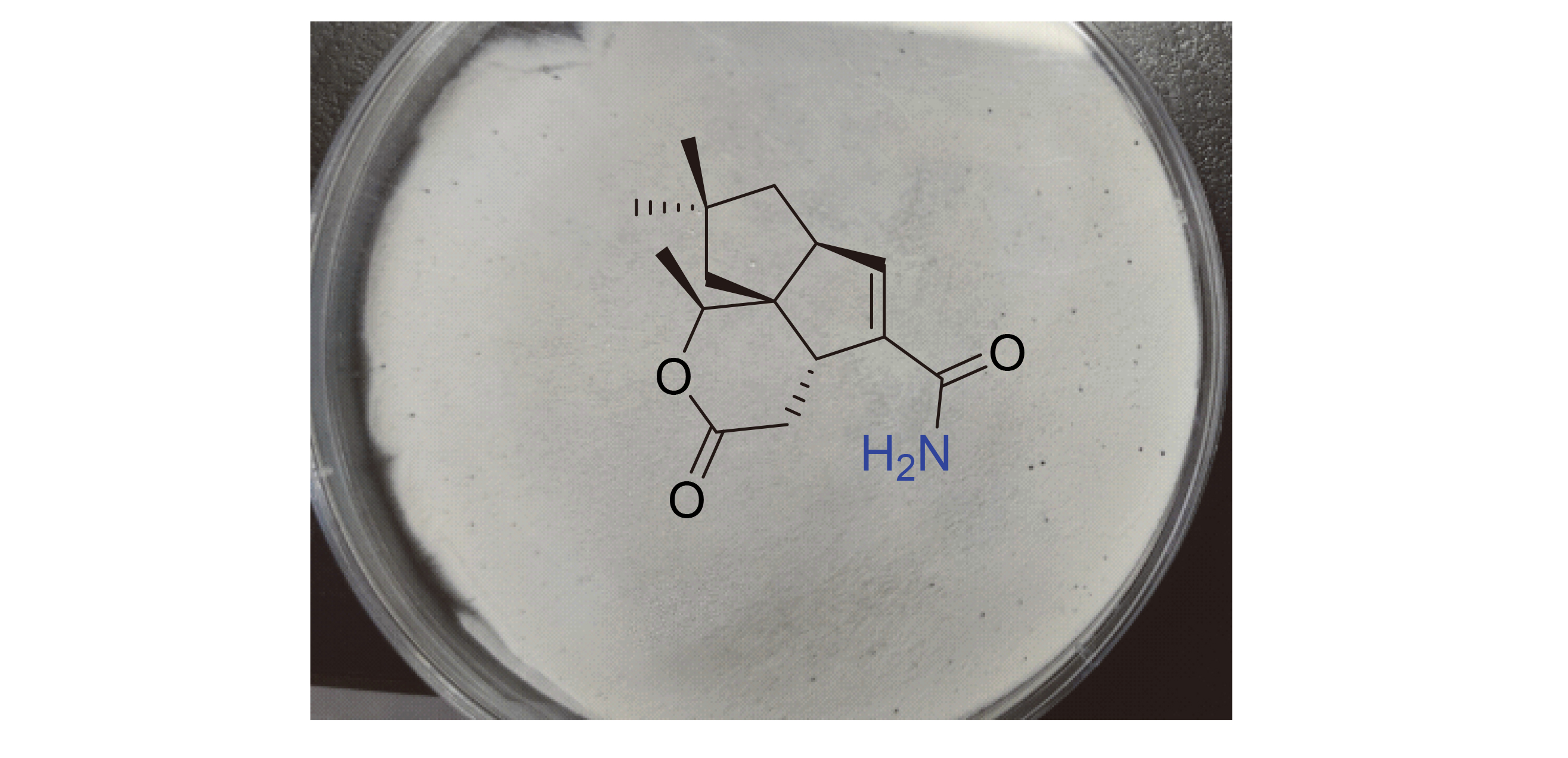
As pentalenolactone-type compounds exhibit antimicrobial action against bacteria, fungi, and protozoa through reaction of their electrophilic epoxy moiety with the active site cysteine of glyceraldehyde-3-phosphate dehydrogenase.[22] Subsequently, compound 1 was evaluated for possible antibacterial activity against Gram-positive bacteria (Staphylococcus aureus and Bacillus subtilis) and Gram-negative bacteria (Escherichia coli), with kanamycin as positive control. The experimental results indicated that the compound 1 exhibited no detectable antibacterial activity, which is consistent with the lacking of the epoxide pharmacophore required for the inactivation of GAPDH by pentalenolactones.
3 Conclusions
In our investigation of natural product diversity from Streptomyces aureus SP-371, neopentalenolactone D1, an amide derivative of neopentalenolactone D, was isolated and identified. It might be a shunt metabolite in the neopentalenolactone biosynthesis pathway. Its possible gene cluster psu was identified from genome of SP-371 by bioinformatic analysis. Notably, the amidation of C-6 carboxylic acid in 1 was likely catalyzed by amidotransferase like CerD,[23] TtmN[24] in Cerulenin and thiotetroamide C biosynthesis respectively. However, there are no amidotransferase-encoding gene within psu cluster. The dedicated enzyme for amidation will be investigated in the future.
4 Experimental section
4.1 General experimental procedures
Mass Spectrometer Thermo Scientific LCQ Fleet Ion Trap, High-Performance Liquid Chromatograph Dionex Ultimate 3000, High-Resolution Mass Spectrometer Agilent Technologies 6230 TOF LC/MS, Nuclear Magnetic Resonance Spectrometer Bruker AVANCE DRX-500, Analytical Chromatographic Column Grace AlltimaTM (5 μmol•L-¹, C18 Column, 4.6 mm×250 mm), Semi-prepara- tive Column YMC-Pack ODS-AQ (5 μm, C18 Column, 10 mm×250 mm). Biological reagents were purchased from New England Biolab, Thermo, and Takara. Chemical reagents were sourced from Shanghai Pharmaceutical Group Co., Ltd., J&K Scientific Ltd. Imported chemical reagents were purchased from Sigma-Aldrich and TCI. Deuterated reagents were obtained from Cambridge Isotope Laboratories. Streptomyces aureus SP-371 was provided by Professor Tao Liming from East China University of Science and Technology.
4.2 Fermentation, isolation and identification
S. aureus were grown in tryptic soy broth (TSB, 3%) at 30 ℃ for 24 h as a seed culture, then 5 mL of seeding culture suspension was transferred into a 500-mL flask containing 100 mL fermentation broth (1% soy bean, 1% peanut meal, 5% glucose, 2% corn starch, 0.6% NH4NO3, 0.3% NaCl, 0.6% CaCO3, pH=7.2) and the flask was cultured at 30 ℃ and 220 r/min for an additional 5 d. At last, the culture broth was centrifuged to yield a supernatant and a mycelium cake. The supernatant was extracted three times with an equal volume of ethyl acetate and then evaporated to dryness. The mycelium was extracted twice with acetone, followed by evaporation of the acetone. The remaining aqueous phase was extracted with an equal volume of ethyl acetate at least three times. The ethyl acetate soluble extract was subjected to silica gel column chromatography (CHCl3- MeOH, V∶V 100∶1, 50∶1, 20∶1, 10∶1) to yield five fractions (Fr.1~Fr.5), Fr.3 was separated by Sephadex LH20 (MeOH) semi-preparative HPLC (10 mm×250 mm, 5 µm, MeCN-H2O 42:58, 4 mL/min) successfully to yield neopentalenolactone D1 (1) (7 mg). White amorphous powder; 1H NMR and 13C NMR see Table 1; HRESIMS calcd for C15H22NO3 [M+H]+ 264.1600, found 264.1594.
4.3 Petri dish diffusion assays
Filter paper disks impregnated with varying concentrations of 1 were placed onto MS solid media, that had been pre-inoculated with Staphylococcus aureus, Bacillus subtilis and Escherichia coli (DH5α). Following incubation at 30 ℃ for 36 h, the antimicrobial potency of 1 was assessed by measuring the diameters of the inhibition zones formed around the disks.
Acknowledgments We thank Professor Tao Liming from East China University of Science and Technology for providing the S. aureus SP-371 strain.
Supporting Information HR-ESI-MS and NMR spectra of compound 1. The Supporting Information is available free of charge via the Internet at http://sioc-journal.cn.
(Zhao, C.)