


1 Introduction
In recent years, a large number of new fluorescent dyes with excellent photo-physical and photo-chemical properties have been developed in the fields of basic and applied research, which are used in ion probes,[1] fluorescent sensor,[2] information encryption,[3] and photo dynamic therapy.[4] These dyes include alkoxy-substituted fluorescent dyes,[5] bithiophene-based fluorophore derivatives,[6] bisarylic methanone derivative,[7] and compounds with 1,8-naphthalimide portion,[8] anthracene fragment,[9] fluorene skeleton,[10] triphenylamine group,[11-12] carbazole unit,[13] and difluoroboron moiety[14] and so on. Among them, difluoroboron compounds have been received more and more attention due to their easily modifiable molecular structure, convenient synthesis, high chemical stability, and adjustable optical performance.[15-17] However, most of fluorescent dyes emit strong fluorescence in the solution but weak luminescence in the solid or aggregated states owing to their rigid molecular structure and excellent molecular planarity,[18] which is known as aggregation-caused quenching (ACQ). It greatly limits the application of fluorescent dyes.
To overcome this difficulty, researchers introduce twi- sted conformational groups into the molecular skeleton to obtain aggregation-induced emission (AIE) molecules. After years of continuous research, AIE molecules have been applied in diverse fields including anti-counterfeiting and latent fingerprint (LFP) imaging. In the field of LFP detection, Lee and his co-workers[19] synthesized an AIE- active difluoroboron compound (LFP-1) with tetraphen- ylene (TPE) unit, which prepared developer (ethanol, 25 μmol/L) could clearly detect the characteristic details of LFP, for example, core, bifurcation, island, ridge ending, and pore and so forth. Lu et al.[20] reported that N,O-chelated difluoroboron complex (TCPB) could be made LFP visualization on the surface of glass under a 365 nm UV lamp by spraying powder. Compared with the powder method, which can easily compromise LFP integrity during powder removal, the solution imaging method, where the difluoroboron compound is dissolved in a mixture of good and poor solvents exploiting its AIE property, is widely used for LFP imaging. Furthermore, the imaging mechanism of LFP is widespread acceptance to hydrophobic-hydrophobic interaction between organic dyes and the residue of LFP. The components of residue mainly include inorganic salt (NaCl), amino acids mainly composed of glycine, grease mainly composed of oleic acid, urea, cholesterol, lactic acid, and glucose.[21] According to Tu’s[22] and Wang’s[23] reports, the developers based on the terpyridine zinc pincer complex [Zn(tpy-NMe2)] and TPE-based pyridinium salt (TPE-2Py-2OH), respectively, only had strong fluorescence responses in the presence of oleic acid. However, Ma and Zhu’s group[21] declared that the obtained TPE-naphthalimide-N-hydroxysuccinimide (TPE- NI-NHS) had excellent fluorescence responses to glycine, lysozyme, oleic acid, and cholesterol. For difluoroboron compounds, it is rarely explored and reported. On the other hand, mechanofluorochromism (MFC) behavior based on strong fluorescence in the solid is also investigated. Especially, the practical application in the field of anticounter- feiting can be further developed.
In this work, we designed and synthesized a novel difluoroboron compound TPA-2 featuring a triphenylamine (TPA) unit as the electron donor and a pyrazole- difluoroboron moiety as the electron acceptor. This donor- acceptor architecture not only facilitates intramolecular charge transfer (ICT) but also introduces a twisted molecular conformation, which is crucial for suppressing ACQ and enabling both AIE and MFC. The incorporation of the pyrazole ring further enhances the electron-accepting ability and stabilizes the aromatic system,[24] thereby promoting solid-state luminescence and stimuli-responsive behavior. The ability of compound TPA-2 to serve as a rewritable security material and a highly sensitive developer for LFP imaging on both porous and non-porous substrates represents a significant advancement in the development of multifunctional fluorescent materials.
2 Results and discussion
2.1 Molecular synthesis and characterization
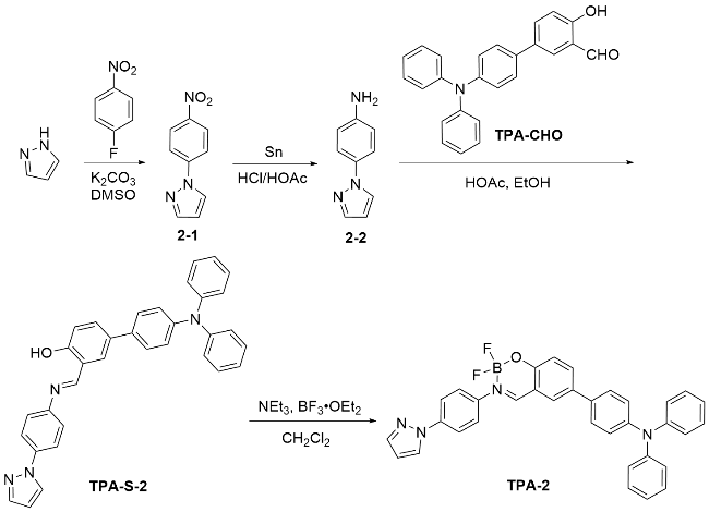
Schiff base compound TPA-2 was synthesized through nucleophilic condensation reaction between compounds 2-2 and TPA-CHO, then the target difluoroboron compound TPA-2 was obtained through coordination reaction between compound TPA-S-2 and boron trifluoride ether. The detailed synthesis processes were outlined below, and the synthetic route was illustrated in Scheme 1. NMR spectra and HRMS result could be found in Figures S1~S3 of Electronic Supplementary Information (ESI).
2.2 UV-vis absorption and fluorescent emission spectra in solutions
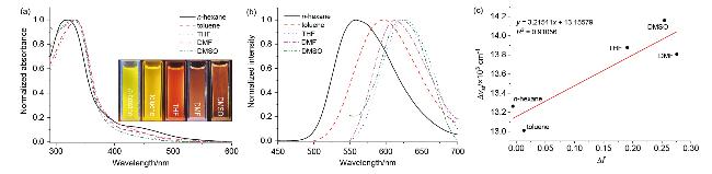
The UV-vis absorption and fluorescence emission spectra of compound TPA-2 in solutions were measured, and the corresponding data were listed in Table S1 (ESI). As seen from Figure 1a, only one dominant absorption band was observed around at 330 nm, ascribing to the ICT transition between the electron donator (TPA group) and the electron acceptor (difluoroboron core and pyrazole moiety). And they displayed minimal spectral shifts as the polarity of the solvent increases. However, significant change in the emission spectra were observed: a red-shift from 559 nm in non-polar n-hexane to 597 nm in weakly polar toluene, and a further red-shift to 626 nm in moderately polar THF, as shown in Figure 1b. This behavior suggests ICT character in the excited state.[25] Additionally, the solvatochromism with naked eyes was observed under a 365 nm UV lamp. However, the emission band was blue-shifted to 613 nm (in dimethylformamide) when the polarity of the solvent further increases, indicating less conjugation degree owing to the larger dihedral angles in high polar organic solvent.[26] As in higher polarity dimethyl sulfoxide (DMSO) solvent, the emission band was again red-shifted to 623 nm. To better elucidate solvent-dependent behaviors, Lippert-Mataga fitting curve was plotted, which the orientational polarizabilities (Δf) were as the abscissa, and the Stokes shifts (Δvst) were as ordinate, as shown in Figure 1c. It was found that the curve had a positive slope and good linear correlation, illustrating that it possessed larger dipole moment of the ICT excited state due to the substantial charge redistribution, and the solvent-dependent fluorescence shifts were predominantly caused by the dipole- dipole interaction between compound TPA-2 molecules and organic solvents.[27]
2.3 AIE property
The AIE properties were investigated in DMSO/water mixtures owing to good dissolved in DMSO but poor solubility in water. As shown in Figure 2a, the profiles of absorption were almost unchanged when the volume fractions of water (fw) were increased from 0% to 50%, meanwhile, the changes of emission intensities were not significant, and the emission colors underwent discernible chan- ges, as shown in Figures 2b and 2c. At low fw, TPA-2 molecules were dispersed. The free rotation of the aryl units facilitated non-radiative relaxation of the excited states,[28] while increased solvent polarity also contributed to emission quenching.[29] Consequently, fluorescence was significantly weakened or effectively quenched. The absorption spectrum displayed level-off tail in the long wavelength region when fw reached 60% due to the Mie scattering effect, which suggested that the nano-aggregates were formed. Concomitantly, the broad and strong emission peaks were conspicuous at about 600 nm. It was attributed that the mutual restriction between the formed nano-aggregates restricted intramolecular rotation around the aryl units, which suppressed the non-radiative transition of the excited state energy.[30] Besides, the Фf in pure DMSO and the aggregate state (fw=60%) were tested, which were 0.0539% and 0.757%, respectively, as shown in Figure S4 (ESI). Therefore, the phenomena of emission increasing were observed. It was so-called AIE. The emission intensity continued to increase to its maximum when fw reached 100%, emitting pink luminescence under the UV lamp, as shown in Figure 2c. Meanwhile, the water- induced aggregation behavior was also investigated in CH3CN/water mixtures, as shown in Figure S5 (ESI). It was observed that the emission intensity reached maximum at fw=90%. There were the discrepant maxima of emission intensities for compound TPA-2 in different mixtures. It might be that it was related to selected different organic solvents.
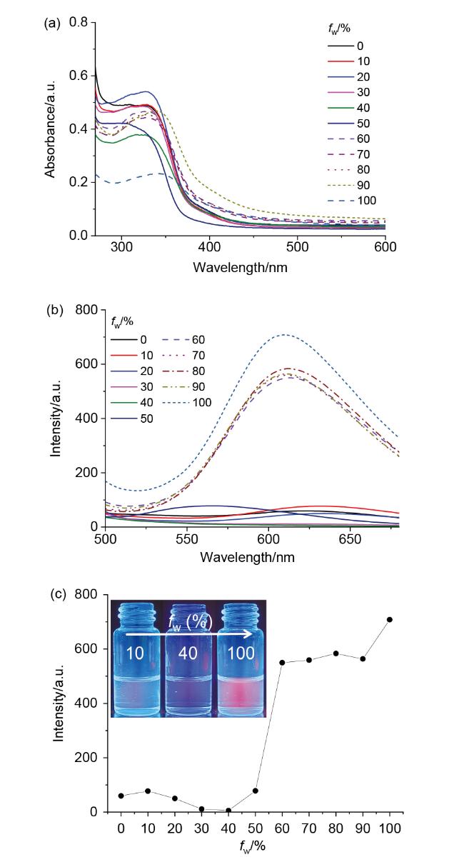
However, the tendencies of the AIE behaviors in DMSO/water and CH3CN/water mixtures were differences, due to the different solvent polarities and viscosities of DMSO and CH3CN. As is well known, the solvent polarity is largest for water (ε=80.1, μ=1.85 D) in three solvents, and that is smallest for CH3CN (ε=37.5, μ=3.92 D) and that is moderate for DMSO (ε=46.7, μ=3.96 D). The viscosities are 0.89, 1.99~2.20, and 0.34~0.37 mPa• s (25 ℃) for water, DMSO and CH3CN, respectively. In the strong polar DMSO/water mixture, the emission peak was located at 622 nm (excited at 370 nm) as fw=0%, and that was located at 613 nm as fw=60% (the aggregate state), respectively. In the weak polar CH3CN/water mixture, the emission peak was located at 627 nm (excited at 390 nm) as fw=0%, and that was located at 538 nm as fw=70% (the aggregate state). The blue-shift in the emission wavelength and enhancement in the emission intensity were induced by hydrophobic environment and the restricted intramolecular rotation in the aggregate state.[31] And compared with in pure solvent, the intensity exhibited nine times increase in the aggregate state of the DMSO/ water mixture, whereas it showed two times increase of the CH3CN/water mixture, indicating that AIE property had a significantly change with solvent polarity.[32] Furthermore, the viscosities were 0.89, 1.99~2.20, and 0.34~0.37 mPa•s (25 ℃) for water, DMSO and CH3CN, respectively. The increased viscosity was further hindrance the free motions of single bond within molecules, which rigidified their conformation and decreased their nonradiative energy loss rate. The significant fluorescence intensity increase appeared at fw=60% in DMSO/water mixture, while it occurred at fw=70% in CH3CN/water mixture. This was because the viscosity in DMSO/water mixture was larger than that in CH3CN/water mixture under the same other condition.[33]
To acquire more information about the formed aggregates in DMSO/water mixture, the morphology in an aggregated microenvironment was also investigated by scanning electron microscope (SEM), as shown in Figure S6 (ESI). It was found that although the solvent polarities and viscosities of DMSO/water and CH3CN/water mixtures were differences, the aggregates were all self-assembly as irregular cylindrical shape in aggregate states. It was played a crucial role in LFP imaging for the DMSO/water mixture of fw=60%.
2.4 MFC behavior
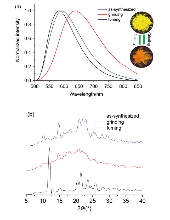
To verify the MFC behavior, the emission spectra of different solid states were tested. As shown in Figure 3, the solid-state emission wavelength was located at 583 nm for TPA-2, and the solid-state luminescence was yellow color. Moreover, the τ was measured to be 8.313 ns, as shown in Figure S7 (ESI). After grinding in a mortar for 30 min using pestle, the emission peak was red-shift to 638 nm. That was to said, the spectra underwent a 55 nm shifts. And the τ was changed to 5.349 ns. The more pronounced decrease in τ after grinding implied that TPA-2 exhibited higher contrast in emission color change upon mechanical stimulation, consistent with Tang’s[34] previous report. And the increased Фf was observed from 0.273% to 0.307%, as shown in Figure S8 (ESI), which indicated that compound TPA-2 was endowed with more compact packing after grinding. After fuming by using CH2Cl2 vapor for 30 s, the fluorescence color of ground TPA-2 could be converted to that of its as-synthesized state, although its emission spectrum was not restored to the initial state. The above resulted indicated that TPA-2 displayed reversibly MFC phenomenon. To better explain the mechanism underlying the MFC behavior, the powder XRD patterns in various solid states were analyzed. As shown in Figure 3b, it was found that the as-synthesized powder of TPA-2 displayed several sharp and intense diffraction peaks, indicating a well-or- dered crystalline state nature. The intensities of diffraction peaks weakened or even disappeared upon grinding, indicating a transition to a disordered amorphous state, because mechanical force destroyed the crystalline state and then the molecular packing underwent a transformation. Although the luminescent color was restored after fuming, the incomplete recovery of emission spectrum was originated from that some molecular packing arrangements were not fully revert to the original crystalline state, as shown in Figure 3b. This could be attributed to residual amorphous domains or slight conformational changes in the twisted TPA units that were not entirely reversible under mild fuming conditions. Such partial irreversibility highlighted the complexity of the molecular packing and the subtle balance between crystalline and amorphous phases in MFC materials.
2.5 Theoretical calculation
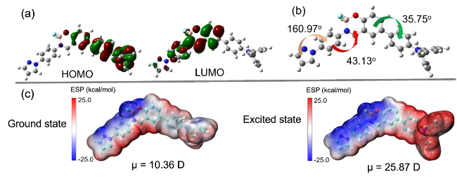
To gain deeper insights into the AIE property and me- chanochromism phenomenon of compound TPA-2, the frontier molecular orbital and optimized structural in the ground state, the electrostatic potential (ESP) surfaces and dipole moments (μ) in the ground and excited states were calculated. The computational details were provided in the ESI. As shown in Figure 4a, the calculated highest occupied molecular orbital (HOMO) was delocalized at TPA group, implying the electron-donating property of TPA unit. However, the lowest unoccupied molecular orbital (LUMO) was localized on around difluoroboron moiety and its surrounding group anywhere near pyrazole moiety, inferring their electron-withdrawing property. The clearly separate charge distribution was beneficial for the ICT process, which was consistent with the above-mentioned result of Figure 1. According to our time-dependent density functional theory (TD-DFT) calculations, the emission wavelengths in vacuum and DMSO were 460 and 481 nm, respectively. The theoretical 481 nm was closer to the experimental 623 nm when considering solvent effect. However, the discrepancy between theoretical and experimental values is probably due to the fact that some transitions violate Kasha’s rule. Furthermore, the oscillator strength (f) for S0→S1 transition and major orbital contributions were also calculated, as shown in Figure S9 and Table S2 (ESI). It was found that the f for S0→S1 transition was 0.2563 and the major orbital contribution from HOMO to LUMO was 57.1%. As shown in Figure 4b, the dihedral angle between five-membered N-heterocyclic ring and the bridged phenyl was 160.97º, and that of between the bridged phenyl and the coordinated difluoroboron ring was 43.13. At the same time, the dihedral angles between the phenyl adjacent to difluoroboron ring and the phenyl of TPA group was 35.75°. In the bargain, it was widely known that TPA group was a propeller-like conformation. Consequently, TPA-2 possessed twisted nonplanar configuration, which was propitious to intermolecular loose packing and weak π-π interaction. It is endowed with its potential property in the aspect of AIE and solid-state luminescence. Furthermore, the μ in the ground state was 10.36 D, as shown in Figure 4c. Compared to the ground state, the negative ESP region (blue color) expanded in the excited state, the μ in the excited state increased to 25.87 D. It implied that the molecular polarity in the excited state was larger than that of the ground state, and the change value was 15.52 D. It was a possibility that the relatively large μ and an obvious ICT effect were endowed with the significant MFC effect.[35]
2.6 Application in anti-counterfeiting and LPF detection
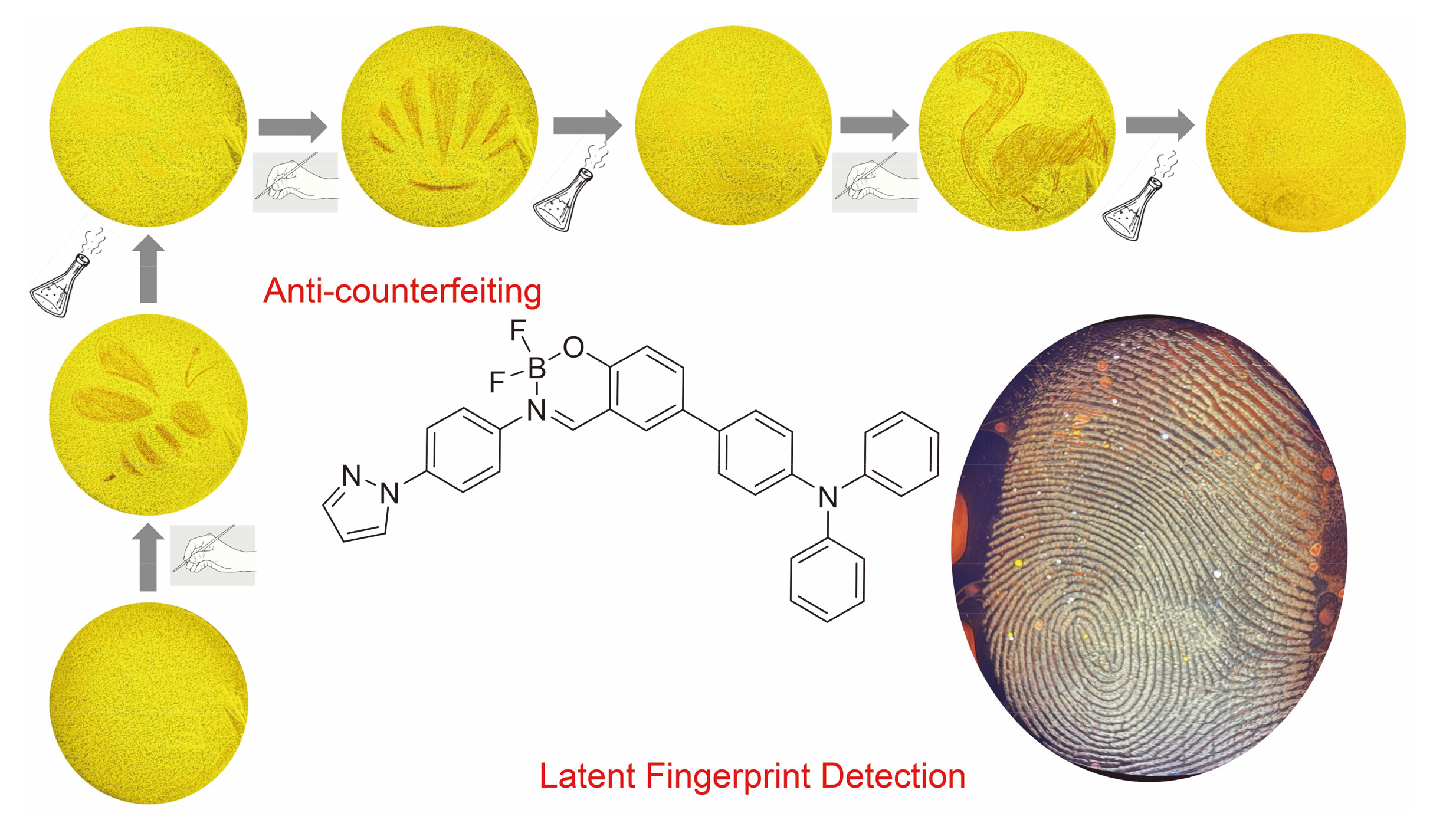
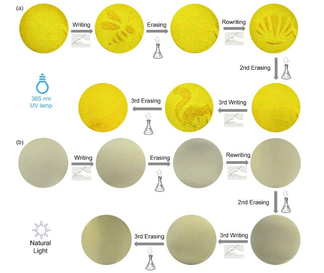
According to the reversible MFC phenomenon of compound TPA-2, its practical application in the field of anti-counterfeiting was explored. Firstly, the commercial Whatman filter paper was immersed into 10 mL of cyclohexane dissolving 10 mg of solid powder in the beaker, loading TPA-2 onto filter paper. And then filter paper was taken out using tweezers after 5 min and dried in the air. Finally, the so-called safety paper was obtained, and the luminescence color under a 365 nm lamp was closely matched its pristine solid-state fluorescence color. As shown in Figure 5a, the safety paper loaded with TPA-2 emitted yellow fluorescence. It was covered by a hollow out pattern of bee, and then slightly crushed using a spatula. A bee with emitting red light on the yellow background was observed, which was called the writing process. Afterward, the red bee disappeared after fumigation using CH2Cl2 vapor for 10 s. This process was called as erasing process. Subsequently, a hollowed-out scallop pattern was written again on the safety paper, which also emitted red fluorescence, and was then erased again using CH2Cl2 vapor. In the third attempt, a squirrel pattern emitting red fluorescence was written and then erased in the same way. Collectively, these results demonstrated that the safety paper prepared by TPA-2 was repeatedly rewritable. By contrast, the prepared safety paper was not significant change under natural light, as shown in Figure 5b, suggesting that it possesses good confidentiality in daily life. Furthermore, six cycles of ink-free writing were also implemented, as shown in Figure S10 (ESI). It was indicated that the safety paper prepared by TPA-2 had cycle durability.
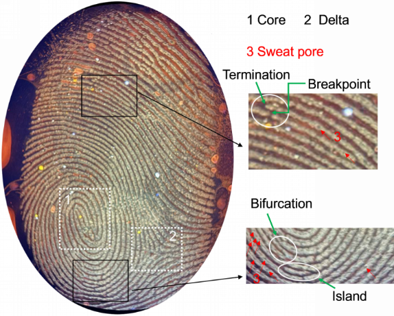
Not only that, its practical application in the field of LFP imaging was explored according to the AIE phenomenon. On this basis, the developer was prepared using DMSO/H2O mixture after dissolving TPA-2 (c=1×10⁻⁵ mol/L, fw=60%). LFP was deposited on the surfaces of tinfoil, glass plate, blade, leather, and cover paper by pressing a right thumb (pre-washed and rubbed on the forehead) onto each substrate. The developer was applied to the LFP area and removed after 10 min. The LFP image was captured using a Huawei mate30 phone, and the clear fingerprint patterns of LFP were observed under 365 nm UV lamp, as shown in Figure 6a. Whether on the surfaces of non-porous (tinfoil, glass plate and blade) or porous (leather and cover paper) substrates, the LFP image areas exhibited more conspicuous fluorescence signals of ridge due to the presence of sweat secretions. Some factors, such as the reflection of light and the hydrophobicity arising from the different surfaces, as well as the charge characteristics of molecule, were contributed to the LFP graphics displaying different luminescence colors.[19,22,36-37] As shown in Figure 6b, LFP without developer treatment appeared as vague gray traces, making specific details difficult to distinguish. Compared with the original LFP, the fluorescence signals were much brighter and more accurate to visualize after using developer, and the characteristic details of fingerprint were also easier to identify and analyze with the naked-eyes under a UV lamp. Taking tinfoil as an example, as shown in Figure 7, the fingerprint details of right thumb could be clearly identified, including the level I (core and delta), level II (termination, breakpoint, bifurcate and island), and level III (sweat pore). Among them, the level I and level II were vein and macroscopic characteristics, respectively, but the level III was microscopic characteristics. In daily life, especially at the crime scene, LFP is usually incomplete and/or damaged, and level III features can provide quantitative data for individual identification.[38] Compared with commercial methods (such as ninhydrin visualization method, as shown in Figure S11, ESI),[39] LFP visualization using the developer of TPA-2 is clearer and easier to identify from on the surface of the tinfoil. Moreover, it displays high sensitivity, good resolution and more complete fingerprint contour.
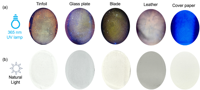
Figure 6 Fluorescence images of LPF on different substrates developed by compound TPA-2 dissolving in DMSO/H2O mixture (a) under a 365 nm UV lamp and (b) the original LFP without using developers under natural light, respectively |
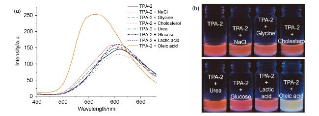
To evaluate the stability of LFP imaging, the real-time fluorescence imaging experiment was explored, as shown in Figure 8. It was found that the furrows and ridges could be clearly distinguished in photographs for 72 h, suggesting that the prepared developer exhibited excellent photostability. The luminescence colors with chromatic aberration were possibly derived from the evaporation of sweat secretions, and/or the role of oxygen and trace water upon exposure to air. Furthermore, the plausible mechanism of the developer based on TPA-2 for visualization of LFP was explored. As shown in Figure 9a, only the sample containing oleic acid resulted in the enhancement of fluorescence intensity and red-shift of emission wavelength. And its fluorescent color was clearly different from other components by comparison, as shown in Figure 9b. According to previous reports,[40-41] it could be originated from the lipophilicity and hydrophobicity of TPA-2, and low polarity and high viscosity of oleic acid. This inhibited the twisted ICT process in TPA-2.[38] The restricted twisted ICT inside the molecules and hydrophobicity (or lipophilicity) interaction between the molecules and oleic acid were proposed as possible mechanisms for LFP visualization n.
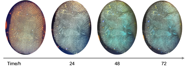
Figure 8 Photographs of LFP undergoing different aging times on the surface of tinfoil under 365 nm UV lamp |


3 Conclusions
A triphenylamine-based difluoroboron compound with an electron donor-acceptor molecular configuration has been successfully synthesized and characterized by NMR and HRMS spectra. The photophysical measurement exhibited its ICT process and solvatochromism in five organic solvents, as well as AIE performance in DMSO/H2O mixtures with different fw. They could be well explained by theoretical calculation results. Moreover, it possessed reversible MFC behavior due to the transformation of stacking mode during grinding-fuming cycle. In practical applications, the prepared safety paper displayed a repeatedly rewritable ink-free writing capability. It could visualize LPF using DMSO/H2O mixture (fw=60%, c=1×10⁻⁵ mol/L) on the surfaces of five substrates. The LFP imaging main stemmed from interaction between oleic acid in sweat secretions and TPA-2 molecules. The results indicated that TPA-2 could be not only applied to personal identification, but also used in field of anti-counterfeiting. As is well known that the optimization of the molecular structure could enhance the fluorescence quantum yield and MFC contrast, which will be a key direction for future research. Furthermore, the long-term stability and environmental adaptability of the developed security paper and fingerprint developers under real-world conditions warrant further investigation.
4 Experimental section
4.1 General experimental information
Compounds 2-1, 2-2, and TPA-CHO were synthesized according to the literature.[24,42] Triethylamine (w=99.5%), boron trifluoride ether solution [BF3 (w=48%), MkSeal], HOAc (w=99.5%), NaCl (w=99.5%), glycine (w=98.5%), cholesterol (w=99%), urea (w=99%), glucose (w=98%), and oleic acid (w=99%) were purchased from Shanghai Macklin Bio-Chem Technology Co., Ltd. Glucose (w=98%) was purchased from Shanghai Baika Che- mical Technology Co., Ltd. Lactic acid (w=90%) was purchased from Shanghai Titan Technology Co., Ltd. Anhydrous CH2Cl2 was purified by drying with CaCl2 followed by atmospheric distillation over CaH2. All other reagents and solvents were utilized without further purification.
All 1H, 13C, and 19F nuclear magnetic resonance (NMR) spectra were recorded using a Bruker Advance II-400 spectrometer with DMSO-d6 as solvents, respectively and tetramethylsilane (TMS) was employed as the internal standard. The melting point was determined using an RY- 1G melting point apparatus. UV-vis absorption and fluorescence emission spectra were obtained with a Shimadzu UV-2450 visible spectrophotometer and Agilent carry eclipse fluorescence spectrophotometer, respectively. Pow- der XRD patterns were acquired on a Bruker D2 Phaser X-ray diffractometer using Cu Kα radiation (λ=0.1506 nm, 30 kV). The fluorescence lifetimes (τ) of the as-syn- thesized powder and grinding states were measured using an Edinburgh FS5 spectrofluorometer. And this equipment was used to measure the absolutely emission quantum yields (Φf) of compound TPA-2 in DMSO solution, aggregated state, and solid state (both before and after grinding). The aggregate behavior of compound TPA-2 were investigated via scanning electron microscopy (SEM, Hitachi, S-3400N). High-resolution mass spectrometry (HR- MS) data were collected using an Agilent 6200 Series TOF/6500 Series Q-TOF LC/MS System mass spectrometer.
4.2 Synthesis
4.2.1 Synthesis of 3-(((4-(1H-pyrazol-1-yl)phenyl)- imino)methyl)-4'-(di-phenylamino)-[1,1'-biphenyl]-4-ol (TPA-S-2)
A mixture of compound 2-2 (0.43 g, 2.66 mmol), TPA-CHO (0.94 g, 2.58 mmol), EtOH (40 mL) and HOAc (1 mL) in a flask was heated to 80 ℃ for 24 h in a nitrogen atmosphere. After cooling to room temperature, the solvent was removed to obtain the crude product. It was purified by column silica gel chromatography with a mixture of ethyl acetate and petroleum ether (V∶V=1∶5) as eluent to obtain 0.922 g (68.3% yield) of TPA-S-2. 1H NMR (400 MHz, DMSO-d6) δ: 13.03 (s, 1H, OH), 9.10 (s, 1H, N=CH), 8.58 (d, J=2.4 Hz, 1H, ArH), 7.99~7.95 (m, 3H), 7.78 (d, J=1.6 Hz, 1H, ArH), 7.74 (dd, J=8.8 Hz, 2.4 Hz, 1H, ArH), 7.61 (q, J=3.6 Hz, 4H, ArH), 7.35 (t, J=8.4 Hz, 4H, ArH), 7.08 (t, J=7.6 Hz, 9H, ArH), 6.58 (t, J=2.0 Hz, 1H, ArH); 13C NMR (101 MHz, DMSO-d6) δ: 163.68, 159.93, 147.59, 146.73, 146.30, 141.63, 138.90, 133.95, 131.61, 131.28, 130.48, 130.06, 128.27, 127.55, 124.41, 124.25, 123.57, 123.03, 120.05, 119.70, 117.69, 108.53.
4.2.2 Synthesis 4-(3-(4-(1H-pyrazol-1-yl)phenyl)-2,2- difluoro-2H-2λ4,3λ4-benzo[e][1,3,2]oxazaborinin-6-yl)- N,N-diphenylaniline (TPA-2)
The boron trifluoride ether solution (1 mL, 7.9 mmol) was dropwise added to the mixture of TPA-S-2 (0.42 g, 0.825 mmol) and anhydrous CH2Cl2 (25 mL) in the presence of triethylamine (1 mL, 7.2 mmol) after 10 min. The mixture was extracted with saturated NaHCO3 solution (40 mL×3) after 24 h, and the solvent was removed. The crude product was purified by column silica gel chromatography with a mixture of a mixture of ethyl acetate and petroleum ether (V∶V=1∶4) as eluent to obtain 0.252 g (55.2% yield) of TPA-2. m.p. 205 ℃; 1H NMR (500 MHz, DMSO-d6) δ: 9.29 (s, 1H, N=CH), 8.62 (s, 1H, ArH), 8.12 (s, 1H, ArH), 8.08 (d, J=6.8 Hz, 3H, ArH), 7.84 (d, J=7.6 Hz, 3H, ArH), 7.62 (d, J=6.4 Hz, 2H, ArH), 7.36 (t, J=6.0 Hz, 4H, ArH), 7.22 (d, J=6.8 Hz, 1H, ArH), 7.10 (t, J=6.8 Hz, 8H, ArH), 6.62 (d, J=14.8 Hz, 1H, ArH); 13C NMR (126 MHz, DMSO-d6) δ: 166.57, 162.80, 147.54, 142.07, 140.38, 140.15, 137.53, 130.07, 128.58, 127.63, 125.26, 124.64, 124.42, 123.98, 123.75, 119.57, 108.84; 19F NMR (471 MHz, DMSO-d6) δ: -132.65; HRMS calcd for C34H25BF2N4O 554.2084, found 554.2086.
Supporting Information 1H NMR and 13C NMR spectra, as well as HRMS results of all compounds, the AIE behavior of compound TPA-2 in CH3CN/water mixtures with different fw, the τ of compound TPA-2 in the solid-states, SEM images of aggregates in the developers of compound TPA-2, photophysical data and computational details of compound TPA-2. The Supporting Information is available free of charge via the Internet at http://sioc-journal.cn/.
(Cheng, F.)