
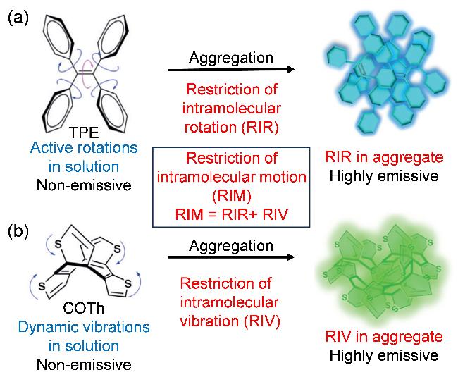
图1 (a)由于分子内旋转受限(RIR)在溶液中不发光而在聚集状态发光的四苯基乙烯(TPE)以及(b)由于分子内振动受限(RIV)在聚集态表现出AIE性质的环八四噻吩(COTh)[17]Figure 1 (a) Non-emissive behavior of tetraphenylethene (TPE) in solution and its emissive behavior upon aggregation due to the restriction of intramolecular rotations (RIR) and (b) AIE activity of cyclooctatetrathiophene (COTh) in the aggregate-state due to the restriction of intramolecular vibration (RIV) Reproduced with permission.[17] |
1 AIE光敏剂结构及其抗菌效果
1.1 小分子光敏剂
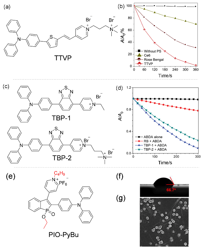
图3 (a) TTVP分子结构及(b)其光动力性能[27]; (c) TBP-1和TBP-2分子结构及其(d)光动力性能[29]; (e) PIO-PyBu分子结构, (f) PIO-PyBu滴涂膜的静态水接触角, 及(g) PIO-PyBu的水相悬浊液[10 μmol/L, 含体积分数为1%的二甲基亚砜(DMSO)]窑干膜的SEM图像[32]Figure 3 (a) Molecular structure of TTVP and (b) its photodynamic properties[27]; (c) Molecular structure of TBP-1 and TBP-2 and (d) their photodynamic properties[29]; (e) Molecular structure of PIO-PyBu; (f) Static water contact angles on the drop-casted films of PIO-PyBu, and (g) SEM images of kiln-dried films from the aqueous solution/suspension [10 μmol/L, with 1% dimethyl sulfoxide (DMSO)] of PIO-PyBu[32] |
1.2 金属配合物光敏剂
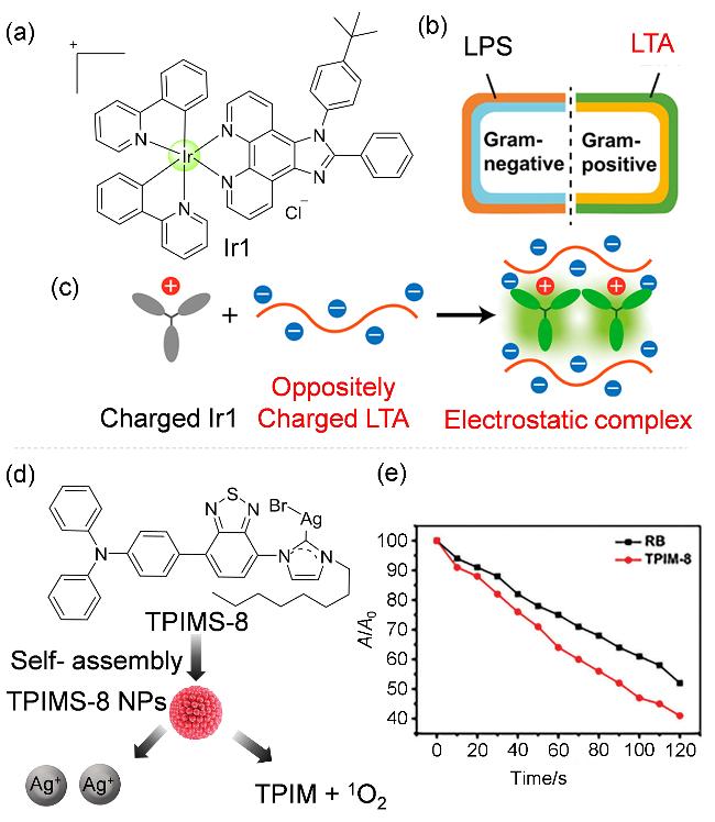
图4 (a) Ir1分子结构、(b)含LTA的革兰阳性菌和含LPS的革兰阴性菌的膜结构、(c)光敏剂对于G(+)细菌膜的作用[46]、(d) TPIMS-8纳米粒子光动力示意图及(e)在白光照射下(60 mW•cm-2), 以9,10-蒽二基-双(亚甲基)二丙二酸(ABDA)作为1O2产生指示剂, 含体积分数为1% DMSO的磷酸缓冲溶液(PBS)中, TPIM-8 (10 μmol/L)或RB (10 μmol/L)中A/A0与照射时间的关系图[47]Figure 4 (a) Molecular structure of Ir1; (b) Structure of Gram-Positive Bacteria Containing LTA and Gram-Negative Bacteria Containing LPS; (c) Effect of Ir1 on G(+) bacterial membrane[46]; (d) Schematic illustration of TPIMS-8 nanoparticle photodynamic effect; (e) Plots of A/A0 versus irradiation time for the 1O2 generation of TPIM-8 (10 μmol/L) and RB (10 μmol/L) in phosphate buffered solution (PBS) with 1% DMSO using ABDA as indicator under white light irradiation (60 mW• cm-2)[47] |
1.3 聚合物光敏剂
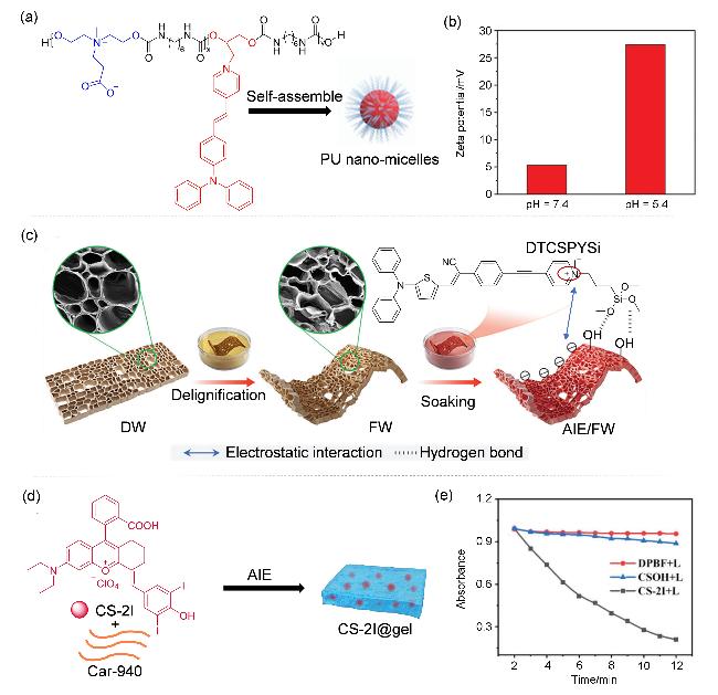
图5 (a)两性离子聚氨酯纳米粒子的合成路线、(b) pH=7.4和pH=5.4下纳米胶束的Zeta电位[51]、(c) AIE/FW FW的制备方法和皮肤伤口修复机制[52]、(d) CS-2I@gel的制备示意图及(e) CS-2I (10 μmol/L, 𝜆ex=660 nm, 10 mW•cm-2)存在时1,3-二苯基异苯并呋喃(DPBF)在425 nm处的吸光度随辐照时间的变化曲线[52]Figure 5 (a) Synthetic route for the zwitterionic polyurethane; (b) Zeta potentials of nanomicelles at pH=7.4 and pH=5.4[51]; (c) Processing strategy and skin wound repair mechanism of AIE/FW[52]; (d) Schematic diagram of the preparation of CS-2I@ge; (e) Plots of the absorbance of 1,3-diphenylisobenzofuran (DPBF) at 425 nm versus irradiation time in the presence of CS-2I[53] (10 μmol/L, 𝜆ex=660 nm, 10 mW•cm−2) |
2 针对性抗菌策略
2.1 对于细菌的抗菌策略
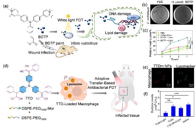
图6 (a) BDTP的化学结构及其抗菌治疗示意图、(b)光照下BDTP对创伤弧菌的抗菌活性研究(15 mW•cm-2, 30 min)、(c)感染后伤口愈合率[58]、(d) TTD的分子结构及其抗菌光动力治疗过程、(e)巨噬细胞与TTDm NPs孵育12 h以及与LysoTracker孵育30 min的荧光共聚焦显微镜图、(f)在白光照射(400~780 nm, 300 mW•cm-2)10 min的条件下TLMs与TTD NPs和生理盐水相比的抗菌活性[60]Figure 6 (a) Chemical structure and schematic illustration on antibacterial treatment of BDTP, (b) antibacterial activity study of BDTP against V. vulnificus with light irradiation (15 mW•cm-2, 30 min); (c) Wound healing rate after infection[58]; (d) Chemical structure of TTD and its antibacterial photodynamic therapy (APDT) processs; (e) Macrophages incubated with TTDm NPs for 12 h and then incubated with LysoTracker for 30 min; (f) Antibacterial activity of TLMs compared to TTD NPs and saline in the S. aureus infected mice under white light irradiation (400~780 nm, 300 mW•cm-2) for 10 min[60] |
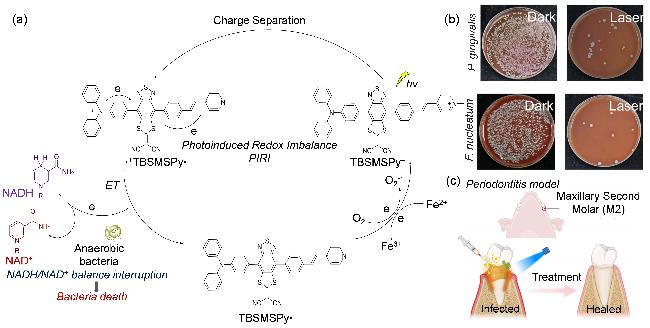
图7 (a)光诱导的厌氧菌氧化还原失衡示意图、(b)含有羊血和牙龈卟啉单胞菌或具核梭状芽孢杆菌的BHI琼脂平板在照射和不照射下经TBSMSPy+处理的照片及(c)牙周炎建模和治疗示意图[63]Figure 7 (a) Schematic showing the proposed photo-induced redox imbalance in anaerobic bacteria; (b) Photograph of a BHI agar plate containing sheep blood and biocidal activity of P. gingivalis and F. nucleatum with TBSMSPy+ treatment under irradiation and without irradiation; (c) Schematic illustration of periodontitis modeling and treatment[63] |
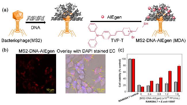
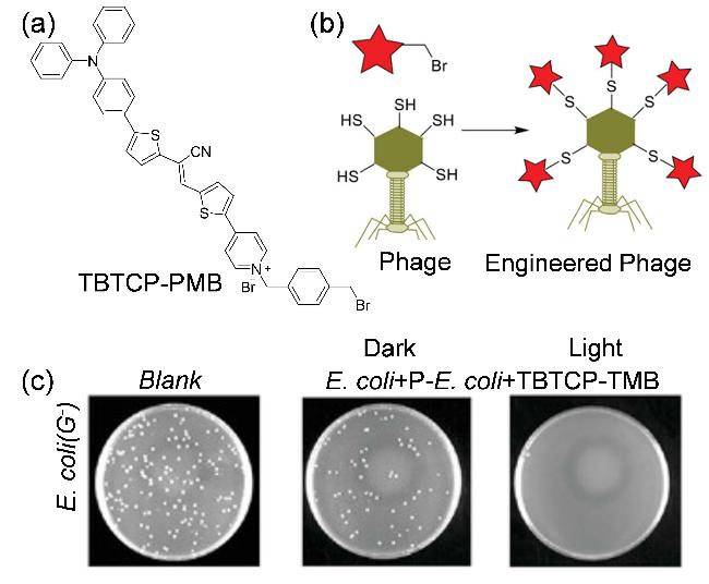
图8 (a)噬菌体纳米生物结合物(MS2-DNA-AIEgen)的示意图、(b) RAW264.7巨噬细胞经宿主大肠杆菌15597 (DAPI蓝色)和MS2- DNA-AIEgen(红色发射)处理后的共聚焦成像(箭头表示重叠的细菌和纳米结合物呈紫色)及(c)被E.coli-15597 (2.0×10⁶ CFU•mL−1)感染RAW264.7细胞存活率[65]Figure 8 (a) Schematic illustration of the bacteriophage nanobioconjugate (MS2-DNA-AIEgen); (b) Confocal images of RAW264.7 macrophages after infected by host E. coli-15597 (stained by blue-colored DAPI) and treatment by MS2-DNA-AIEgen (red-colored emission), sequentially (the arrows indicate the overlapped bacteria and nanoconjugates that showed a purple color); (c) Cell viability tests of RAW264.7 infected by host E. coli-15597[65] (2.0×10⁶ CFU•mL−1) |
2.2 对于真菌的抗菌策略
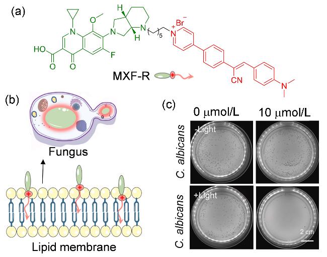
图9 (a) MXF-R的化学结构、(b) MXF-R的亲疏水部分分析及MXF-R在脂膜中的分布示意图及(c) MXF-R对白念珠菌有无白光照射(90 mW•cm-2) 30 min的杀伤作用[67].Figure 9 (a) Chemical structure of MXF-R; (b) Analysis of hydrophilic and hydrophobic parts of MXF-R and schematic diagram of MXF-R’s distribution in the lipid membrane; (c) Killing effects of MXF-R against C. albicans with or without white light irradiation (90 mW•cm-2) for 30 min[67] |
3 联合治疗
3.1 多模态治疗
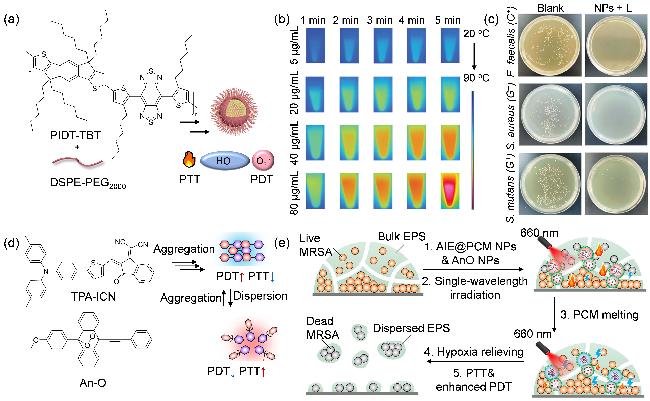
图10 (a) PIDT-TBT纳米粒子示意图、(b)不同浓度下PIDT-TBT NPs的红外(IR)热图像、(c)在808 nm激光照射(1 W•cm−2)下用PIDT-TBT NPs (120 μg•mL-1)处理5 min的E. faecalis、S. aureus和S. mutans的照片[71]、(d)聚集态和分散态光敏剂的开关能量耗散路径及(e) AIE@PCM NPs对MRSA的杀灭机制[72]Figure 10 (a) Schematic diagram of PIDT-TBT nanoparticles; (b) Infrared (IR) thermal images of PIDT-TBT NPs at different concentrations; (c) Photographs of E. faecalis, S. aureus, and S. mutans treated with PIDT-TBT NPs under 808 nm laser irradiation (1 W•cm−2) for 5 min; (d) Switched energy dissipation pathways of the AIE phototheranostic agent in the aggregated and dispersed states; (e) Bactericidal mechanism of AIE@PCM NPs against MRSA[72] |
3.2 其它抗菌成分联用
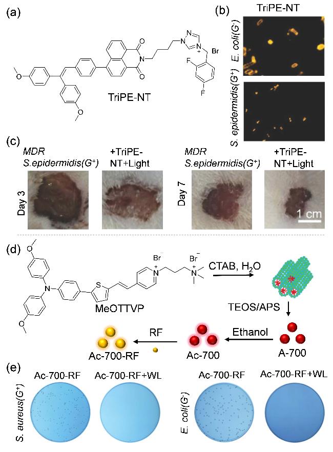
图11 (a) TriPE-NT的化学结构式、(b)大肠杆菌和表皮葡萄球菌与10×10-6 mol/L TriPE NT (橙色)孵育的荧光图像、(c) 受伤后不同时间段用TriPE NT加白光照射(4 mW•cm-2)治疗的伤口照片[74]、(d) Ac-700-RF制备过程示意图及(e)有/无白光照射(WL)条件下(24 mW•cm-2, 10 min)分别用Ac-700-RF处理的S. aureus和E. coli琼脂平板(S. aureus: Ac-700-RF 4.0 µg•mL-1; E. coli: Ac-700-RF 16.0 µg•mL-1)[75]Figure 11 (a) Chemical structure of TriPE-NT; (b) Fluorescent images of E. coli and S. epidermidis incubated with 10×10-6 mol/L of TriPE-NT (orange); (c) Photographs of wounds treated by TriPE-NT plus white-light irradiation (4 mW•cm−2) after injury for different time periods;[74] (d) Schematic illustration of the preparing process of Ac-700-RF; (e) Agar plates of S. aureus and E. coli treated with Ac-700-RF with/without WL (24 mW•cm-2, 10 min) (S. aureus: Ac-700-RF 4 µg•mL−1; E. coli: Ac-700-RF 16.0 µg•mL-1)[75] |
4 其他应用
4.1 空气中病原菌的阻断
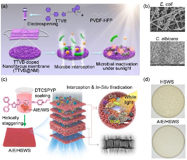
图13 (a)通过静电纺丝制备的TTVB@NM及其进行微生物拦截和阳光下对微生物灭活的示意图[81]、(b) TTVB@NM表面拦截不同微生物的SEM图像、(c) AIE/FW的自上而下制备过程及(d)白光(16 mW•cm-2)照射10 min下, 分别通过HSWS和AIE/HSWS拦截的S. aureus气溶胶培养的菌落照片[82]Figure 13 (a) Schematic illustration of the preparation of TTVB@NM through electrospinning for microbe interception and microbial inactivation under sunlight;[81] (b) SEM images of different microbes intercepted on the surface of TTVB@NM; (c) Top-down assembly process of AIE/FW; (d) Photographs of S. aureus colonies incubated from S. aureus aerosols intercepted by HSWS and AIE/HSWS, respectively, upon irradiation with white light (16 mW•cm-2) for 10 min[82] |