


1 Introduction
The plasma membrane (PM), a dynamic phospholipid bilayer, functions as an essential biophysical barrier demarcating the intracellular microenvironment from the extracellular milieu. This phospholipid bilayer not only maintains ionic homeostasis via selective permeability mechanisms but also coordinates critical cellular functions such as transmembrane signal transduction, vesicular trafficking, and intercellular communication. Pathological alterations in plasma membrane integrity, which may arise from structural destabilization (e.g., lipid peroxidation) or functional dysregulation (e.g., ion channel malfunction), can trigger progressive depolarization cascades. This ultimately leads to cytoplasmic contents efflux and necroptotic cell death.[1] Accordingly, understanding the spatiotemporal organization and biophysical properties of cell membranes is of paramount importance for advancing research in precision medicine and molecular diagnostics.[2] Leveraging their noninvasiveness, specificity, and real-time detection capability, fluorescence imaging with PM-targeted probes has emerged as a powerful technique for dynamic tracking and therapeutic monitoring.[3-4] While substantial breakthroughs have been achieved in the design of membrane- specific fluorescent probes, clinical translation remains hindered by challenges in the biocompatibility and photophysical properties of probes. For instance, the established cyanine dyes (DiD, DiI, DiO and DiR), despite their widespread utilization in membrane imaging, display critical performance limitations encompassing aqueous insolubility, inadequate membrane retention efficacy, photostability deficits, and aggregation-caused fluorescence quenching.[5-6] These intrinsic technical limitations have spurred the evolution of rationally designed fluorescent probes with enhanced PM imaging performance.[7-9]
Excimer-based fluorochromes exhibit distinct photophysical properties, including red-shifted emission, broadened spectra, short fluorescence lifetimes, and large Stokes shifts. These advantages have driven rapid advancements in excimer probes,[10] including applications in pH-sensitive diagnostics,[11] deep-tissue imaging,[12] and enzyme detection.[13-16] Notably, dual-emission systems integrating monomeric and excimeric fluorophores enable ratiometric sensing and dual-channel fluorescence imaging, achieving enhanced target specificity through wavelength-dependent signal amplification in biological environments.[17-20] Pyrene, a common polycyclic aromatic hydrocarbon made of four fused benzene rings, demonstrates remarkable photoluminescence efficiency and superior thermal stability, attributable to its extended π-conjugated system and rigid planar architecture. Notably, this structural rigidity facilitates intermolecular π-interactions, establishing pyrene as an ideal candidate for excimer formation.[21] Pyrene-based excimer probes have been extensively employed across diverse scientific domains,[22-24] such as biolabeling applications,[25-27] recognition of ds-DNA, RNA, and proteins,[28] visualization of acidic organelles,[29] and enzymatic activity monitoring.[30-31]
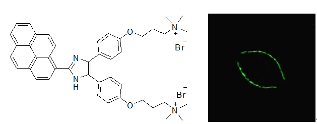
In this study, an amphipathic fluorescent dye (BAPI) featuring two hydrophilic ammonium groups and a hydrophobic pyrene core was successfully synthesized. The amphiphilic character of BAPI enables selective lipid membrane targeting. It exhibits microenvironment-sensitive and concentration-dependent fluorescence behavior characterized by monomer/excimer equilibrium. In aqueous solution, weak monomer emission dominates at concentrations below 5 μmol/L, while excimer fluorescence becomes predominant at concentrations above 5 μmol/L. Nevertheless, in sodium dodecyl sulfate (SDS) micelles, the dye shows only excimer emission. Leveraging the microenvironment- controlled monomer/excimer emission, BAPI was employed as a wash-free cell membrane fluorescent probe demonstrating multiple advantages such as high fluorescence quantum yield, a large Stokes shift, good water solubility, low cytotoxicity and rapid cellular internalization.
2 Results and discussion
2.1 Design and synthesis
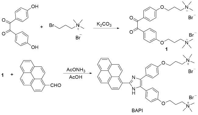
Capitalizing on the highly efficient photoluminescence of imidazole-pyrene systems,[32-33] we developed a dication-grafted fluorophore (BAPI) for bioimaging purposes. Owing to its intrinsic amphiphilic architecture, BAPI is expected to specifically target the plasma membrane, positioning it as a promising probe for live-cell imaging. Because of its dicationic nature, BAPI remains soluble and exhibits monomer emission in aqueous solution within a certain concentration range. In contrast, when incorporated into lipid vesicles, BAPI molecules are prone to adopt a densely packed arrangement, leading to excimer formation and excimer emission. This monomer-to-excimer transition is applicable to wash-free imaging of the cell membrane. BAPI was efficiently synthesized using a straightforward two-step protocol, as outlined in Scheme 1.
2.2 Dynamic excimer in solutions
The formation of dynamic excimers is closely related to solution concentration: the monomer dominates at low concentrations, while excimer becomes predominant, driven by the increased intermolecular collision probability with rising concentration. Consequently, the monomer-to- excimer emission intensity ratio of dynamic excimers shows concentration-dependent variation.[34-35] To investigate the kinetic behavior of excimer formation, we measured the emission spectra of BAPI in dimethyl sulfoxide (DMSO) at various concentrations. As illustrated in Figure 1A, the emission profile undergoes a transition from monomer-dominated to excimer-dominated character with increasing concentration. At an extremely low concentration (0.15 μmol/L), the spectrum exhibits four weak peaks between 400 and 600 nm. As the concentration increases, the emission band at 505 nm experiences significant enhancement. The triplet peaks at 412, 435, and 470 nm are consistent with the vibrational fine structure of the monomeric species. In contrast, the 505 nm band displays characteristic excimer emission, as evidenced by its broad, featureless profile, a large Stokes shift (129 nm), and a relatively short fluorescence lifetime (3~4 ns) (The fluorescence lifetimes of BAPI were determined to be 3.31 ns in DMSO and 3.41 ns in sodium dodecyl sulfate solution.).
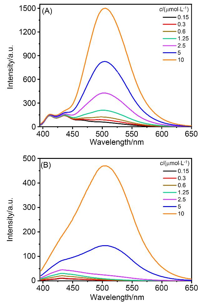
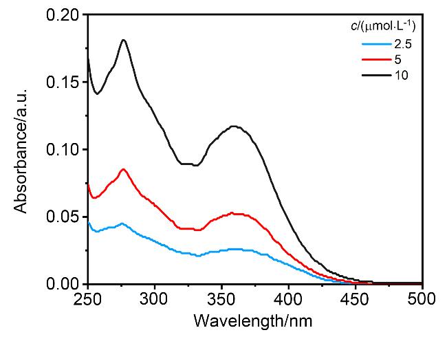
The emission spectra of BAPI in aqueous solution are similar to those in DMSO (Figure 1B): monomer emission (432 nm) predominates at low concentrations, with excimer emission (505 nm) becoming dominant as the concentration rises. However, notable differences exist between them: (1) In aqueous solution, monomer emission intensity increases with rising concentration, whereas in DMSO it remains constant; (2) The monomer emission in DMSO displays a well-resolved vibrational fine structure, which is smeared out in aqueous solution; (3) A higher concentration is required for predominant excimer emission in water (5 μmol/L) than in DMSO (1.25 μmol/L). Additionally, no Tyndall effect was observed in either DMSO or aqueous solution, suggesting the absence of nanoparticles or micelles. This absence of nanostructures in both solutions ensures that excimer emission arises solely from dynamic excimer formation via diffusion-controlled molecular collisions. The absorption spectra of BAPI in aqueous solution further confirmed dynamic excimer formation. As shown in Figure 2, the absorption spectra of BAPI at different concentrations (2.5, 5, 10 μmol/L) are nearly identical in shape, with the intensity being the only concentration-dependent parameter. In contrast, a clear monomer-to-excimer transition is observed in the emission spectra across this concentration range. These results indicate that while BAPI molecules behave independently in the ground state, their excited-state behavior is concentration-dependent: they exist as monomers at 2.5 μmol/L but form excimers at 10 μmol/L.
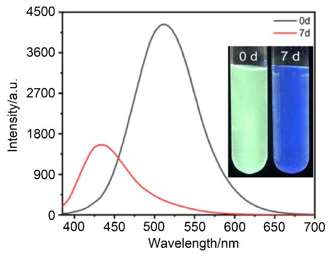
Notably, the excimer emission of BAPI in water is less stable over time. As shown in Figure 3, the freshly prepared aqueous solution of BAPI (10 μmol/L) displays a green excimer emission that gradually transitions to a blue mono- mer emission upon standing at ambient temperature for seven days. This spectral shift can be attributed to the slow equilibrium involving intermolecular interactions. During the initial dissolution phase, π-π stacking between BAPI molecules predominates, which facilitates excimer formation and is responsible for the green emission. In contrast, following a 7-day period, ion-dipole interactions between BAPI and water molecules become dominant, thus resulting in monomer emission. By contrast, no time-depen- dent spectral changes are observed in DMSO. Consequently, the excimers formed in aqueous solution are kinetically favored but thermodynamically unstable.
2.3 Static excimer in solid and aggregated states
To explore the propensity of BAPI to form static excimer, its solid-state emission and aggregation-induced emission (AIE) spectra were measured. As shown in Figure 4A, solid BAPI exhibits green fluorescence centered at 489 nm under 370 nm UV excitation, confirming the formation of excimers in the solid state. The AIE properties of BAPI were investigated in tetrahydrofuran (THF)/EtOAc mixtures with increasing volume fractions of EtOAc (fEtOAc) (THF, a good solvent; EtOAc, a poor solvent). As shown in Figure 4B, as fEtOAc increased, a continuous rise in excimer fluorescence intensity at 485 nm was observed, whereas the monomer emission (<450 nm) showed little and irregular change. This result indicates the formation of molecular aggregates. The aggregated particles exhibited a mean hydrodynamic diameter of 820 nm, as determined by dynamic light scattering (DLS). The AIE process was demonstrated by photographs taken upon 360 nm UV excitation at fEtOAc values of 10% and 90% (Figure 4C). Notably, the emission wavelength of BAPI excimer in solid and aggregation state is shorter than that in DMSO and water (505 nm), which should be ascribed to the degree of intramolecular charge transfer (ICT) under different environments. Pyrene derivatives generally exhibit aggregation-caused quenching (ACQ) due to non-radiative relaxation arising from successive π-π stacking architectures.[36] In the case of BAPI, excimer emission is observed in solution, in solid state, and in aggregated nanoparticles, demonstrating its pronounced tendency for dimeric excimer formation.
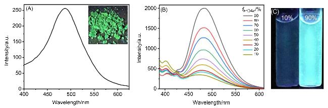
Figure 4 (A) Emission spectra of solid-state BAPI excited at 370 nm and the corresponding photos under 365 nm UV irradiation; (B) Emission spectra of 10 μmol/L BAPI in THF/EtOAc mixtures with increasing volume fractions of EtOAc (fEtOAc) excited at 370 nm: (C) Photos under 365 nm UV irradiation when fEtOAc is 10% and 90% |
2.4 Excimer emission in surfactant systems
Because BAPI has an amphiphilic nature that enables its potentially ordered assembly in micellar/vesicular systems, we screened it against four types of surfactants, namely sodium dodecyl sulfate (SDS), cetyltrimethylammonium bromide (CTAB), cocamidopropyl betaine (CAPB), and Pluronic F127 (F127), to assess their effects on BAPI excimer formation. The investigation began with an analysis of the emission spectra of BAPI across a range of surfactant concentrations. Excimer emissions were exclusively observed in all surfactant-containing aqueous solutions, and the emission intensity showed no dependence on surfactant concentration, except for CAPB, which led to a continuous increase in excimer emission with increasing surfactant concentration. Based on the concentration-intensity relationships, 20 mmol/L SDS, 100 mmol/L CTAB, 50 mmol/L CAPB, and 100 mmol/L F127 were selected to compare their effects on excimer emission under identical experimental conditions. As shown in Figure 5A, SDS induced the most intense excimer emission, peaking at approximately 494 nm. F127 produced the second strongest excimer emission with a slightly shorter wavelength (486 nm) compared to SDS. In the CTAB and CAPB systems, BAPI also exhibited considerable excimer fluorescence with a peak around 503 nm.
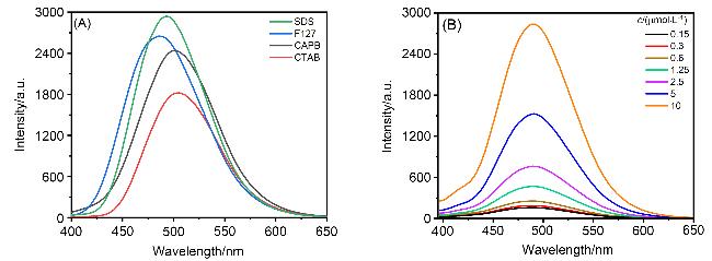
Figure 5 (A) Emission spectra of 5 μmol/L BAPI in aqueous solutions of SDS (20 mmol/L), CTAB (100 mmol/L), CAPB (50 mmol/L) and F127 (100 mmol/L) under 370 nm UV irradiation; (B) Emission spectra of BAPI at various concentrations in 20 mmol/L SDS solution under 370 nm UV irradiation |
To investigate the dynamic process of excimer formation in SDS solution, the emission spectra of BAPI at increasing concentrations were measured. As shown in Figure 5B, the excimer emission around 494 nm exhibited a continuous increase as the concentration increased, whereas no monomer emission was detected within the tested concentration range. In contrast, monomer emission was observed in either DMSO or aqueous solution under low-concentration conditions. This phenomenon can be attributed to the spontaneous enrichment and ordered arrangement of BAPI molecules within SDS micelles, which significantly enhances the probability of excimer formation. Therefore, BAPI molecules undergo a monomer-to-excime conversion when they migrate from an aqueous environment into micelles under low-concentration conditions (<2.5 μmol/L), suggesting its potential for wash-free PM imaging.
2.5 Cell membrane imaging
In addition to the monomer-excimer transition, BAPI showed a high fluorescence quantum yield of 75.66% in DMSO and 64.42% in SDS micelles, which make it a promising candidate for PM imaging. To translate this potential into application, we first assessed its cytotoxicity with a thiazolyl blue tetrazolium bromide (MTT) assay. As shown in Figure 6, the survival rates of the HeLa cells were 99.2%, 90.7% and 88.2% after 24 h of incubation with 0, 2 and 5 μmol/L BAPI, respectively. Even at a concentration of 20 µmol/L, cell viability remained above 86.1%, indicating its low cytotoxicity and good biocompatibility. Cell imaging was conducted according to a previously established protocol.[9] As shown in Figure 7, cells were stained with 3 μmol/L BAPI for 5 min and then imaged using confocal laser scanning microscopy without removing the extracellular dye. The green fluorescence was exclusively localized to the plasma membrane, indicating that BAPI specifically stains the plasma membrane. Colocalization imaging with the commercial dye DiI showed a high spatial correlation between the green emission from BAPI and the red fluorescence from DiI, further confirming the plasma membrane targeting specificity of BAPI. Colocalization analysis with Hoechst clearly delineated the cell membrane and the nucleus. As shown in Figure 8, cells treated with BAPI produced high-quality images regardless of whether the extracellular dye was removed. In contrast, the imaging quality of DiI was substantially inferior in the absence of a washing step compared with the washed control, as evidenced by increased background fluorescent spots.
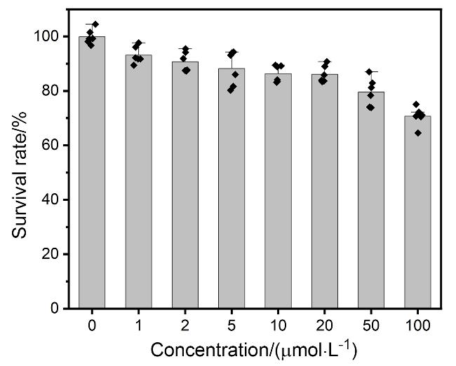
Figure 6 Viability of HeLa cells treated with different concentrations of BAPI for 24 h |
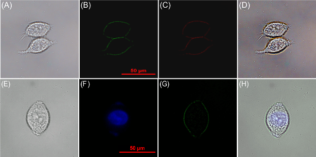
Figure 7 Confocal images of HeLa cells co-incubated with (A~D) BAPI (3.0 μmol/L, 20 min) and DiI (3.0 μmol/L, 15 min) and (E~H) Hoechst 33342 (20 min) and BAPI (3.0 μmol/L, 20 min)(A) Brightfield, (B) green channel (λex=488 nm), (C) red channel (λex=561 nm), and (D) merge of A~C. (E) brightfield, (F) blue channel (λex=405 nm), (G) green channel (λex=488 nm), and (H) merge of E~G. |
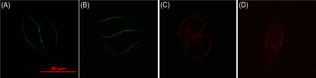
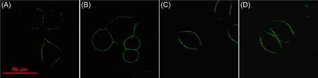
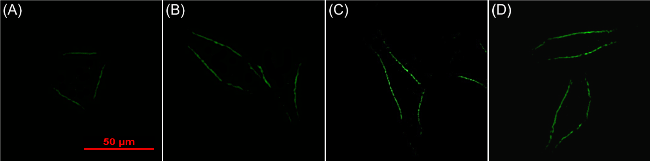
The efficiency of the staining process is crucial for real-time monitoring. Therefore, the imaging quality of BAPI in HeLa cells was next evaluated across a range of concentrations and incubation durations. As shown in Figure 9, treatment with 5.0 μmol/L BAPI for only 1 min resulted in dim and discontinuous membrane boundaries. In contrast, a well-defined membrane staining pattern was observed after 5~30 min of incubation. At a fixed 5-min incubation, 1 μmol/L BAPI produced distinctly weak fluorescence, while concentrations above 2 μmol/L resulted in clear membrane boundaries (Figure 10). These findings confirm the excellent imaging performance of BAPI.


3 Conclusions
In summary, we have synthesized a pyrene-based amphiphilic fluorescent probe (BAPI) and investigated its application in cell membrane imaging. It shows concentra-tion-dependent monomer-excimer conversion in dimethylsulfoxide or aqueous solutions. However, exclusive excimer emission was observed in various surfactant-containing aqueous solutions, suggesting its potential in cell membrane imaging. Capitalizing on its monomer-to-excimer conversion upon migrating from the aqueous phase into micelles, BAPI was developed as a wash-free cell membrane-target- ing probe with low cytotoxicity, rapid cellular internalization (5 min), a large Stokes shift (129 nm), and high fluorescence efficiency (Φ=75.66% in DMSO).
4 Experimental section
4.1 Apparatus and chemicals
All chemicals were purchased from commercial suppliers and used without further purification. Deionized water and analytical-grade organic solvents were employed for spectral measurements. 1H NMR spectra were recorded on a Bruker Av400 NMR spectrometer. Fluorescence spectra were acquired using a Hitachi F-7000 fluorescence spectrometer. Absorption spectra were taken using a Hitachi U-3900/3900H UV-Vis spectrophotometer. Absolute fluorescence quantum yields and fluorescence lifetimes were measured using an Edinburgh FLS1000 fluorescence spectrometer. Confocal imaging was performed on a Nikon A1 microscope.
4.2 Synthesis of 3,3'-(4,4'-(2-(pyren-1-yl)-1H-imida- zole-4,5-diyl)bis(4,1-phenylene))bis(oxy)bis(N,N,N-trimethylpropan-1-aminium) bromide (BAPI)
4,4'-Dihydroxybenzil (242.0 mg, 1.0 mmol), (3-bromo- propyl)trimethylammonium bromide (575.0 mg, 2.2 mmol), and potassium carbonate (553 mg, 4.0 mmol) were added to 3.0 mL of acetonitrile. The mixture was stirred at 80 ℃ until thin layer chromatography (TLC) indicated completion (approximately 7 h). After filtration to remove potassium carbonate, 9.0 mL of ethyl acetate was added to the filtrate, which was then refrigerated for 3 h. The resulting precipitate was collected by suction filtration to afford compound 1 as a light yellow solid (488.0 mg, 81% yield). Compound 1 decomposed at 320 ℃. 1H NMR (400 MHz, DMSO-d6) δ: 7.87 (d, J=8.8 Hz, 4H), 7.18 (d, J=8.8 Hz, 4H), 4.21 (t, J=6.0 Hz, 4H), 3.59~3.55 (m, 4H), 3.16 (s, 18H), 2.27~2.20 (m, 4H); 13C NMR (100 MHz, DMSO-d6) δ: 193.9, 164.0, 132.4, 125.7, 115.8, 65.8, 63.1, 52.7, 22.7.
Compound 1 (301.0 mg, 0.5 mmol), 1-pyrenecarbox- aldehyde (138 mg, 0.6 mmol), and ammonium acetate (154.2 mg, 2.0 mmol) were added to 3.0 mL of acetic acid. The mixture was stirred at 110 ℃ for 5 h. Then, 9.0 mL of ethyl acetate was added to the mixture, which was subsequently refrigerated for 5 h. The resulting precipitate was collected by suction filtration to afford BAPI as a yellow-green solid (634.0 mg, yield 78%). BAPI decomposed at 260 ℃. 1H NMR (400 MHz, DMSO-d6) δ: 9.58 (d, J=9.4 Hz, 1H), 9.54 (d, J=8.1 Hz, 1H), 8.40 (d, J=8.1 Hz, 1H), 8.35 (d, J=2.8 Hz, 1H), 8.33 (d, J=2.8 Hz, 1H), 8.30 (d, J=9.4 Hz, 1H), 8.25~8.24 (m, 2H), 8.12 (t, J=7.6 Hz, 1H), 7.58 (d, J=8.6 Hz, 4H), 7.02 (d, J=8.6 Hz, 4H), 4.12 (t, J=5.9 Hz, 4H), 3.59~3.55 (m, 4H), 3.16 (s, 18H), 2.26~2.19 (m, 4H); 13C NMR (100 MHz, DMSO-d6) δ: 174.1, 170.2, 157.8, 145.9, 131.5, 131.1, 130.9, 129.6, 129.5, 128.3, 128.3, 127.8, 127.1, 127.0, 126.5, 126.0, 125.6, 125.3, 125.3, 124.9, 124.3, 114.9, 65.3, 63.4, 52.8, 23.8. HRMS calcd for 1/2C43H48N4O2 [M2+]/2 326.1883, found 326.1879.
Supporting Information 1H NMR, 13C NMR spectra, particle size distribution, fluorescence lifetime, fluorescence quantum yield, HRMS spectra, and some emission spectra. The Supporting Information is available free of charge via the Internet at http://sioc-journal.cn.
(Zhao, C.)