研究论文

新型含反式双键哈尔碱衍生物的设计、合成及抗癌活性评价

  • 胡冬燕 a, b ,
  • 卢雨彤 a ,
  • 韩广田 a ,
  • 余莎 c ,
  • 李喜安 a ,
  • 任华忠 a ,
  • 肖吉 , d, * ,
  • 易东 , c, *
展开
  • a 乐山职业技术学院生物医药学院 乐山市药物开发工程技术研究中心 四川乐山 614000
  • b 甘肃农业大学植物保护学院 甘肃兰州 730070
  • c 西南医科大学药学院 泸州市绿色制药技术重点实验室 四川泸州 646000
  • d 四川卫生康复职业学院 基础医学院 四川自贡 643000

收稿日期: 2025-12-16

  修回日期: 2026-03-16

  网络出版日期: 2026-05-14

基金资助

天然产物化学与小分子催化四川省高校重点实验室开放课题(TRCWYXFZCH2025B03)

乐山市科技计划(23SZD002)

乐山市科技计划(23SZD028)

Design, Synthesis, and Anticancer Activity Evaluation of trans-Double Bond-Containing Harmine Derivatives

  • Dongyan Hu a, b ,
  • Yutong Lu a ,
  • Guangtian Han a ,
  • Sha Yu c ,
  • Xi'an Li a ,
  • Huazhong Ren a ,
  • Ji Xiao , d, * ,
  • Dong Yi , c, *
Expand
  • a Drug Development Engineering Technology Research Center of Leshan, College of Biomedicine, Leshan Vocational and Technical College, Leshan, Sichuan 614000
  • b College of Plant Protection, Gansu Agricultural University, Lanzhou, Gansu 730070
  • c Green Pharmaceutical Technology Key Laboratory of Luzhou City, School of Pharmacy, Southwest Medical University, Luzhou, Sichuan 646000
  • d Department of Basic Medical Sciences, Sichuan Vocational College of Health and Rehabilitation, Zigong, Sichuan 643000

Received date: 2025-12-16

  Revised date: 2026-03-16

  Online published: 2026-05-14

Supported by

Opening Project of Sichuan Province Key Laboratory of Natural Products and Small Molecule Synthesis(TRCWYXFZCH2025B03)

Science and Technology Plan of Leshan(23SZD002)

Science and Technology Plan of Leshan(23SZD028)

Copyright

© 2026 Shanghai Institute of Organic Chemistry, Chinese Academy of Sciences

摘要

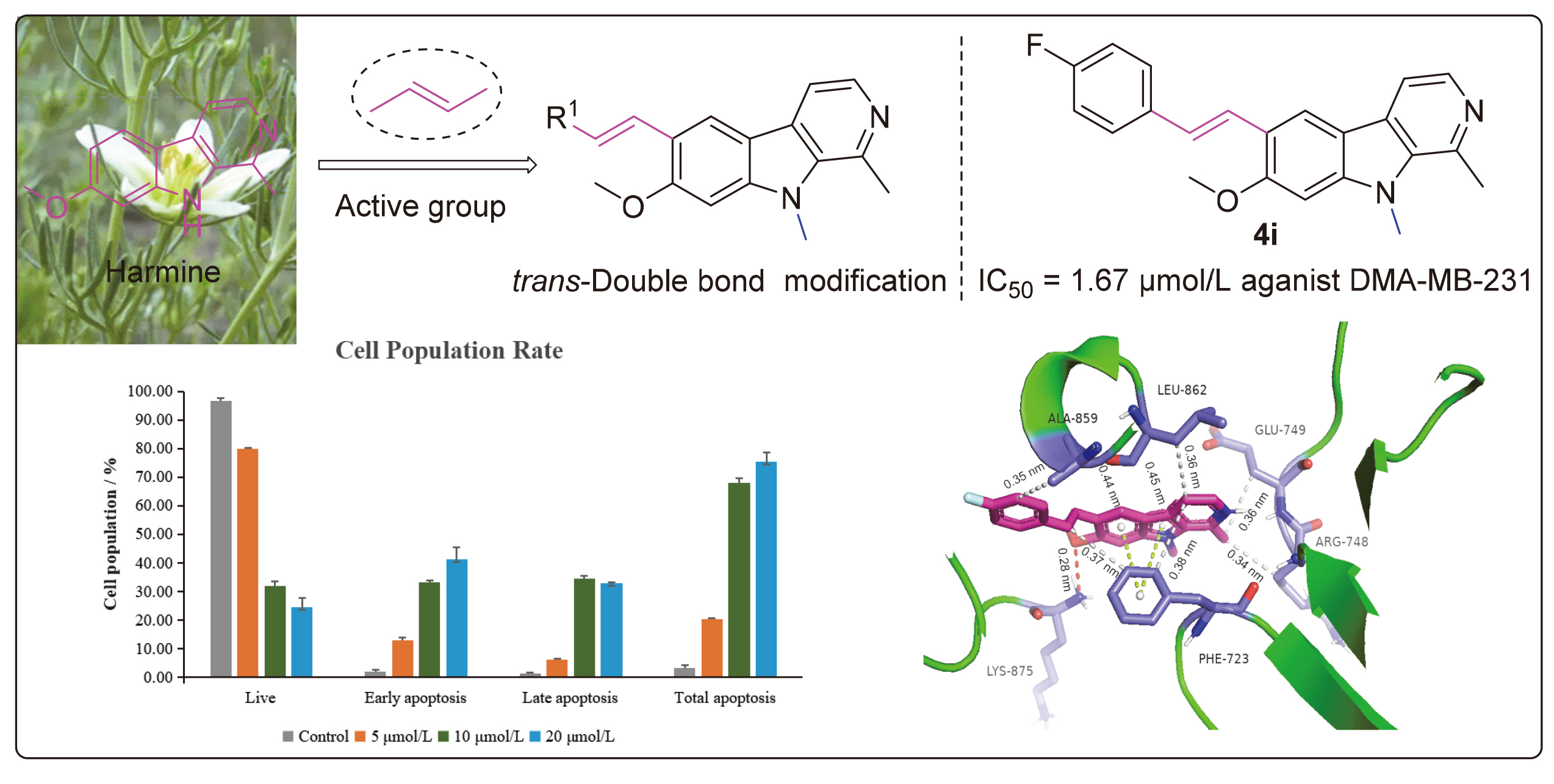
为探究反式双键引入对哈尔碱抗癌活性的影响, 本研究合成了19种新型6位被反式双键取代的哈尔碱衍生物, 并测试了其对A549、HepG2、MDA-MB-231细胞系的抑制活性. 结果显示, 大部分衍生物的抗癌活性强于母体哈尔碱. 其中, 反-6-(4-氟苯乙烯基)-7-甲氧基-1,9-二甲基-9H-吡啶并[3,4-b]吲哚(4i)展现出最强的抗增殖活性, 对MDA-MB-231细胞的IC50值为1.67 μnol/L. 进一步机制研究显示, 该化合物能以浓度依赖性方式诱导细胞凋亡, 同时将MDA- MB-231细胞的细胞周期阻滞于G2/M期. 此外, 分子对接研究显示, 化合物4i通过氢键、疏水作用和π-π相互作用, 与表皮生长因子受体酪氨酸激酶(EGFR)活性位点表现出强结合力. 针对EGFR激酶下游信号通路关键基因的RT-qPCR分析表明, 化合物4i可能是一个EGFR抑制剂. 值得注意的是, 化合物4i在正常人肺上皮细胞BEAS-2B与癌细胞MDA-MB-231之间呈现出良好的选择性(SI=3.06), 具备进一步研究的潜力.

本文引用格式

胡冬燕 , 卢雨彤 , 韩广田 , 余莎 , 李喜安 , 任华忠 , 肖吉 , 易东 . 新型含反式双键哈尔碱衍生物的设计、合成及抗癌活性评价[J]. 有机化学, 2026 , 46(8) : 3189 -3201 . DOI: 10.6023/cjoc202512020

Abstract

To investigate the impact of introducing a trans-double bond on the anticancer activity of harmine, nineteen novel harmine derivatives substituted with a trans-double bond at the 6-position were designed and synthesized. Their inhibitory activities against A549, HepG2, and MDA-MB-231 cell lines were then evaluated. The results indicated that the majority of the derivatives exhibited higher activities than the parent compound harmine. Among these derivatives, compound (E)-6-(4-fluorostyryl)-7-methoxy-1,9-dimethyl-9H-pyrido[3,4-b]indole (4i) demonstrated the most potent anti-proliferative activity, with an IC50 value of 1.67 μmol/L against MDA-MB-231 cells. Further mechanistic studies revealed that this compound could induce apoptosis in a concentration-dependent manner and arrest the cell cycle of MDA-MB-231 at the G2/M phase. Furthermore, molecular docking studies indicated that compound 4i exhibited a strong binding affinity to the epidermal growth factor receptor (EGFR) active site through hydrogen bonding, hydrophobic interactions, and π-π stacking. RT-qPCR analysis of the key genes in the downstream signaling pathway of EGFR kinase suggested that 4i might indeed serve as an EGFR inhibitor. Notably, compound 4i exhibited favorable selectivity (SI=3.06) between normal lung epithelial cells BEAS-2B and cancer cells MDA-MB-231, highlighting its potential for further investigation.

1 Introduction

Despite significant advancements in cancer treatment and prevention have been made, cancer remains a formidable global threat in the 21st century.[1-2] In addition to ongoing efforts in cancer prevention and diagnosis, the creation of novel, more effective, and less toxic anticancer drugs has emerged as a critical challenge for medicinal chemists in the fight against cancer proliferation. Historically, bioactive natural products have played a crucial role in the discovery of new anticancer drugs.[3-4] β-Carboline compounds and their derivatives have attracted considerable interest in anticancer research, owing to their unique biological and pharmacological properties.[5-6] Harmine, a naturally occurring β-carboline alkaloid, has been demonstrated to exert its antitumor effects by targeting a range of molecular targets, including DNA,[7] EGFR,[8] Twist1,[9] TAZ,[10] FAK/AKT,[11] and PI3K/Akt.[12]
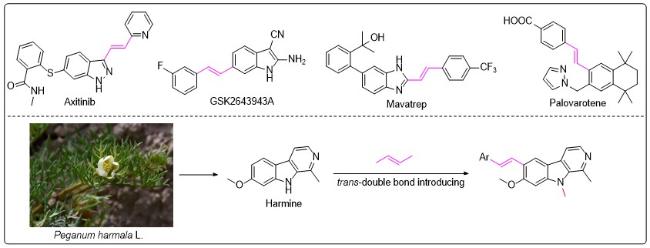
In organic chemistry, it is widely acknowledged that trans-double bond groups exhibit greater stability compared to their cis counterparts.[13] As a privileged structural motif, the trans-double bond is commonly employed in drug design. Firstly, in contrast to the bent conformation of cis-double bonds, the linearity of trans-double bond exerts a significant influence on the overall molecular conformation.[14] Through covalent or non-covalent interactions with proteins, trans-double bonds can enhance their binding to target proteins, thereby increasing activity.[15-16] Secondly, the incorporation of a trans-double bond can effectively reduce toxic side effects.[17] Lastly, the introduction of a trans-double bond may decrease in vivo metabolic clearance rates, thereby enhancing metabolic stability.[18] Due to their unique reactivity and structural properties, trans-double bonds are frequently incorporated into the molecular structures of various pharmaceuticals (Figure 1). These include selective tyrosine kinase inhibitor, such as axitinib, which is utilized in renal cell carcinoma treatment,[19] as well as GSK2643943A, a compound exhibiting potential anticancer effects.[20]
Figure 1 Representative drugs containing trans-double bonds and the design rationale of this study
In the structural modification of harmine, the modification at the 6-position remains a less-studied area. Since 2022, our group[21-24] has initiated research on the 6-posi- tion modifications of harmine. The modification approach included the introduction of an alkyne group via Sonogashira coupling, the establishment of a C—C bond through Suzuki coupling, and the formation of a C—N bond via nucleophilic substitution. Experimental results verified the effectiveness of the 6-position modifications. Based on the above findings and considering the important role of the trans-double bond in the pharmaceutical field, a series of 6-alkenylated harmine derivatives were designed and synthesized through molecular hybridization. The objective of this study was to explore the influence of introducing a trans-double bond at the 6-position of harmine on its anticancer activity. The specific design strategy is presented in Figure 1.

2 Results and discussion

2.1 Synthesis of the target compounds

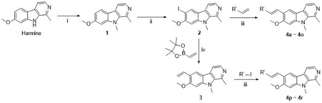
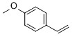
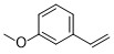
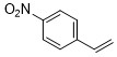
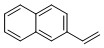
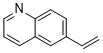
Compounds 1 and 2 were prepared following the methods described in our previous work.[21-22] Compound 1 was dissolved in acetic acid and then reacted with N-iodo-succinimide (NIS), an iodinating agent, to yield the iodinated compound 2. Subsequently, compound 2 underwent a Heck reaction with aromatic alkenes, resulting in the formation of 6-alkenylated derivatives 4a~4o (Scheme 1). It is worth noting that the terminal alkene- containing compound 3 was synthesized as an intermediate for aromatic alkenes unavailable commercially. Initially, the alkenyl boronate underwent a Suzuki coupling with compound 2 to form compound 3. Compound 3 was then subjected to a Heck reaction with aromatic iodides, yielding the target compounds 4p~4r (Scheme 1). All synthesized compounds were characterized and verified using 1H NMR, 13C NMR, and HRMS.
Scheme 1 Synthetic route of compounds 3, 4a~4r

Reagents and conditions: (i) NaH, CH3I, dry DMF, 0 ℃ to r.t., 2 h; (ii) NIS, AcOH, r.t., overnight; (iii) Pd(OAc)2, tri(o-tolyl)phosphine, NEt3, CH3CN, N2, 70 ℃, 10 h; (iv) Pd(OAc)2, X-phos (564483-18-7), K3PO4, 1,4-dioxane, H2O, N2, 100 ℃, 8 h

2.2 Anticancer activity of target compounds in vitro

Initially, a terminal alkenyl moiety was installed at the 6-position of harmine to afford compound 3. To assess the impact of introducing an alkenyl group on the inhibition effect of cancer cell proliferation, 3-(4,5-dimethylthiazol- 2-yl)-5-(3-carboxymethoxyphenyl)-2-(4-sulfophenyl)-2H-tetrazolium (MTS) assay was applied to evaluate the anti- proliferative activity of compound 3 toward A549, HepG2, and MDA-MB-231 cells. Cisplatin was used as a positive control, and the IC50 values were summarized in Table 1. Compared to the parent compound harmine, compound 3 exhibited greater inhibitory activities against these three cancer cell lines. Particularly for HepG2 and MDA-MB- 231 cells, its efficacy surpassed that of the positive control cisplatin. However, the magnitude of anti-proliferative activity improvement was not substantial. Our previous studies have demonstrated that introducing an aromatic ring at the 6-position of harmine can enhance the anti-proliferative activity.[21-23] Consequently, we further investigated the impact of introducing an aromatic ring onto the vinyl group on anti-proliferative activity.
Table 1 Antiproliferative activities of target compounds 3 and 4a~4r against three cancer cell linesa
Compound Alkenyl IC50b/(μmol•L-1)
A549 HepG2 MDA-MB-231
Harmine 26.03±0.34 15.27±0.63 9.93±0.63
3 20.59±0.29 5.91±0.25 5.99±0.03
4a 4.68±0.04 4.15±0.08 2.58±0.11
4b 20.94±0.04 8.96±0.11 3.22±0.17
4c 3.81±0.03 12.44±0.39 7.42±1.19
4d 6.09±0.13 12.87±0.40 2.93±0.20
4e >50 40.57±1.70 >50
4f 3.79±0.01 4.60±0.01 5.47±0.19
4g 5.68±0.15 6.34±0.04 20.99±0.86
4h 5.29±0.22 22.16±0.14 9.76±0.82
4i 3.98±0.05 4.19±0.04 1.67±0.15
4j 4.82±0.05 4.38±0.09 4.67±0.24
4k 21.73±0.58 16.98±0.66 5.59±0.04
4l 3.89±0.04 17.12±0.23 2.42±0.12
4m 4.75±0.02 15.27±0.69 4.66±0.11
4n 13.82±0.37 22.78±0.65 5.43±0.15
4o 20.29±0.23 20.28±0.16 4.56±0.13
4p 3.95±0.06 4.42±0.05 2.95±0.44
4q 4.41±0.04 19.30±0.34 2.90±0.09
4r 21.81±0.22 22.28±0.28 11.75±0.32
Cisplatinc 18.99±0.77 15.72±0.24 28.82±0.87

a The operations were performed in triplicate and the data displayed as mean±SD. b IC50 (μmol/L) values were determined by Reed and Muench method. c Used as a positive control.

As indicated in Table 1, compounds 4a~4r were obtained when various aromatic groups were appended to the terminal alkene of compound 3. The majority of these compounds 4a~4r demonstrated potent anti-proliferative activities, surpassing that of compound 3. The IC50 values for compounds 4a, 4c, 4d, 4f, 4g~4j, 4l~4m, 4p, and 4q against A549 cells ranged from 3.79 μmol/L to 6.09 μmol/L, demonstrating significant improvement of inhibitory effects than compound 3 (IC50=20.59 μmol/L against A549). Compared with compound 3 (IC50=5.91 μmol/L against HepG2), the inhibitory activity of compounds 4a~4r against HepG2 cells did not exhibit significant enhancement; in some cases, they even promoted cancer cells proliferation. The IC50 values of compounds 4a, 4b, 4d, 4f, and 4i~4g on MDA-MB-231 cells ranged from 1.67 μmol/L to 5.59 μmol/L, suggesting a stronger inhibitory effect compared to compound 3 (IC50=6.00 μmol/L). Notably, compound 4i (IC50=1.67 μmol/L) exhibited the highest potency in inhibiting cell proliferation in MDA- MB-231 cells.
According to the aforementioned data, the preliminary structure-activity relationship could be summarized as follows: (1) Compounds featuring quinoline or iso-quinoline rings (4p~4q), which contain nitrogen atoms, showed superior anti-proliferative activity compared to compounds with naphthalene rings and benzothiophene rings containing sulfur atoms (4n~4o, 4r). This may be related to the fact that nitrogen atoms in the structure can act as hydrogen bond acceptors, thereby forming more interaction forces with the target protein. (2) The introduction of aromatic groups (4a~4r) into the vinyl moiety (3) did not significantly improve the inhibition of HepG2 cells proliferation. (3) It appears that regardless of whether it is an electron donating (OCH3) or electron withdrawing (F) substituent, the para substitution on the benzene ring seems to be more conducive to the improvement of anticancer activity than the meta and ortho substitution. This can be seen from the comparison of compounds 4c~4e and 4i~4k. For the three types of tumor cells, compounds 4c (except for MDA-MB-231) and 4i have significantly higher anti-proliferative activity than those of compounds 4d~4e and 4j~4k. Finally, compound 4i, bearing a para-F substituent, exhibited the most performing anti-proliferative activity against MDA-MB-231 cells with an IC50 value of 1.67 μmol/L. Based on these findings, compound 4i and the MDA-MB-231 cells were chosen for further mechanistic studies.

2.3 Cell cycle analysis of compound 4i

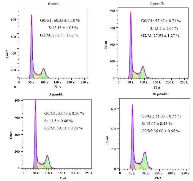
The cell cycle is an ordered set of events that ultimately leads to cell growth and division.[25] Blocking the normal progression of the cell cycle can prevent cancer cells from transitioning between different growth phases, thereby affecting their proliferation. To investigate whether 6- alkenyl harmine derivatives can induce cell cycle arrest, compound 4i, which demonstrated the most promising, was selected for cell cycle analysis. The analysis was conducted using flow cytometry with propidium iodide (PI) staining. As illustrated in Figure 2, when different concentrations (2, 5 and 10 μmol/L) of 4i were incubated with the MDA-MB-231 cell for 24 h, compared with the control group (0 μmol/L of 4i), the proportion of cells in the G2/M phase increased from 27.17% to 36.00% in the experimental group. In contrast, the proportion of cells in the G0/G1 phase decreased, whereas the proportion of cells in the S phase remained relatively constant. These findings corroborated that 4i could disrupt the normal cell cycle of tumor cells and induce cell cycle arrest at the G2/M phase, thereby inhibiting the proliferation of MDA-MB-231 cells.
Figure 2 Compound 4i induced G2/M arrest in MDA-MB-231 cancer cells

MDA-MB-231 cells were incubated with different concentrations of 4i (0, 2, 5 and 10 μmol/L) for 24 h.

2.4 Cell apoptosis analysis and cell viability analysis of compound 4i

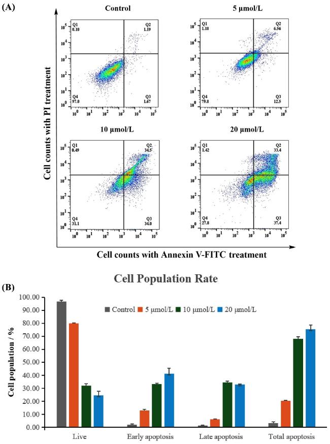
Apoptosis is a physiological, programmed cell death process that is crucial for embryonic development, tissue and organ formation, aging, and the clearance of pathological cells.[26-27] Environmental and genetic factors may disrupt the normal process of cell apoptosis, leading to the evasion of programmed cell death and the acquisition of almost infinite proliferation capacity, which results in the formation of tumor cells.[28] Therefore, inducing or promoting apoptosis has emerged as a crucial mechanism through which many anticancer drugs exert their therapeutic effects. To verify whether our compound possesses the ability to induce apoptosis, a double-staining assay using fluorescein isothiocyanate (FITC) Annexin V/PI was conducted, and flow cytometry was employed to detect the apoptosis of MDA-MB-231 cells induced by compound 4i. As illustrated in Figure 3, the viability of MDA-MB-231 cells progressively diminished as the concentration of 4i increased. Meanwhile, the early apoptosis rate increased from 1.67% in the control group to 12.5%, 34.0%, and 37.4%, while the late apoptosis rate rose from 1.19% to 6.56%, 34.5%, and 33.4%. Overall, the total apoptosis rate increased from 2.96% in the vehicle control group to 20.24%, 68.99%, and 72.22%, respectively. The experimental results indicated that compound 4i promotes the apoptosis of MDA-MB-231 cells in a concentration-depen- dent manner.
Figure 3 Effect of 4i on induction of apoptosis in MDA-MB-231 cells

Each data point represents the average of three independent experiments. (A) MDA-MB-231 cells were incubated with different concentrations of 4i (0, 5, 10 and 20 μmol/L) for 24 h, then stained with Annexin V-FITC/PI, and the apoptosis was detected by flow cytometry. Q1: Necrotic cells, Q2: Late apoptosis, Q3: Early apoptosis, Q4: Live cells. (B) The percentage of cell distribution was shown in the histograms.

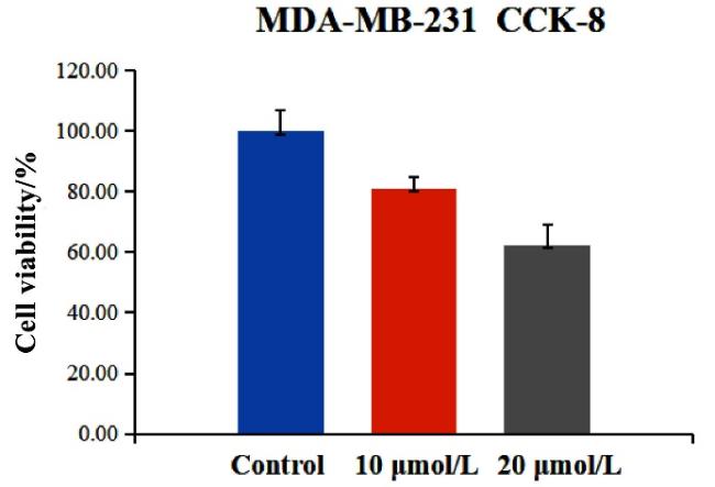
Due to the observation that there was no significant statistical difference in apoptosis induction rate between 10 μmol/L and 20 μmol/L of compound 4i. In order to further clarify whether this apoptotic phenomenon was specifically triggered by the compound mechanism of action, a cell viability assay was conducted. As shown in Figure 4, the cell viability in the 10 μmol/L treatment group was 81.15%, exceeding the 80% tolerance threshold. This suggests that the apoptosis observed at 10 μmol/L was mainly attributed to the specific pharmacodynamic effect of the compound 4i. In contrast, the cell viability in the 20 μmol/L treatment group declined to 62.50%, falling below the 80% tolerance threshold. This indicates that compound 4i at the higher concentration of 20 μmol/L might induce apoptosis primarily through non-specific cytotoxic effects.
Figure 4 Effect of 4i on cell viability in MDA-MB-231 cells

MDA-MB-231 cells were incubated with different concentrations of 4i (0, 10 and 20 μmol/L) for 24 h. Each data point represents the average of five independent experiments.

2.5 Molecular docking simulation

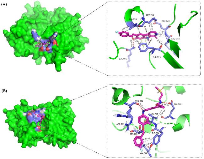
As a key member of the HER family, the epidermal growth factor receptor kinase (EGFR kinase) is closely associated with cell proliferation, growth, survival, and drug resistance. Literature suggests that the EGFR protein exhibited high expression levels in MDA-MB-231 cells, and inhibiting EGFR could potentially suppress tumor cell proliferation.[29] Compound 4i demonstrated potent inhibitory effect against MDA-MB-231 cells. To investigate the interaction between compound 4i and EGFR kinase, molecular docking experiment was conducted. The docking was performed using AutoDock Tools (ADT) version 1.5.7 software on the EGFR kinase domain (PDB ID: 3W2S). Figure 5A illustrated the theoretical binding mode of compound 4i with EGFR. The maximal binding energy between compound 4i and EGFR was -6.95 kJ/mol. Compound 4i was inserted firmly into the active pocket of EGFR, where the oxygen atom of the 7-methoxy group formed a hydrogen bond (0.28 nm) with the Lys-875 residue of EGFR. The carboline ring, the trans double bond, and the 6-position phenyl ring, as components of polycyclic aromatic hydrocarbons, formed hydrophobic interactions with Phe-723, Arg-748, Glu-749, Ala-859, and Leu- 862 residues. Moreover, the carboline ring scaffold of compound 4i exhibited a π-π interaction with the amino acid residue (Phe-723).
Figure 5 Predicted binding model of compound 4i (A) and Lapatinib (B) with EGFR (PDB code: 3W2S)
To explore the similarities or differences of EGFR binding patterns between compound 4i and other EGFR kinase inhibitor, docking studies of Lapatinib with EGFR protein were also conducted (Figure 5B). The maximum binding energy of Lapatinib to EGFR was -7.51 kJ/mol. The NH group of Lapatinib established hydrogen bonds with residues Arg-831 and Glu-762, and the oxygen atom of the ether bond formed a hydrogen bond with residue Ser-768. Additionally, the furan ring and the fluorine-sub- stituted benzene ring displayed hydrophobic interactions with residues Leu-703, Tyr-764, and Val-765. In regard to binding sites, compound 4i demonstrated differences from Lapatinib; nevertheless, their binding energies were com-parable, indicating that their binding affinities to EGFR were substantially similar.

2.6 RT-qPCR analysis of EGFR downstream signa- ling pathway-related genes

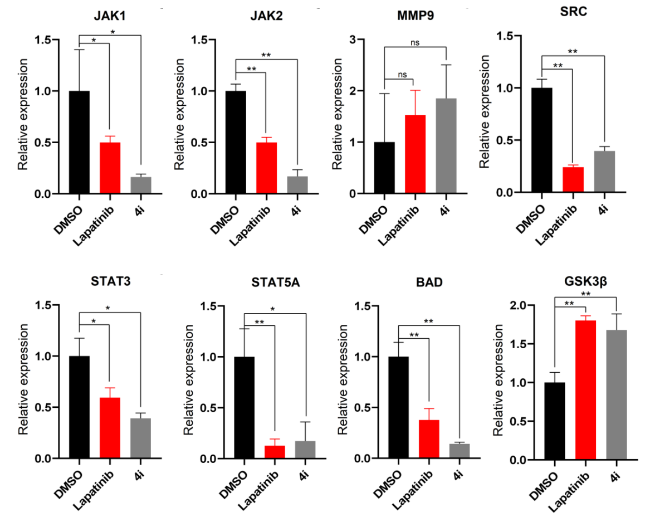
To experimentally validate whether compound 4i serves as an EGFR inhibitor, Lapatinib and dimethyl sulfoxide (DMSO) were respectively employed as positive and negative controls to investigate the impacts on key genes within the EGFR downstream signaling pathway. As depicted in Figure 6, among the eight genes under test, MMP9 and GSK3β displayed up-regulation, whereas the remaining six genes presented down-regulation. Notably, both compound 4i and Lapatinib manifested highly congruent trends in gene regulation. Considering that these eight genes are distributed across diverse EGFR downstream signaling pathways, it is rational to deduce that the mode of action of compound 4i on MDA-MB-231 cells is similar to that of Lapatinib. These results preliminarily indicate that compound 4i might function as an EGFR kinase inhibitor.
Figure 6 Effects of 4i and Lapatinib on key genes related to EGFR downstream signaling pathways

Statistical differences were analyzed via Student’s t-test, *: p≤0.05, **: p≤0.01, ns (not significant): P>0.05

2.7 Preliminary toxicity evaluation of compound 4i

In the pharmaceutical field, both efficacy and toxicity are critical factors that need to be taken into account. To preliminarily evaluate the toxicity profile of these 6- alkenylated harmine derivatives, the inhibitory effect of compound 4i on BEAS-2B cells (normal lung epithelial cells) was compared with that of cisplatin. According to Table 2, the IC50 value of compound 4i for BEAS-2B cells was 5.11 μmol/L. Furthermore, the selectivity index (SI), which serves as a significant parameter for evaluating the balance between drug efficacy and safety, was calculated for both compound 4i and the positive control drug cisplatin. Compared to cisplatin (SI=1.45), compound 4i exhibited higher selectivity (SI=3.06).
Table 2 In vitro assessment of the anti-proliferative effects of compound 4i on MDA-MB-231 cells and normal BEAS-2B cells along with its selectivity index a
Compound IC50b/(μmol/L) Selective index (SI)c
MDA-MB-231 BEAS-2B
4i 1.67±0.15 5.11±0.27 3.06
Cisplatind 28.82±0.87 41.83±0.96 1.45

a The operations were performed in triplicate and the data displayed as mean±SD. b IC50 values were determined by Reed and Muench method. c SI=IC50 non-cancerous cell/IC50 cancer cell. d Used as a positive control.

3 Conclusions

In conclusion, a series of novel harmine derivatives (4a~4r) featuring a trans-double bond were designed and synthesized. The structures of these newly developed compounds were confirmed via 1H NMR, 13C NMR, and HRMS. Anti-proliferative activity assays indicated that the majority of the target compounds displayed favorable inhibitory activity against three tumor cell lines. Notably, compound 4i demonstrated stronger inhibitory activity against all the tested tumor cells in comparison to the positive control, cisplatin. Specifically, it exhibited the highest inhibitory activity on the MDA-MB-231 cell line, with an IC50 value of 1.67 μmol/L.
Furthermore, this research also investigated the potential anti-proliferative mechanism of 4i by conducting cell cycle and apoptosis experiments. The findings indicated that 4i was capable of arresting the MDA-MB-231 cell cycle at the G2/M phase and triggering cell apoptosis in a concentration-dependent manner. Molecular docking studies revealed that compound 4i formed robust binding interactions with the EGFR kinase domain via hydrogen bonds, hydrophobic forces, and π-π stacking. Based on the high similarity in the effects of compound 4i and the positive control Lapatinib on key genes in the EGFR downstream signaling pathway, it can be inferred that 4i may be an EGFR inhibitor. Preliminary toxicity evaluations indicated that compound 4i exhibited a relatively low toxicity profile and demonstrated favorable selectivity between cancerous and non-cancerous cell lines. These findings offer valuable insights for subsequent compound modification research.

4 Experimental section

4.1 Chemistry

4.1.1 Materials and methods

Harmine was purchased from Nanjing Dolon Biotechnology Co., Ltd. Other chemical reagents and solvents were purchased from Shanghai Haohong Scientific Co., Ltd and Shanghai Titan Scientific Co., Ltd. All chemicals and solvents were purchased from commercially available sources and used without further purification, unless otherwise specified. 1H NMR and 13C NMR spectra were collected in CDCl3 or DMSO-d6 on a Bruker Avance 400. Mass analysis data were acquired using a SCIEX UPLC (EXion)-Q-TOF (X500R). All reactions were monitored using thin-layer chromatography (TLC) plates and observed under UV light. The products were purified via flash column chromatography using silica gel (200~300 mesh).

4.1.2 Preparation of compound 1

Compound 1 was synthesized according to the method in the literature,[22] and the NMR characterization data were consistent with the literature.

4.1.3 Preparation of compound 2

Compound 2 was synthesized according to the method in the literature,[21] and the NMR characterization data were consistent with the literature.

4.1.4 General procedure for preparation of compounds 4a~4o

Protected by N2, compound 2 (176 mg, 0.5 mmol) was dissolved in CH3CN (10 mL), followed by the addition of tri(o-tolyl)phosphine (31 mg, 0.1 mmol), Pd(OAc)2 (11 mg, 0.05 mmol), Et3N (140 μL, 1 mmol), and aromatic alkenes (0.75 mmol). The reaction mixture was stirred at 70 ℃ for 8 h. Upon completion of the reaction, the mixture was concentrated under reduced pressure using a rotary evaporator. The residue was then purified by column chromatography using a gradient elution of dichloromethane/methanol (VV=50∶1~20∶1) to yield compounds 4a~4o.
(E)-7-Methoxy-1,9-dimethyl-6-styryl-9H-pyrido[3,4-b]- indole (4a): White solid, yield 96%. m.p. 205~206 ℃; 1H NMR (400 MHz, CDCl3) δ: 8.26 (d, J=5.2 Hz, 1H), 8.17 (s, 1H), 7.70 (d, J=5.2 Hz, 1H), 7.58~7.54 (m, 3H), 7.38~7.34 (m, 2H), 7.26~7.22 (m, 1H), 7.14 (d, J=16.4 Hz, 1H), 6.64 (s, 1H), 3.95 (s, 6H), 2.99 (s, 3H); 13C NMR (100 MHz, CDCl3) δ: 158.5, 143.0, 141.0, 138.3, 138.2, 135.9, 129.1, 128.6, 127.5, 127.1, 126.4, 123.8, 120.5, 118.7, 114.5, 112.3, 90.8, 55.8, 32.3, 23.5; HRMS (ESI) calcd for C22H21N2O [M+H] 329.1649, found 329.1637.
(E)-7-Methoxy-1,9-dimethyl-6-(4-methylstyryl)-9H-pyrido[3,4-b]indole (4b): White solid, yield 86%. m.p. 231~232 ℃; 1H NMR (400 MHz, CDCl3) δ: 8.26 (d, J=5.2 Hz, 1H), 8.17 (s, 1H), 7.70 (d, J=5.2 Hz, 1H), 7.51 (d, J=16.4 Hz, 1H), 7.46 (d, J=7.6 Hz, 2H), 7.17 (d, J=7.6 Hz, 2H), 7.12 (d, J=16.4 Hz, 1H), 6.64 (s, 1H), 3.96 (s, 3H), 3.95 (s, 3H), 2.99 (s, 3H), 2.36 (s, 3H); 13C NMR (100 MHz, CDCl3) δ: 158.5, 142.9, 141.0, 138.3, 136.9, 135.9, 135.4, 129.4, 129.1, 127.5, 126.3, 122.8, 120.7, 118.6, 114.5, 112.3, 90.8, 55.8, 32.3, 23.4, 21.3; HRMS (ESI) calcd for C23H23N2O [M+H] 343.1805, found 343.1795.
(E)-7-Methoxy-6-(4-methoxystyryl)-1,9-dimethyl-9H-pyrido[3,4-b]indole (4c): White solid, yield 95%. m.p. 260~261 ℃; 1H NMR (400 MHz, CDCl3) δ: 8.25 (d, J=5.2 Hz, 1H), 8.15 (s, 1H), 7.69 (d, J=5.2 Hz, 1H), 7.51~7.49 (m, 2H), 7.42 (d, J=16.4 Hz, 1H), 7.09 (d, J=16.4 Hz, 1H), 6.91~6.89 (m, 2H), 6.63 (s, 1H), 3.95 (s, 6H), 3.83 (s, 3H), 2.99 (s, 3H); 13C NMR (100 MHz, CDCl3) δ: 158.9, 158.4, 142.8, 141.0, 138.3, 135.9, 131.0, 129.1, 127.6, 127.1, 121.7, 120.8, 118.4, 114.5, 114.1, 112.3, 90.8, 55.8, 55.3, 32.3, 23.5; HRMS (ESI) calcd for C23H23N2O2 [M+H] 359.1754, found 359.1739.
(E)-7-Methoxy-6-(3-methoxystyryl)-1,9-dimethyl-9H-pyrido[3,4-b]indole (4d): White solid, yield 74%. m.p. 253~254 ℃; 1H NMR (400 MHz, CDCl3) δ: 8.26 (d, J=5.2 Hz, 1H), 8.16 (s, 1H), 7.69 (d, J=5.2 Hz, 1H), 7.55 (d, J=16.4 Hz, 1H), 7.30~7.26 (m, 1H), 7.17~7.08 (m, 3H), 6.82~6.79 (m, 1H), 6.63 (s, 1H), 3.95 (s, 3H), 3.94 (s, 3H), 3.86 (s, 3H), 2.98 (s, 3H); 13C NMR (100 MHz, CDCl3) δ: 159.7, 158.6, 143.1, 141.1, 139.7, 138.3, 136.0, 129.6, 129.2, 127.4, 124.2, 120.4, 119.2, 118.9, 114.6, 112.7, 112.3, 111.7, 90.9, 55.8, 55.3, 32.4, 23.5; HRMS (ESI) calcd for C23H23N2O2 [M+H] 359.1754, found 359.1745.
(E)-7-Methoxy-6-(2-methoxystyryl)-1,9-dimethyl-9H-pyrido[3,4-b]indole (4e): White solid, yield 87%. m.p. 233~234 ℃; 1H NMR (400 MHz, CDCl3) δ: 8.28 (s, 1H), 8.27 (d, J=5.2 Hz, 1H), 7.75 (d, J=5.2 Hz, 1H), 7.69 (dd, J=7.6, 1.6 Hz, 1H), 7.60~7.50 (m, 2H), 7.26~7.22 (m, 1H), 7.00~6.97 (m, 1H), 6.93~6.89 (m, 1H), 6.69 (s, 1H), 4.01 (s, 3H), 3.97 (s, 3H), 3.92 (s, 3H), 3.02 (s, 3H); 13C NMR (100 MHz, CDCl3) δ: 158.6, 156.7, 143.0, 141.0, 138.3, 136.0, 129.3, 128.2, 127.3, 126.2, 124.1, 122.0, 121.2, 120.8, 118.8, 114.6, 112.4, 110.9, 90.8, 55.8, 55.6, 32.4, 23.5; HRMS (ESI) calcd for C23H23N2O2 [M+H] 359.1754, found 359.1738.
(E)-7-Methoxy-1,9-dimethyl-6-(4-nitrostyryl)-9H-pyri- do[3,4-b]indole (4f): yellow solid, yield 75%. m.p. 255~256 ℃; 1H NMR (400 MHz, CDCl3) δ: 8.32~8.20 (m, 4H), 7.79~7.65 (m, 4H), 7.27~7.20 (m, 1H), 6.79 (s, 1H), 4.10 (s, 3H), 4.06 (s, 3H), 3.06 (s, 3H); 13C NMR (100 MHz, CDCl3) δ: 158.9, 146.2, 144.9, 143.7, 141.3, 138.8, 136.2, 129.1, 128.7, 126.6, 124.9, 124.2, 119.6, 119.3, 114.8, 112.3, 91.0, 55.9, 32.5, 23.6; HRMS (ESI) calcd for C22H20N3O3 [M+H] 374.1499, found 374.1505.
(E)-6-(4-Chlorostyryl)-7-methoxy-1,9-dimethyl-9H-py-rido[3,4-b]indole (4g): White solid, yield 77%. m.p. 240~241 ℃; 1H NMR (400 MHz, CDCl3) δ: 8.27 (d, J=5.2 Hz, 1H), 8.16 (s, 1H), 7.70 (d, J=5.2 Hz, 1H), 7.52 (d, J=16.4 Hz, 1H), 7.46 (d, J=8.4 Hz, 2H), 7.31 (d, J=8.4 Hz, 2H), 7.07 (d, J=16.4 Hz, 1H), 6.68 (s, 1H), 3.99 (s, 3H), 3.98 (s, 3H), 3.01 (s, 3H); 13C NMR (101 MHz, CDCl3) δ: 158.6, 143.2, 141.1, 138.5, 136.7, 136.0, 132.5, 129.1, 128.7, 127.5, 126.2, 124.5, 120.2, 118.9, 114.7, 112.3, 90.9, 55.8, 32.4, 23.5; HRMS (ESI) calcd for C22H20ClN2O [M+H] 363.1259, found 363.1254.
(E)-6-(4-Bromostyryl)-7-methoxy-1,9-dimethyl-9H-py- rido[3,4-b]indole (4h): White solid, yield 92%. m.p. 255~256 ℃; 1H NMR (400 MHz, CDCl3) δ: 8.26 (d, J=5.2 Hz, 1H), 8.16 (s, 1H), 7.70 (d, J=5.2 Hz, 1H), 7.53 (d, J=16.4 Hz, 1H), 7.46 (d, J=8.4 Hz, 2H), 7.40 (d, J=8.4 Hz, 2H), 7.05 (d, J=16.4 Hz, 1H), 6.67 (s, 1H), 3.98 (s, 3H), 3.98 (s, 3H), 3.00 (s, 3H); 13C NMR (100 MHz, CDCl3) δ: 158.6, 143.2, 141.1, 138.4, 137.2, 136.0, 131.7, 129.1, 127.8, 126.2, 124.6, 120.7, 120.1, 118.9, 114.6, 112.3, 90.9, 55.8, 32.3, 23.5; HRMS (ESI) calcd for C22H20BrN2O [M+H] 407.0754, found 407.0740.
(E)-6-(4-Fluorostyryl)-7-methoxy-1,9-dimethyl-9H-py- rido[3,4-b]indole (4i): White solid, yield 90%. m.p. 223~225 ℃; 1H NMR (400 MHz, CDCl3) δ: 8.26 (d, J=5.2 Hz, 1H), 8.16 (s, 1H), 7.70 (d, J=5.2 Hz, 1H), 7.53~7.49 (m, 2H), 7.47 (d, J=16.4 Hz, 1H), 7.09 (d, J=16.4 Hz, 1H), 7.07~7.02 (m, 2H), 6.67 (s, 1H), 3.98 (s, 3H), 3.97 (s, 3H), 3.01 (s, 3H); 13C NMR (100 MHz, CDCl3) δ: 162.1 (d, JC-F=244.9 Hz), 158.5, 143.0, 141.1, 138.4, 136.0, 134.4 (d, JC-F=3.3 Hz), 129.1, 127.8 (d, JC-F=7.8 Hz), 126.3, 123.6 (d, JC-F=2.5 Hz), 120.3, 118.7, 115.5 (d, JC-F=21.5 Hz), 114.6, 112.3, 90.8, 55.8, 32.3, 23.5; HRMS (ESI) calcd for C22H20FN2O [M+H] 347.1554, found 347.1547.
(E)-6-(3-fluorostyryl)-7-methoxy-1,9-dimethyl-9H-py-rido[3,4-b]indole (4j): White solid, yield 81%. m.p. 217~218 ℃; 1H NMR (400 MHz, CDCl3) δ: 8.27 (d, J=5.2 Hz, 1H), 8.17 (s, 1H), 7.70 (d, J=5.2 Hz, 1H), 7.55 (d, J=16.4 Hz, 1H), 7.31~7.24 (m, 3H), 7.09 (d, J=16.4 Hz, 1H), 6.95~6.90 (m, 1H), 6.68 (s, 1H), 3.99 (s, 3H), 3.98 (s, 3H), 3.01 (s, 3H); 13C NMR (100 MHz, CDCl3) δ: 163.2 (d, JC-F=243.1 Hz), 158.6, 143.2, 141.1, 140.6 (d, JC-F=7.7 Hz), 138.4, 136.0, 130.0 (d, JC-F=8.4 Hz), 129.1, 126.2 (d, JC-F=2.6 Hz), 125.2, 122.4 (d, JC-F=2.6 Hz), 119.9, 119.0, 114.6, 113.8 (d, JC-F=21.4 Hz), 112.5 (d, JC-F=21.7 Hz), 112.3, 90.8, 55.8, 32.3, 23.5; HRMS (ESI) calcd for C22H20FN2O [M+H] 347.1554, found 347.1541.
(E)-6-(2-Fluorostyryl)-7-methoxy-1,9-dimethyl-9H-py-rido[3,4-b]indole (4k): White solid, yield 75%. m.p. 211~213 ℃; 1H NMR (400 MHz, CDCl3) δ: 8.27 (d, J=5.2 Hz, 1H), 8.22 (s, 1H), 7.73~7.67 (m, 2H), 7.62 (d, J=16.8 Hz, 1H), 7.32 (d, J=16.4 Hz, 1H), 7.23~7.05 (m, 3H), 6.66 (s, 1H), 3.98 (s, 3H), 3.97 (s, 3H), 3.00 (s, 3H); 13C NMR (100 MHz, CDCl3) δ: 160.3 (d, JC-F=247.1 Hz), 158.6, 143.2, 141.1, 138.4, 136.0, 129.2, 128.2 (d, JC-F=8.2 Hz), 126.8 (d, JC-F=3.8 Hz), 126.1, 125.9 (d, JC-F=4.2 Hz), 124.2 (d, JC-F=3.4 Hz), 120.4, 119.3 (d, JC-F=4.0 Hz), 119.0, 115.7 (d, JC-F=22.1 Hz), 114.6, 112.3, 90.8, 55.8, 32.3, 23.4; HRMS (ESI) calcd for C22H20FN2O [M+H] 347.1554, found 347.1538.
(E)-7-Methoxy-1,9-dimethyl-6-(2-(pyridin-3-yl)vinyl)-9H-pyrido[3,4-b]indole (4l): White solid, yield 30%. m.p. 237~238 ℃; 1H NMR (400 MHz, CDCl3) δ: 8.76 (s, 1H), 8.47 (dd, J=4.8, 1.6 Hz, 1H), 8.29 (d, J=5.2 Hz, 1H), 8.24 (s, 1H), 7.89 (dt, J=8.0, 1.6 Hz, 1H), 7.75 (d, J=5.2 Hz, 1H), 7.63 (d, J=16.8 Hz, 1H), 7.30~7.27 (m, 1H), 7.13 (d, J=16.8 Hz, 1H), 6.75 (s, 1H), 4.06 (s, 3H), 4.02 (s, 3H), 3.04 (s, 3H); 13C NMR (100 MHz, CDCl3) δ: 158.6, 148.5, 148.0, 143.3, 141.2, 138.5, 136.1, 133.9, 132.5, 129.1, 126.1, 123.6, 123.5, 119.8, 119.2, 114.7, 112.3, 90.9, 55.8, 32.4, 23.5; HRMS (ESI) calcd for C21H20N3O [M+H] 330.1601, found 330.1592.
(E)-7-Methoxy-1,9-dimethyl-6-(2-(thiophen-2-yl)vinyl)-9H-pyrido[3,4-b]indole (4m): White solid, yield 74%. m.p. 224~225 ℃; 1H NMR (400 MHz, CDCl3) δ: 8.26 (d, J=5.2 Hz, 1H), 8.09 (s, 1H), 7.68 (d, J=5.2 Hz, 1H), 7.38~7.27 (m, 2H), 7.17 (d, J=4.8 Hz, 1H), 7.07 (dd, J=3.6, 1.2 Hz, 1H), 7.01 (dd, J=4.8, 3.6 Hz, 1H), 6.65 (s, 1H), 3.97 (s, 3H), 3.97 (s, 3H), 3.00 (s, 3H); 13C NMR (100 MHz, CDCl3) δ: 158.5, 144.0, 143.0, 141.0, 138.3, 136.0, 129.1, 127.6, 125.3, 123.8, 123.7, 120.8, 120.1, 118.9, 114.6, 112.3, 90.9, 55.8, 32.3, 23.4; HRMS (ESI) calcd for C20H19N2OS [M+H] 335.1213, found 335.1202.
(E)-7-Methoxy-1,9-dimethyl-6-(2-(naphthalen-2-yl)-vinyl)-9H-pyrido[3,4-b]indole (4n): White solid, yield 93%. m.p. 244~245 ℃; 1H NMR (400 MHz, CDCl3) δ: 8.29 (d, J=5.2 Hz, 1H), 8.27 (s, 1H), 7.87 (s, 1H), 7.83~7.79 (m, 4H), 7.75 (d, J=5.2 Hz, 1H), 7.70 (d, J=16.4 Hz, 1H), 7.49~7.41 (m, 2H), 7.32 (d, J=16.4 Hz, 1H), 6.73 (s, 1H), 4.02 (s, 6H), 3.03 (s, 3H); 13C NMR (100 MHz, CDCl3) δ: 158.7, 143.2, 141.1, 138.4, 136.1, 135.7, 133.8, 132.8, 129.3, 128.2, 127.9, 127.7, 127.6, 126.2, 126.18, 125.6, 124.2, 123.7, 120.6, 118.9, 114.7, 112.4, 90.9, 55.9, 32.4, 23.5; HRMS (ESI) calcd for C26H23N2O [M+H] 379.1805, found 379.1793.
(E)-7-Methoxy-1,9-dimethyl-6-(2-(naphthalen-1-yl)-vinyl)-9H-pyrido[3,4-b]indole (4o): White solid, yield 43%. m.p. 258~259 ℃; 1H NMR (400 MHz, CDCl3) δ: 8.30~8.28 (m, 3H), 7.92 (d, J=16.0 Hz, 1H), 7.86 (dd, J=8.0, 1.6 Hz, 1H), 7.81~7.76 (m, 3H), 7.60 (d, J=16.0 Hz, 1H), 7.56~7.46 (m, 3H), 6.69 (s, 1H), 3.97 (s, 6H), 3.01 (s, 3H); 13C NMR (100 MHz, CDCl3) δ: 158.7, 143.1, 141.1, 138.4, 136.0, 135.8, 133.8, 131.4, 129.2, 128.6, 127.6, 127.0, 125.9, 125.8, 125.7, 124.6, 123.9, 123.4, 120.9, 119.3, 114.6, 112.4, 90.9, 55.8, 32.4, 23.5; HRMS (ESI) calcd for C26H23N2O [M+H] 379.1805, found 379.1795.

4.1.5 General procedure for preparation of compound 3

Under the protection of N2, compound 2 (700 mg, 2.0 mmol) was dissolved in a 1,4-dioxane/H2O (11 mL, VV=10∶1) mixture solvent, then pinacol vinylboronate (CAS: 75927-49-0, 616 mg, 4.0 mmol), X-phos (CAS: 564483-18-7, 144 mg, 0.3 mmol), Pd(OAc)2 (36 mg, 0.16 mmol) and K3PO4 (1.27 g, 6.0 mmol) were added. The reaction mixture was stirred at 100 ℃ for 8 h and monitored by TLC. After the reaction was completed, the reaction mixture was concentrated by a rotatory evaporator, and the residue was purified by column chromatography [V(dichloromethane)∶V(methanol)=50∶1 to 20∶1] to obtain (E)-7-methoxy-1,9-dimethyl-6-styryl-9H-pyrido[3, 4-b]indole (3), white solid, yield 73%. m.p. 192~193 ℃; 1H NMR (400 MHz, CDCl3) δ: 8.25 (d, J=5.2 Hz, 1H), 8.10 (s, 1H), 7.69 (d, J=5.2 Hz, 1H), 7.14 (dd, J=17.6, 11.2 Hz, 1H), 6.66 (s, 1H), 5.79 (dd, J=17.6, 1.2 Hz, 1H), 5.26 (dd, J=11.2, 1.2 Hz, 1H), 3.99 (s, 3H), 3.95 (s, 3H), 3.01 (s, 3H); 13C NMR (101 MHz, CDCl3) δ: 158.4, 143.1, 141.0, 138.3, 135.9, 132.0, 129.1, 120.9, 119.0, 114.4, 112.9, 112.2, 90.7, 55.7, 32.3, 23.5; HRMS (ESI) calcd for C16H17N2O [M+H] 253.1336, found 253.1326.

4.1.6 General procedure for preparation of compounds 4p~4r

Except for replacing compound 2 with compound 3 and aromatic alkenes with iodinated compounds, the other operations were the same as those in Section 4.1.4.
(E)-7-Methoxy-1,9-dimethyl-6-(2-(quinolin-6-yl)vinyl)-9H-pyrido[3,4-b]indole (4p): White solid, yield 62%. m.p. 271~272 ℃; 1H NMR (400 MHz, CDCl3) δ: 8.84 (dd, J=4.0, 1.6 Hz, 1H), 8.29 (d, J=5.2 Hz, 1H), 8.26 (s, 1H), 8.11~8.03 (m, 3H), 7.92~7.71 (m, 3H), 7.72 (d, J=11.4 Hz, 1H), 7.37 (dd, J=8.0, 4.0 Hz, 1H), 7.30 (d, J=16.4 Hz, 1H), 6.72 (s, 1H), 4.03 (s, 3H), 4.02 (s, 3H), 3.03 (s, 3H); 13C NMR (100 MHz, CDCl3) δ: 158.6, 149.8, 147.9, 143.2, 141.1, 138.5, 136.5, 136.0, 135.9, 129.6, 129.2, 128.7, 127.4, 126.5, 125.5, 125.3, 121.4, 120.1, 118.9, 114.7, 112.3, 90.9, 55.9, 32.4, 23.5; HRMS (ESI) calcd for C25H22N3O [M+H] 380.1758, found 380.1749.
(E)-6-(2-(Isoquinolin-5-yl)vinyl)-7-methoxy-1,9-dime-thyl-9H-pyrido[3,4-b]indole (4q): White solid, yield 63%. m.p. 287~288 ℃; 1H NMR (400 MHz, CDCl3) δ: 9.27 (s, 1H), 8.58 (d, J=5.2 Hz, 1H), 8.34 (s, 1H), 8.31 (d, J=5.2 Hz, 1H), 8.07 (d, J=6.0 Hz, 1H), 8.03 (d, J=7.2 Hz, 1H), 7.90~7.80 (m, 3H), 7.69~7.61 (m, 2H), 6.81 (s, 1H), 4.10 (s, 3H), 4.05 (s, 3H), 3.07 (s, 3H); 13C NMR (100 MHz, CDCl3) δ: 158.8, 153.1, 143.4, 143.1, 141.2, 138.6, 134.8, 133.9, 129.2, 128.0, 127.2, 127.0, 126.9, 122.6, 120.5, 119.6, 116.9, 114.7, 112.4, 91.0, 55.9, 32.5, 23.6; HRMS (ESI) calcd for C25H22N3O [M+H] 380.1758, found 380.1749.
(E)-6-(2-(Benzo[b]thiophen-3-yl)vinyl)-7-methoxy-1,9-dimethyl-9H-pyrido[3,4-b]indole (4r): Light yellow solid, yield 40%. m.p. 259~260 ℃; 1H NMR (400 MHz, CDCl3) δ: 8.29 (d, J=5.2 Hz, 1H), 8.28 (s, 1H), 8.10~8.06 (m, 1H), 7.90~7.88 (m, 1H), 7.78 (d, J=5.2 Hz, 1H), 7.64 (d, J=16.4 Hz, 1H), 7.59 (s, 1H), 7.48~7.37 (m, 3H), 6.76 (s, 1H), 4.05 (s, 3H), 4.02 (s, 3H), 3.05 (s, 3H); 13C NMR (100 MHz, CDCl3) δ: 158.7, 143.2, 141.1, 140.5, 138.4, 138.0, 136.1, 135.0, 129.2, 125.5, 124.5, 124.2, 122.9, 122.1, 121.0, 120.7, 119.7, 119.0, 114.7, 112.4, 90.9, 55.9, 32.5, 23.5; HRMS (ESI) calcd for C24H21N2OS [M+H] 385.1369, found 385.1375.

4.2 Biology

4.2.1 Materials and methods

Flow cytometer (FACSCelesta model) and FITC Annexin V Apoptosis Detection kit were purchased from BD Biosciences. Propidium iodide (PI) and 3-(4,5-dimethyl- thiazol-2-yl)-5-(3-carboxymethoxyphenyl)-2-(4-sulfophen- yl)-2H-tetrazolium (MTS) were purchased from Sigma (St. Louis, MO). Cell lines A549 (No. CRM-CCL-185), Hep- G2 (No. CBP60199), MDA-MB-231 (No. CRM-HTB-26) were purchased from ATCC. These cell lines were cultured in RPMI 1640 medium (GIBCO-BRL, Grand Island, NY) supplemented with 10% fetal calf serum (FCS) by Kunming Institute of Botany, Chinese Academy of Sciences.

4.2.2 Anti-cancer cell proliferation assay

The anti-cancer cell proliferation assay was conducted in accordance with literature.[22] An MTS assay was conducted on A549, HepG2, and MDA-MB-231 cells to assess the in vitro anti-proliferative effects of target compounds 3 and 4a~4r, using cisplatin as the positive control. Logarithmic-phase cells were prepared as single-cell suspensions in RPMI-1640 medium containing 10% fetal bovine serum and then seeded into 96-well plates at 3000 to 15000 cells per well. The cells were cultured at 37 ℃ in 5% CO2 for 24 h. Then, the test compounds were added and incubated for 48 h at 50, 10, 2, 0.4, and 0.08 μmol/L, with three replicate wells per concentration. After 48 h incubation, 20 μL of MTS solution and 100 μL of culture medium were added to each well, followed by an incubation period of 2 to 4 h before measuring light absorption. The absorbance at 492 nm was measured using a multifunctional microplate reader. After data analysis, the IC50 value was calculated using the Reed and Muench method.

4.2.3 Cell cycle assay

The cell cycle assay was conducted following literature.[22] MDA-MB-231 cells were cultured in 6-well plates and treated with compound 4i at 0, 2, 5, and 10 μmol/L for 24 h. Then, the cells were trypsinized, rinsed with phosphate buffered saline (PBS), centrifuged, and fixed with 70% ethanol. Prior to staining, the fixative was removed by washing. Subsequently, the cells were treated with 100 μL of RNase A solution and incubated in a 37 ℃ water bath for 30 min. Next, PI (400 μL) was added for staining, and the samples were stored in the dark at room temperature for 15 min. Finally, the DNA content distribution was analyzed using a flow cytometer and FlowJo software.

4.2.4 Cell apoptosis assay

The cell apoptosis assay was conducted as described in the literature.[22] MDA-MB-231 cells were cultured overnight in 6-well plates and then exposed to different concentrations (0, 5, 10, and 20 μmol/L) of compound 4i for 24 h. After trypsinization and washing with PBS, the cells were collected by centrifugation. Subsequently, the cells were resuspended in 100 μL of binding buffer. Then, 5 μL of Annexin V-FITC and 5 μL of PI were added sequentially, and the mixture was incubated in the dark at room temperature for 15 min. Cell apoptosis was ultimately detected using flow cytometry.

4.2.5 CCK-8 assay employed to evaluate the cytotoxicity of compound 4i towards MDA-MB-231 cells

MDA-MB-231 cells in the logarithmic growth phase were prepared into a cell suspension with a density of 5×10⁴ cells/mL. A total of 100 μL cell suspension (5000 cells per well) was added to each well of a 96-well plate. Blank wells containing only medium without cells were established for background subtraction. The plates were incubated overnight at 37 ℃ in a 5% CO2 incubator to facilitate cell adhesion. The medium was aspirated the following day. The experimental group was supplemented with 100 μL of fresh complete medium containing 10 μmol/L or 20 μmol/L compound 4i, whereas the control group received complete medium containing an equivalent volume of DMSO. Each group consisted of 5 replicates, and the incubation was continued for 48 h. After incubation, the drug-containing medium was aspirated, and 100 μL of fresh complete medium along with 10 μL of CCK-8 stock solution was added to each well. The plate was gently mixed to prevent the formation of air bubbles, followed by incubation in the incubator for 1 h. The absorbance at a wavelength of 450 nm was measured using a microplate reader (OD value). The mean OD value of each group was calculated and subtracted from the OD value of blank wells. The cell survival rate (%) was calculated as (experimental group OD-blank OD)/(control group OD-blank OD)×100%. The toxic effects of compound 4i on MDA- MB-231 cells were evaluated by comparing the cell survival rates across different concentration groups.

4.2.6 Molecular docking of target compound 4i with EGFR

Molecular docking was performed using Auto Dock Tools (version 1.5.7). The target EGFR protein crystal structure (PDB ID: 3W2S) was downloaded from the RCSB PDB library. Chem3D Ultra was used to construct a 3D structure of compound 4i and saved in SDF format. Then, compound 4i was imported into Auto dock, set as a ligand after adding hydrogens, Gasteiger partial atomic charges and assigning rotatable bonds. The protein structure was set as a receptor after adding hydrogens and deleting water molecules and the original ligand. AutoDock Tools (V 1.5.7) was utilized to define the grid box parameters for molecular docking. After docking runs, the highest-ranked pose among the top 10 docking poses of the ligand was selected for further analysis and integration into the crystal structure. Ultimately, the docking results were visualized using Pymol software.

4.2.7 Detection of the effects of 4i and Lapatinib on EGFR downstream signaling pathway genes using RT- qPCR method.

MDA-MB-231 cells in the logarithmic growth phase were seeded at a density of 2×10⁵ cells per well in a 6- well plate and incubated overnight at 37 ℃ in a 5% CO2 incubator to facilitate cell adhesion. On the next day, the old medium was discarded. The experimental group was supplemented with fresh complete medium containing 10 μmol/L 4i, the positive control group with medium containing 10 μmol/L Lapatinib, and the control group with medium containing an equal volume of DMSO. The cells were then cultured for an additional 24 h. After the treatment period, the medium was removed. The cells were rinsed with PBS, and 500 μL of TRIzol replacement reagent was added to each well for cell lysis. The cell lysate was collected for the extraction of total RNA, and the concentration and purity of the RNA were determined. Equal volumes of RNA were utilized for reverse transcription to synthesize cDNA, which was employed as templates for amplification on a real-time fluorescent quantitative PCR system to detect the expression levels of genes downstream of the EGFR signaling pathway, with GAPDH serving as the internal reference gene. The relative expression levels of each gene were calculated using the 2-ΔΔCt method, and the differences between 4i group, the DMSO control group, and the positive control group were compared to assess the regulatory effect of 4i on genes downstream of EGFR signaling pathway. Three replicates were established for each sample to ensure the reliability of the data and statistical significance.

4.2.8 Preliminary toxicity evaluation of compound 4i

The cytotoxic activity of compound 4i on BEAS-2B (human normal lung epithelial cells) was evaluated using an MTS assay with cisplatin serving as the positive control. The procedure was identical to that described in Section 4.2.2, except that A549, HepG2, and MDA-MB-231 cancer cell lines were replaced with BEAS-2B human normal cells.
Supporting Information Primer design for RT-qPCR analysis, and 1H NMR and 13C NMR spectra of compounds 3 and 4a~4r. The Supporting Information is available free of charge via the Internet at http://sioc-journal.cn/.
(Cheng, F.)
[1]
Jemal, A.; Center, M. M.; DeSantis, C.; Ward, E. M. Cancer Epidemiol., Biomarkers Prev. 2010, 19, 1893.

[2]
Thun, M. J.; DeLancey, J. O.; Center, M. M.; Jemal, A.; Ward, E. M. Carcinogenesis 2010, 31, 100.

[3]
Newman, D. J.; Cragg, G. M. J. Nat. Prod. 2020, 83, 770.

[4]
Atanasov, A. G.; Waltenberger, B.; Pferschy-Wenzig, E.-M.; Linder, T.; Wawrosch, C.; Uhrin, P. Biotechnol. Adv. 2015, 33, 1582.

[5]
Tshikhudo, P. P.; Mabhaudhi, T.; Koorbanally, N. A.; Mudau, F. N.; Avendaño Caceres, E. O.; Popa, D.; Calina, D.; Sharifi-Rad, J. Chem. Biodiversity 2024, 21, e202301263.

[6]
Luo, B.; Song, X. Q. Eur. J. Med. Chem. 2021, 224, 113688.

[7]
Sharma, S.; Yadav, M.; Gupta, S. P.; Pandav, K.; Kumar, S. Chem.- Biol. Interact. 2016, 260, 256.

[8]
Gao, J.; Zhu, H.; Wan, H.; Zou, X.; Ma, X.; Gao, G. Oncol. Rep. 2017, 38, 2927.

[9]
Nafie, E.; Lolarga, J.; Lam, B. PLoS One 2021, 16, e0247652.

[10]
Ding, Y.; He, J.; Huang, J.; Yu, T.; Shi, X.; Zhang, T.; Yan, G.; Chen, S.; Peng, C. Int. J. Oncol. 2019, 54, 1995.

[11]
Zhu, Y.-G.; Lv, Y.-X.; Guo, C.-Y.; Xiao, Z.-M.; Jiang, Q.-G.; Kuang, H. Life Sci. 2021, 270, 119112.

[12]
Baldini, E.; Cardarelli, S.; Campese, A. F.; Lori, E.; Fallahi, P.; Virili, C.; Forte, F.; Pironi, D.; Di Matteo, F. M.; Palumbo, P.; Costanzo, M. L.; DAndrea, V.; Centanni, M.; Sorrenti, S.; Antonelli, A.; Ulisse, S. Int. J. Mol. Sci. 2024, 25, 1121.

[13]
Kwasniewski, S. P.; Claes, L.; François, J.-P.; Deleuze, M. S. J. Chem. Phys. 2003, 118, 7823.

[14]
Rummela, L.; Hankea, K.; Beckerb, J.; Schreiner, P. R. Synlett 2023, 34, 1129.

[15]
Koolaji, N.; Abu-Mellal, A.; Tran, V. H.; Duke, R. K.; Duke, C. C. Eur. J. Med. Chem. 2013, 63, 415.

[16]
Gao, Y.-Z.; van Haren, M. J.; Buijs, N.; Innocenti, P.; Zhang, Y.-R.; Sartini, D.; Campagna, R.; Emanuelli, M.; Parsons, R. B.; Jespers, W.; Gutiérrez-de-Terán, H.; van Westen, G. J. P.; Martin, N. I. J. Med. Chem. 2021, 64, 12938.

[17]
Wang, Y.-F.; Peiffer, B. J.; Su, Q.; Liu, J.-O. ACS Med. Chem. Lett. 2019, 10, 1279.

[18]
Mao, J.-L.; Yuan, H.; Wang, Y.-H.; Wan, B.-J.; Pieroni, M.; Huang, Q.-Q.; van Breemen, R. B.; Kozikowski, A. P.; Franzblau, S. G. J. Med. Chem. 2009, 52, 6966.

[19]
Huang, J.-W.; Wang, Y.-M.; Xu, F.; Wang, Z.-Y.; Wu, G.-Y.; Kong, W.; Cheoklong, N. G.; Tricard, T.; Wu, X.-R.; Zhai, W.; Zhang, W.; Zhang, J.-Y.; Zhang, D.; Chen, S.-Y.; Lian, Y.-Q.; Chen, Y.-H.; Zhang, J.; Huang, Y.-R.; Xue, W. J. Immunother. Cancer 2024, 12, e008475.

[20]
Lu, R.-T.; Wu, G.-X.; Chen, M.-L.; Ji, D.-M.; Liu, Y.-H.; Zhou, G. G. Y.; Fu, W.-M. Mol. Ther.: Oncol. 2021, 23, 477.

[21]
Hu, D.-Y.; Han, G.-T.; Yu, S.; Ren, H.-Z.; Li, X.-A.; Xu, J.; Feng, J.-F.; Yi, D. Chem. Biodiversity 2025, 22, e00130.

[22]
Hu, D.-Y.; Han, G.-T.; Ren, H. Z.; Li, X. W.; Li, X. A.; Yue, L.-R.; Xu, J.; Feng, J. F.; Guo, L. Fitoterapia 2022, 163, 105329.

[23]
Hu, D.-Y.; Han, G.-T.; Li, X.-A.; Ren, H.-Z.; Yue, L.-R.; Guo, L.; Feng, J.-F. Chin. J. Org. Chem. 2022, 42, 1863 (in Chinese).

(胡冬燕, 韩广田, 李喜安, 任华忠, 岳李荣, 郭力, 封家福, 有机化学, 2022, 42, 1863.)

[24]
Liu, Z.-W.; Hu, D.-Y.; Han, G.-T.; Yang, S.-Y. J. Agric. Food Chem. 2026, 74, 1688.

[25]
Wakelin, L. P. G.; Bu, X. Y.; Eleftheriou, A.; Parmar, A.; Hayek, C.; Stewart, B. W. J. Med. Chem. 2003, 46, 5790.

[26]
Evan, G. I.; Vousden, K. H. Nature 2001, 411, 342.

[27]
Bello, E. D.; Sian, V.; Bontempi, G.; Zwergel, C.; Fioravanti, R.; Noce, B.; Castiello, C.; Tomassi, S.; Corinti, D.; Passeri, D.; Pellicciari, R.; Mercurio, C.; Varasi, M.; Altucci, L.; Tripodi, M.; Strippoli, R.; Nebbioso, A.; Valente, S.; Mai, A. Eur. J. Med. Chem. 2023, 247, 115022.

[28]
Kamal, A.; Faazil, S.; Malik, M. S. Expert Opin. Ther. Pat. 2014, 24, 339.

[29]
Dhawan, S.; Kerru, N.; Awolade, P.; Singh-Pillay, A.; Saha, S. T.; Kaur, M.; Jonnalagadda, S. B.; Singh, P. Bioorg. Med. Chem. 2018, 26, 5612.

文章导航

/