化学学报 ›› 2024, Vol. 82 ›› Issue (10): 1069-1085.DOI: 10.6023/A24070218 上一篇 下一篇
综述
投稿日期:2024-07-16
发布日期:2024-09-13
作者简介: |
蒋励, 男, 汉族, 2001年出生于浙江温州, 2023年6月毕业于四川大学化学学院, 2023年9月进入复旦大学化学系攻读博士学位, 研究方向为近红外二区多重成像. |
 |
陈子晗, 本科毕业于华东师范大学化学与分子工程学院, 2019年9月进入复旦大学化学系攻读博士研究生. 目前已在Angew. Chem. Int. Ed.、TrAC Trend Anal. Chem.等期刊上发表第一作者论文4篇, 研究方向为稀土纳米材料的合成与近红外二区成像、疾病相关标志物的荧光检测方法与体外多靶标诊断. |
 |
凡勇, 复旦大学化学系教授, 博士生导师, 国家优秀青年基金获得者. 2009年获得西安交通大学理学学士学位, 2015年获得清华大学物理系理学博士学位, 2015~2018年复旦大学化学系博士后, 2019年1月加入复旦大学化学系, 主要研究领域包括功能性近红外荧光纳米材料、荧光介观材料的设计与合成及其在医学成像、疾病诊断和治疗中的应用. |
基金资助:
Li Jiang, Zihan Chen, Yong Fan( )
)
Received:2024-07-16
Published:2024-09-13
Contact:
*E-mail: fan_yong@fudan.edu.cn
Supported by:文章分享

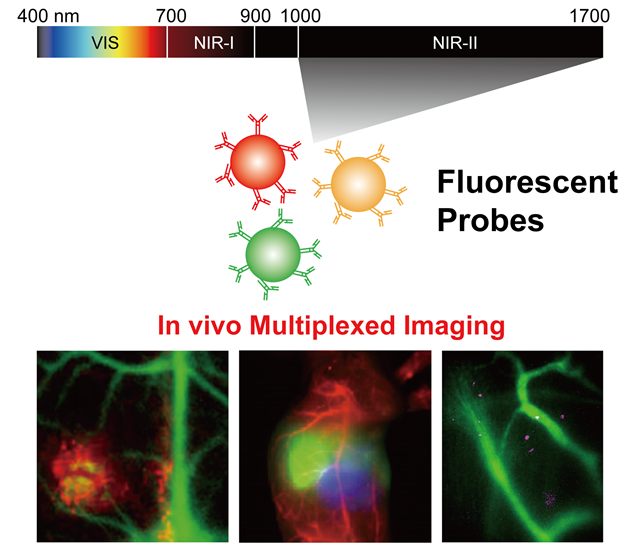
近红外二区(NIR-II, 1000~1700 nm)荧光成像技术, 因生物组织对其低的吸收和散射, 以及接近零的自体荧光背景噪声, 使得其在生物体内具备深的穿透能力以及高的分辨率和信噪比, 目前已经成为活体内研究的一种新型的技术手段, 尤其适用于活体组织内的多重成像. 本综述首先回顾了NIR-II荧光探针的设计与合成, 涵盖稀土基纳米颗粒、量子点及有机荧光分子等, 并探讨了多重成像技术对方法和设备的需求与成像技术进展, 最后分析了它们在血管/肿瘤/淋巴、多器官以及细胞层面的多重成像应用. 同时, 也展望了该新型成像模式在未来发展中的方向以及最终走向临床应用所面临的挑战.
蒋励, 陈子晗, 凡勇. 近红外二区荧光探针在活体多重成像中的研究进展[J]. 化学学报, 2024, 82(10): 1069-1085.
Li Jiang, Zihan Chen, Yong Fan. Research Progress on Fluorescent Probes in the Second Near-Infrared Window for In Vivo Multiplexed Imaging[J]. Acta Chimica Sinica, 2024, 82(10): 1069-1085.


| [1] |
Fan, Y.; Wang, S.; Zhang, F. Angew. Chem. Int. Ed. 2019, 58 13208.
|
| [2] |
Shimomura, O.; Johnson, F. H.; Saiga, Y. J. Cell. Physiol. 1962, 59 223.
|
| [3] |
Hell, S. W.; Wichmann, J. Opt. Lett. 1994, 19 780.
doi: 10.1364/ol.19.000780 pmid: 19844443 |
| [4] |
Reinhardt, S. C. M.; Masullo, L. A.; Baudrexel, I.; Steen, P. R.; Kowalewski, R.; Eklund, A. S.; Strauss, S.; Unterauer, E. M.; Schlichthaerle, T.; Strauss, M. T.; Klein, C.; Jungmann, R. Nature 2023, 617 711.
|
| [5] |
Irkle, A.; Vesey, A. T.; Lewis, D. Y.; Skepper, J. N.; Bird, J. L. E.; Dweck, M. R.; Joshi, F. R.; Gallagher, F. A.; Warburton, E. A.; Bennett, M. R.; Brindle, K. M.; Newby, D. E.; Rudd, J. H.; Davenport, A. P. Nat. Commun. 2015, 6 7495.
|
| [6] |
Helms, G.; Dathe, H.; Kallenberg, K.; Dechent, P. Magn. Reson. Med. 2008, 60 1396.
|
| [7] |
Clough, T. J.; Jiang, L.; Wong, K.-L.; Long, N. J. Nat. Commun. 2019, 10 1420.
|
| [8] |
Jathoul, A. P.; Laufer, J.; Ogunlade, O.; Treeby, B.; Cox, B.; Zhang, E.; Johnson, P.; Pizzey, A. R.; Philip, B.; Marafioti, T.; Lythgoe, M. F.; Pedley, R. B.; Pule, M. A.; Beard, P. Nat. Photonics 2015, 9 239.
|
| [9] |
Pu, K.; Shuhendler, A. J.; Jokerst, J. V.; Mei, J.; Gambhir, S. S.; Bao, Z.; Rao, J. Nat. Nanotechnol. 2014, 9 233.
|
| [10] |
Pratt, E. C.; Skubal, M.; Mc Larney, B.; Causa-Andrieu, P.; Das, S.; Sawan, P.; Araji, A.; Riedl, C.; Vyas, K.; Tuch, D.; Grimm, J. Nat. Biomed. Eng. 2022, 6 559.
|
| [11] |
Hu, L.; Dong, C.; Wang, Z.; He, S.; Yang, Y.; Zi, M.; Li, H.; Zhang, Y.; Chen, C.; Zheng, R.; Jia, S.; Liu, J.; Zhang, X.; He, Y. Aging Cell 2023, 22, e13896.
|
| [12] |
Yang, Y.; Chen, Y.; Pei, P.; Fan, Y.; Wang, S.; Zhang, H.; Zhao, D.; Qian, B.-Z.; Zhang, F. Nat. Nanotechnol. 2023, 18 1195.
|
| [13] |
Ren, F.; Wang, F.; Baghdasaryan, A.; Li, Y.; Liu, H.; Hsu, R.; Wang, C.; Li, J.; Zhong, Y.; Salazar, F.; Xu, C.; Jiang, Y.; Ma, Z.; Zhu, G.; Zhao, X.; Wong, K. K.; Willis, R.; Christopher Garcia, K.; Wu, A.; Mellins, E.; Dai, H. Nat. Biomed. Eng. 2023, 726.
|
| [14] |
Li, C.; Wang, Q. ACS Nano 2018, 12 9654.
|
| [15] |
Ntziachristos, V. Nat. Methods 2010, 7 603.
doi: 10.1038/nmeth.1483 pmid: 20676081 |
| [16] |
Voelker, R. JAMA 2022, 327 27.
|
| [17] |
Zhang, P.; Wu, Q.; Yang, J.; Hou, M.; Zheng, B.; Xu, J.; Chai, Y.; Xiong, L.; Zhang, C. Acta Biomater. 2022, 146 450.
|
| [18] |
Ding, S.; Lu, L.; Fan, Y.; Zhang, F. J. Rare Earths 2020, 38 451.
|
| [19] |
Frangioni, J. V. Curr. Opin. Chem. Biol. 2003, 7 626.
doi: 10.1016/j.cbpa.2003.08.007 pmid: 14580568 |
| [20] |
Lai, Y.; Dang, Y.; Sun, Q.; Pan, J.; Yu, H.; Zhang, W.; Xu, Z. Chem. Sci. 2022, 13 12511.
doi: 10.1039/d2sc05242c pmid: 36349272 |
| [21] |
Feng, Z.; Bai, S.; Qi, J.; Sun, C.; Zhang, Y.; Yu, X.; Ni, H.; Wu, D.; Fan, X.; Xue, D.; Liu, S.; Chen, M.; Gong, J.; Wei, P.; He, M.; Lam, J. W. Y.; Li, X.; Tang, B. Z.; Gao, L.; Qian, J. Adv. Mater. 2021, 33 2008123.
|
| [22] |
Yu, W. Q.; Liu, R.; Zhou, Y.; Gao, H. L. ACS Cent. Sci. 2020, 6 100.
|
| [23] |
Zhang, Z.; Du, Y.; Shi, X.; Wang, K.; Qu, Q.; Liang, Q.; Ma, X.; He, K.; Chi, C.; Tang, J.; Liu, B.; Ji, J.; Wang, J.; Dong, J.; Hu, Z.; Tian, J. Nat. Rev. Clin. Oncol. 2024, 21 449.
doi: 10.1038/s41571-024-00892-0 pmid: 38693335 |
| [24] |
Peloso, A.; Franchi, E.; Canepa, M. C.; Barbieri, L.; Briani, L.; Ferrario, J.; Bianco, C.; Quaretti, P.; Brugnatelli, S.; Dionigi, P.; Maestri, M. HPB 2013, 15 928.
|
| [25] |
Cousins, A.; Thompson, S. K.; Wedding, A. B.; Thierry, B. Biotechnol. Adv. 2014, 32 269.
doi: 10.1016/j.biotechadv.2013.10.011 pmid: 24189095 |
| [26] |
Hong, G.; Antaris, A. L.; Dai, H. Nat. Biomed. Eng. 2017, 1 0010.
|
| [27] |
Lifante, J.; Shen, Y.; Ximendes, E.; Martín Rodríguez, E.; Ortgies, D. H. J. Appl. Phys. 2020, 128 171101.
|
| [28] |
Huang, S.; Heikal, A. A.; Webb, W. W. Biophys. J. 2002, 82 2811.
|
| [29] |
Becker, W. J. Microsc. 2012, 247 119.
|
| [30] |
Fan, Y.; Wang, P.; Lu, Y.; Wang, R.; Zhou, L.; Zheng, X.; Li, X.; Piper, J. A.; Zhang, F. Nat. Nanotechnol. 2018, 13 941.
|
| [31] |
Jacques, S. L. Phys. Med. Biol. 2013, 58, R37.
|
| [32] |
Horton, N. G.; Wang, K.; Kobat, D.; Clark, C. G.; Wise, F. W.; Schaffer, C. B.; Xu, C. Nat. Photonics 2013, 7 205.
|
| [33] |
Wang, F.; Ren, F.; Ma, Z.; Qu, L.; Gourgues, R.; Xu, C.; Baghdasaryan, A.; Li, J.; Zadeh, I. E.; Los, J. W. N.; Fognini, A.; Qin-Dregely, J.; Dai, H. Nat. Nanotechnol. 2022, 17 653.
|
| [34] |
Pansare, V. J.; Hejazi, S.; Faenza, W. J.; Prud’homme, R. K. Chem. Mater. 2012, 24 812.
pmid: 22919122 |
| [35] |
Chen, Y.; Wang, S.; Zhang, F. Nat. Rev. Bioeng. 2023, 1 60.
|
| [36] |
Feng, Z.; Tang, T.; Wu, T.; Yu, X.; Zhang, Y.; Wang, M.; Zheng, J.; Ying, Y.; Chen, S.; Zhou, J.; Fan, X.; Zhang, D.; Li, S.; Zhang, M.; Qian, J. Light Sci. Appl. 2021, 10 197.
|
| [37] |
Carr, J. A.; Aellen, M.; Franke, D.; So, P. T. C.; Bruns, O. T.; Bawendi, M. G. Proc. Natl. Acad. Sci. 2018, 115 9080.
|
| [38] |
Feng, Z.; Li, Y.; Chen, S.; Li, J.; Wu, T.; Ying, Y.; Zheng, J.; Zhang, Y.; Zhang, J.; Fan, X.; Yu, X.; Zhang, D.; Tang, B. Z.; Qian, J. Nat. Commun. 2023, 14 5017.
doi: 10.1038/s41467-023-40728-6 pmid: 37596326 |
| [39] |
Koman, V. B.; Bakh, N. A.; Jin, X.; Nguyen, F. T.; Son, M.; Kozawa, D.; Lee, M. A.; Bisker, G.; Dong, J.; Strano, M. S. Nat. Nanotechnol. 2022, 17 643.
|
| [40] |
Ming, J.; Chen, Y.; Miao, H.; Fan, Y.; Wang, S.; Chen, Z.; Guo, Z.; Guo, Z.; Qi, L.; Wang, X.; Yun, B.; Pei, P.; He, H.; Zhang, H.; Tang, Y.; Zhao, D.; Wong, G. K.-L.; Bünzli, J.-C. G.; Zhang, F. Nat. Photonics 2024, DOI: 10.1038/s41566-024-01517-9.
|
| [41] |
Mei, M.; Wu, B.; Wang, S.; Zhang, F. Curr. Opin. Chem. Biol. 2024, 80 102469.
|
| [42] |
Wang, F. F.; Ma, Z. R.; Zhong, Y. T.; Salazar, F.; Xu, C.; Ren, F. Q.; Qu, L. Q.; Wu, A. M.; Dai, H. J. Proc. Natl. Acad. Sci. 2021, 118, e2023888118.
|
| [43] |
Ma, Z. R.; Wang, F. F.; Zhong, Y. T.; Salazar, F.; Li, J. C.; Zhang, M. X.; Ren, F. Q.; Wu, A. N. M.; Dai, H. J. Angew. Chem. Int. Ed. 2020, 59 20552.
|
| [44] |
Zhu, X.; Zhang, H.; Zhang, F. Acc. Mater. Res. 2023, 4 536.
|
| [45] |
Zhao, M.; Sik, A.; Zhang, H.; Zhang, F. Adv. Opt. Mater. 2023, 11 2202039.
|
| [46] |
Ma, Z. R.; Wan, H.; Wang, W. Z.; Zhang, X. D.; Uno, T.; Yang, Q. L.; Yue, J. Y.; Gao, H. P.; Zhong, Y. T.; Tian, Y.; Sun, Q. C.; Liang, Y. Y.; Dai, H. J. Nano Res. 2019, 12 273.
|
| [47] |
Zhu, X.; Wang, X.; Zhang, H.; Zhang, F. Angew. Chem. Int. Ed. 2022, 61, e202209378.
|
| [48] |
Wan, H.; Ma, H. L.; Zhu, S. J.; Wang, F. F.; Tian, Y.; Ma, R.; Yang, Q. L.; Hu, Z. B.; Zhu, T.; Wang, W. Z.; Ma, Z. R.; Zhang, M. X.; Zhong, Y. T.; Sun, H. T.; Liang, Y. Y.; Dai, H. J. Adv. Funct. Mater. 2018, 28 1804956.
|
| [49] |
Zhong, Y. T.; Ma, Z. R.; Zhu, S. J.; Yue, J. Y.; Zhang, M. X.; Antaris, A. L.; Yuan, J.; Cui, R.; Wan, H.; Zhou, Y.; Wang, W. Z.; Huang, N. F.; Luo, J.; Hu, Z. Y.; Dai, H. J. Nat. Commun. 2017, 8 737.
|
| [50] |
Antaris, A. L.; Chen, H.; Cheng, K.; Sun, Y.; Hong, G. S.; Qu, C. R.; Diao, S.; Deng, Z. X.; Hu, X. M.; Zhang, B.; Zhang, X. D.; Yaghi, O. K.; Alamparambil, Z. R.; Hong, X. C.; Cheng, Z.; Dai, H. J. Nat. Mater. 2016, 15 235.
|
| [51] |
Diao, S.; Blackburn, J. L.; Hong, G. S.; Antaris, A. L.; Chang, J. L.; Wu, J. Z.; Zhang, B.; Cheng, K.; Kuo, C. J.; Dai, H. J. Angew. Chem. Int. Ed. 2015, 54 14758.
|
| [52] |
Hong, G. S.; Robinson, J. T.; Zhang, Y. J.; Diao, S.; Antaris, A. L.; Wang, Q. B.; Dai, H. J. Angew. Chem. Int. Ed. 2012, 51 9818.
|
| [53] |
Hong, G. S.; Lee, J. C.; Robinson, J. T.; Raaz, U.; Xie, L. M.; Huang, N. F.; Cooke, J. P.; Dai, H. J. Nat. Med. 2012, 18 1841.
|
| [54] |
Welsher, K.; Sherlock, S. P.; Dai, H. J. Proc. Natl. Acad. Sci. 2011, 108 8943.
|
| [55] |
Liu, Z.; Tabakman, S.; Welsher, K.; Dai, H. J. Nano Res. 2009, 2 85.
|
| [56] |
Xiong, L.; Fan, Y.; Zhang, F. Acta Chim. Sinica 2019, 77 1239 (in Chinese).
doi: 10.6023/A19080305 |
|
(熊麟, 凡勇, 张凡, 化学学报, 2019, 77 1239.)
doi: 10.6023/A19080305 |
|
| [57] |
Welsher, K.; Liu, Z.; Sherlock, S. P.; Robinson, J. T.; Chen, Z.; Daranciang, D.; Dai, H. J. Nat. Nanotechnol. 2009, 4 773.
doi: 10.1038/nnano.2009.294 pmid: 19893526 |
| [58] |
Diao, S.; Hong, G. S.; Antaris, A. L.; Blackburn, J. L.; Cheng, K.; Cheng, Z.; Dai, H. J. Nano Res. 2015, 8 3027.
|
| [59] |
Wang, F.; Zhong, Y.; Bruns, O.; Liang, Y.; Dai, H. Nat. Photonics 2024, 18 535.
|
| [60] |
Chen, G.; Tian, F.; Zhang, Y.; Zhang, Y.; Li, C.; Wang, Q. Adv. Funct. Mater. 2014, 24 2481.
|
| [61] |
Naczynski, D. J.; Tan, M. C.; Zevon, M.; Wall, B.; Kohl, J.; Kulesa, A.; Chen, S.; Roth, C. M.; Riman, R. E.; Moghe, P. V. Nat. Commun. 2013, 4 2199.
doi: 10.1038/ncomms3199 pmid: 23873342 |
| [62] |
Ren, F.; Ding, L.; Liu, H.; Huang, Q.; Zhang, H.; Zhang, L.; Zeng, J.; Sun, Q.; Li, Z.; Gao, M. Biomaterials 2018, 175 30.
|
| [63] |
Wang, R.; Li, X.; Zhou, L.; Zhang, F. Angew. Chem. Int. Ed. 2014, 53 12086.
doi: 10.1002/anie.201407420 pmid: 25196421 |
| [64] |
Li, B.; Zhao, M.; Zhang, F. ACS Mater. Lett. 2020, 2 905.
|
| [65] |
Yu, G. T.; Luo, M. Y.; Li, H.; Chen, S.; Huang, B.; Sun, Z. J.; Cui, R.; Zhang, M. ACS Nano 2019, 13 12830.
|
| [66] |
Wang, T.; Wang, S.; Liu, Z.; He, Z.; Yu, P.; Zhao, M.; Zhang, H.; Lu, L.; Wang, Z.; Wang, Z.; Zhang, W.; Fan, Y.; Sun, C.; Zhao, D.; Liu, W.; Bünzli, J.-C. G.; Zhang, F. Nat. Mater. 2021, 20 1571.
|
| [67] |
Qi, J.; Sun, C.; Zebibula, A.; Zhang, H.; Kwok, R. T. K.; Zhao, X.; Xi, W.; Lam, J. W. Y.; Qian, J.; Tang, B. Z. Adv. Mater. 2018, 30, e1706856.
|
| [68] |
He, A.; Li, X.; Dai, Z.; Li, Q.; Zhang, Y.; Ding, M.; Wen, Z.-F.; Mou, Y.; Dong, H. J. Nanobiotechnol. 2023, 21 236.
|
| [69] |
Chang, Y. L.; Chen, H. R.; Xie, X. Y.; Wan, Y.; Li, Q. Q.; Wu, F. X.; Yang, R.; Wang, W.; Kong, X. G. Nat. Commun. 2023, 14 1079.
|
| [70] |
Chen, Z. H.; Wang, X.; Yang, M.; Ming, J.; Yun, B.; Zhang, L.; Wang, X.; Yu, P.; Xu, J.; Zhang, H.; Zhang, F. Angew. Chem. Int. Ed. 2023, 62, e202311883.
|
| [71] |
Tong, S.; Zhong, J.; Chen, X.; Deng, X.; Huang, J.; Zhang, Y.; Qin, M.; Li, Z.; Cheng, H.; Zhang, W.; Zheng, L.; Xie, W.; Qiu, P.; Wang, K. ACS Nano 2023, 17 3686.
|
| [72] |
Yang, Y.; Jiang, Q.; Zhang, F. Chem. Rev. 2024, 124 554.
doi: 10.1021/acs.chemrev.3c00506 pmid: 37991799 |
| [73] |
Yang, M.; Gong, H.; Yang, D.; Feng, L.; Gai, S.; Zhang, F.; Ding, H.; He, F.; Yang, P. Chin. Chem. Lett. 2024, 35 108468.
|
| [74] |
Ren, F.; Jiang, Z.; Han, M.; Zhang, H.; Yun, B.; Zhu, H.; Li, Z. VIEW 2021, 2 20200128.
|
| [75] |
Fan, Y.; Zhang, F. Adv. Opt. Mater. 2019, 7 1801417.
|
| [76] |
Fan, Y.; Liu, L.; Zhang, F. Nano Today 2019, 25 68.
|
| [77] |
Wu, Z.; Ke, J.; Liu, Y.; Sun, P.; Hong, M. Acta Chim. Sinica 2022, 80 542 (in Chinese).
|
|
(吴志芬, 柯建熙, 刘永升, 孙蓬明, 洪茂椿, 化学学报, 2022, 80 542.)
doi: 10.6023/A21120571 |
|
| [78] |
Nannuri, S. H.; Kulkarni, S. D.; , K, S. C.; Chidangil, S.; George, S. D. RSC Adv. 2019, 9 9364.
|
| [79] |
Jin, G. Q.; Guo, L. J.; Zhang, J.; Gao, S.; Zhang, J. L. Top. Curr. Chem. 2022, 380 31.
|
| [80] |
Liu, Z. Y.; Liu, A. A.; Fu, H.; Cheng, Q. Y.; Zhang, M. Y.; Pan, M. M.; Liu, L. P.; Luo, M. Y.; Tang, B.; Zhao, W.; Kong, J.; Shao, X.; Pang, D. W. J. Am. Chem. Soc. 2021, 143 12867.
|
| [81] |
Li, Y.; Cai, Z.; Liu, S.; Zhang, H.; Wong, S. T. H.; Lam, J. W. Y.; Kwok, R. T. K.; Qian, J.; Tang, B. Z. Nat. Commun. 2020, 11 1255.
|
| [82] |
Zhu, X.; Liu, C.; Hu, Z.; Liu, H.; Wang, J.; Wang, Y.; Wang, X.; Ma, R.; Zhang, X.; Sun, H.; Liang, Y. Nano Res. 2020, 13 2570.
|
| [83] |
Sun, C.; Li, B.; Zhao, M.; Wang, S.; Lei, Z.; Lu, L.; Zhang, H.; Feng, L.; Dou, C.; Yin, D.; Xu, H.; Cheng, Y.; Zhang, F. J. Am. Chem. Soc. 2019, 141 19221.
|
| [84] |
Cosco, E. D.; Spearman, A. L.; Ramakrishnan, S.; Lingg, J. G. P.; Saccomano, M.; Pengshung, M.; Arús, B. A.; Wong, K. C. Y.; Glasl, S.; Ntziachristos, V.; Warmer, M.; McLaughlin, R. R.; Bruns, O. T.; Sletten, E. M. Nat. Chem. 2020, 12 1123.
|
| [85] |
Zhu, S.; Herraiz, S.; Yue, J.; Zhang, M.; Wan, H.; Yang, Q.; Ma, Z.; Wang, Y.; He, J.; Antaris, A. L.; Zhong, Y.; Diao, S.; Feng, Y.; Zhou, Y.; Yu, K.; Hong, G.; Liang, Y.; Hsueh, A. J.; Dai, H. Adv. Mater. 2018, 30, e1705799.
|
| [86] |
Meador, W. E.; Lin, E. Y.; Lim, I.; Friedman, H. C.; Ndaleh, D.; Shaik, A. K.; Hammer, N. I.; Yang, B.; Caram, J. R.; Sletten, E. M.; Delcamp, J. H. Nat. Chem. 2024, 16 970.
|
| [87] |
Cherukuri, P.; Bachilo, S. M.; Litovsky, S. H.; Weisman, R. B. J. Am. Chem. Soc. 2004, 126 15638.
pmid: 15571374 |
| [88] |
Baghdasaryan, A.; Wang, F.; Ren, F.; Ma, Z.; Li, J.; Zhou, X.; Grigoryan, L.; Xu, C.; Dai, H. Nat. Commun. 2022, 13 5613.
doi: 10.1038/s41467-022-33341-6 pmid: 36153336 |
| [89] |
Chang, B.; Li, D.; Ren, Y.; Qu, C.; Shi, X.; Liu, R.; Liu, H.; Tian, J.; Hu, Z.; Sun, T.; Cheng, Z. Nat. Biomed. Eng. 2022, 6 629.
|
| [90] |
Wegner, K. D.; Hildebrandt, N. Chem. Soc. Rev. 2015, 44 4792.
doi: 10.1039/c4cs00532e pmid: 25777768 |
| [91] |
Xie, R.; Rutherford, M.; Peng, X. J. Am. Chem. Soc. 2009, 131 5691.
|
| [92] |
Huang, B.; Tang, T.; Liu, F.; Chen, S.-H.; Zhang, Z.-L.; Zhang, M.; Cui, R. Chin. Chem. Lett. 2024, 109694.
|
| [93] |
Shen, S.; Wang, Q. Chem. Mater. 2013, 25 1166.
|
| [94] |
Zebibula, A.; Alifu, N.; Xia, L.; Sun, C.; Yu, X.; Xue, D.; Liu, L.; Li, G.; Qian, J. Adv. Funct. Mater. 2018, 28 1703451.
|
| [95] |
Zhang, M.; Yue, J.; Cui, R.; Ma, Z.; Wan, H.; Wang, F.; Zhu, S.; Zhou, Y.; Kuang, Y.; Zhong, Y.; Pang, D.-W.; Dai, H. Proc. Natl. Acad. Sci. 2018, 115 6590.
|
| [96] |
Zheng, W.; Gao, G.; Deng, H.; Sun, T. Acta Chim. Sinica 2023, 81 763 (in Chinese).
|
|
(郑文山, 高冠斌, 邓浩, 孙涛垒, 化学学报, 2023, 81 763.)
doi: 10.6023/A23040128 |
|
| [97] |
Jeong, S.; Jung, Y.; Bok, S.; Ryu, Y. M.; Lee, S.; Kim, Y. E.; Song, J.; Kim, M.; Kim, S. Y.; Ahn, G. O.; Kim, S. Adv. Healthcare Mater. 2018, 7, e1800695.
|
| [98] |
Bruns, O. T.; Bischof, T. S.; Harris, D. K.; Franke, D.; Shi, Y.; Riedemann, L.; Bartelt, A.; Jaworski, F. B.; Carr, J. A.; Rowlands, C. J.; Wilson, M. W. B.; Chen, O.; Wei, H.; Hwang, G. W.; Montana, D. M.; Coropceanu, I.; Achorn, O. B.; Kloepper, J.; Heeren, J.; So, P. T. C.; Fukumura, D.; Jensen, K. F.; Jain, R. K.; Bawendi, M. G. Nat. Biomed. Eng. 2017, 1 0056.
|
| [99] |
Jiang, L.; Wang, K.; Qiu, L. Asian J. Pharm. Sci. 2022, 17 924.
|
| [100] |
Yin, B.; Liu, X.; Li, Z.; Ye, Z.; Wang, Y.; Yin, X.; Liu, S.; Song, G.; Huan, S.; Zhang, X.-B. Chin. Chem. Lett. 2024, 110119.
|
| [101] |
Sun, B.; Ma, R.; Wang, X.; Ma, S.; Li, W.; Liu, T.; Zhu, W.; Ji, Z.; Hettie, K. S.; Liu, C.; Liang, Y.; Zhu, S. VIEW 2024, 5 20230097.
|
| [102] |
Gui, Y.; Chen, K.; Sun, Y.; Tan, Y.; Luo, W.; Zhu, D.; Xiong, Y.; Yan, D.; Wang, D.; Tang, B. Z. Chin. J. Chem. 2023, 41 1249.
|
| [103] |
Liu, S.; Li, Y.; Kwok, R. T. K.; Lam, J. W. Y.; Tang, B. Z. Chem. Sci. 2021, 12 3427.
|
| [104] |
Xu, W.; Wang, D.; Tang, B. Z. Angew. Chem. Int. Ed. 2021, 60 7476.
|
| [105] |
Cheng, Y.; Sun, C.; Ou, X.; Liu, B.; Lou, X.; Xia, F. Chem. Sci. 2017, 8 4571.
doi: 10.1039/c7sc00402h pmid: 28626568 |
| [106] |
Hu, Z.; Fang, C.; Li, B.; Zhang, Z.; Cao, C.; Cai, M.; Su, S.; Sun, X.; Shi, X.; Li, C.; Zhou, T.; Zhang, Y.; Chi, C.; He, P.; Xia, X.; Chen, Y.; Gambhir, S. S.; Cheng, Z.; Tian, J. Nat. Biomed. Eng. 2020, 4 259.
|
| [107] |
Carr, J. A.; Franke, D.; Caram, J. R.; Perkinson, C. F.; Saif, M.; Askoxylakis, V.; Datta, M.; Fukumura, D.; Jain, R. K.; Bawendi, M. G.; Bruns, O. T. Proc. Natl. Acad. Sci. 2018, 115 4465.
|
| [108] |
O'Connell, M. J.; Bachilo, S. M.; Huffman, C. B.; Moore, V. C.; Strano, M. S.; Haroz, E. H.; Rialon, K. L.; Boul, P. J.; Noon, W. H.; Kittrell, C.; Ma, J.; Hauge, R. H.; Weisman, R. B.; Smalley, R. E. Science 2002, 297 593.
pmid: 12142535 |
| [109] |
Zhu, S.; Yang, Q.; Antaris, A. L.; Yue, J.; Ma, Z.; Wang, H.; Huang, W.; Wan, H.; Wang, J.; Diao, S.; Zhang, B.; Li, X.; Zhong, Y.; Yu, K.; Hong, G.; Luo, J.; Liang, Y.; Dai, H. Proc. Natl. Acad. Sci. 2017, 114 962.
|
| [110] |
Shi, W.-Q.; Zeng, L.; He, R.-L.; Han, X.-S.; Guan, Z.-J.; Zhou, M.; Wang, Q.-M. Science 2024, 383 326.
doi: 10.1126/science.adk6628 pmid: 38236955 |
| [111] |
Schmidt, E. L.; Ou, Z.; Ximendes, E.; Cui, H.; Keck, C. H. C.; Jaque, D.; Hong, G. Nat. Rev. Methods Primers 2024, 4 23.
|
| [112] |
Zhong, Y.; Ma, Z.; Wang, F.; Wang, X.; Yang, Y.; Liu, Y.; Zhao, X.; Li, J.; Du, H.; Zhang, M.; Cui, Q.; Zhu, S.; Sun, Q.; Wan, H.; Tian, Y.; Liu, Q.; Wang, W.; Garcia, K. C.; Dai, H. Nat. Biotechnol. 2019, 37 1322.
|
| [113] |
Naczynski, D. J.; Sun, C.; Türkcan, S.; Jenkins, C.; Koh, A. L.; Ikeda, D.; Pratx, G.; Xing, L. Nano Lett. 2015, 15 96.
doi: 10.1021/nl504123r pmid: 25485705 |
| [114] |
Cao, X.; Jiang, S.; Jia, M. J.; Gunn, J. R.; Miao, T.; Davis, S. C.; Bruza, P.; Pogue, B. W. J. Biomed. Opt. 2018, 24 1.
doi: 10.1117/1.JBO.24.5.051405 pmid: 30468044 |
| [115] |
Knez, D.; Hanninen, A. M.; Prince, R. C.; Potma, E. O.; Fishman, D. A. Light Sci. Appl. 2020, 9 125.
|
| [116] |
Kim, T.; O'Brien, C.; Choi, H. S.; Jeong, M. Y. Appl. Spectrosc. Rev. 2018, 53 349.
|
| [117] |
Wang, F.; Qu, L.; Ren, F.; Baghdasaryan, A.; Jiang, Y.; Hsu, R.; Liang, P.; Li, J.; Zhu, G.; Ma, Z.; Dai, H. Proc. Natl. Acad. Sci. 2022, 119, e2123111119.
|
| [118] |
Chen, Y.; Yang, Y.; Zhang, F. Nat. Protoc. 2024, 19 2386.
|
| [119] |
Bonis-O'Donnell, J. T. D.; Page, R. H.; Beyene, A. G.; Tindall, E. G.; McFarlane, I. R.; Landry, M. P. Adv. Funct. Mater. 2017, 27 1702112.
|
| [120] |
Huisken, J.; Swoger, J.; Del Bene, F.; Wittbrodt, J.; Stelzer, E. H. Science 2004, 305 1007.
doi: 10.1126/science.1100035 pmid: 15310904 |
| [121] |
Wang, F.; Wan, H.; Ma, Z.; Zhong, Y.; Sun, Q.; Tian, Y.; Qu, L.; Du, H.; Zhang, M.; Li, L.; Ma, H.; Luo, J.; Liang, Y.; Li, W. J.; Hong, G.; Liu, L.; Dai, H. Nat. Methods 2019, 16 545.
|
| [122] |
Pei, P.; Chen, Y.; Sun, C.; Fan, Y.; Yang, Y.; Liu, X.; Lu, L.; Zhao, M.; Zhang, H.; Zhao, D.; Liu, X.; Zhang, F. Nat. Nanotechnol. 2021, 16 1011.
|
| [123] |
Lu, L.; Li, B.; Ding, S.; Fan, Y.; Wang, S.; Sun, C.; Zhao, M.; Zhao, C.-X.; Zhang, F. Nat. Commun. 2020, 11 4192.
|
| [124] |
Zhu, X.; Liu, X.; Zhang, H.; Zhao, M.; Pei, P.; Chen, Y.; Yang, Y.; Lu, L.; Yu, P.; Sun, C.; Ming, J.; Abraham, I. M.; El-Toni, A. M.; Khan, A.; Zhang, F. Angew. Chem. Int. Ed. 2021, 60 23545.
doi: 10.1002/anie.202108124 pmid: 34487416 |
| [125] |
Yang, Y.; Wang, S.; Lu, L.; Zhang, Q.; Yu, P.; Fan, Y.; Zhang, F. Angew. Chem. Int. Ed. 2020, 59 18380.
|
| [126] |
Xu, H.; Yang, Y.; Lu, L.; Yang, Y.; Zhang, Z.; Zhao, C.-X.; Zhang, F.; Fan, Y. Anal. Chem. 2022, 94 3661.
|
| [127] |
Cao, C.; Jin, Z.; Shi, X.; Zhang, Z.; Xiao, A.; Yang, J.; Ji, N.; Tian, J.; Hu, Z. IEEE Trans. Biomed. Eng. 2022, 69 2404.
|
| [128] |
Qu, Q.; Nie, H.; Hou, S.; Wang, F.; He, K.; Deng, P.; Chen, S.; Zhang, Z.; Chi, C.; Hu, Z.; Shan, H.; Tian, J. Eur. J. Nucl. Med. Mol. Imaging 2022, 49 4752.
|
| [129] |
Smith, A. M.; Mancini, M. C.; Nie, S. Nat. Nanotechnol. 2009, 4 710.
|
| [130] |
Huang, J.; Su, L.; Xu, C.; Ge, X.; Zhang, R.; Song, J.; Pu, K. Nat. Mater. 2023, 22 1421.
|
| [1] | 刘童, 皮慧慧, 陈冰昆, 张小玲. 近红外二区硫系银量子点制备方法及癌症诊疗应用研究进展[J]. 化学学报, 2024, 82(9): 1001-1012. |
| [2] | 武虹乐, 郭锐, 迟涵文, 唐永和, 宋思睿, 葛恩香, 林伟英. 喹啉基粘度荧光探针的合成及其检测应用[J]. 化学学报, 2023, 81(8): 905-911. |
| [3] | 贺晓梦, 袁方, 张素雅, 张健健. 基于尼罗红类ONOO–近红外荧光探针的开发及其成像应用[J]. 化学学报, 2023, 81(11): 1515-1521. |
| [4] | 孙丽, 王亚静, 李涛, 郭英姝, 张书圣. 金纳米笼探针用于线粒体成像和光热损伤细胞★[J]. 化学学报, 2023, 81(10): 1301-1310. |
| [5] | 宋思睿, 唐永和, 孙良广, 郭锐, 姜冠帆, 林伟英. 基于香豆素荧光团的新型极性检测荧光探针的开发及其成像应用[J]. 化学学报, 2022, 80(9): 1217-1222. |
| [6] | 吴志芬, 柯建熙, 刘永升, 孙蓬明, 洪茂椿. 稀土近红外二区纳米荧光影像探针及其生物医学应用※[J]. 化学学报, 2022, 80(4): 542-552. |
| [7] | 王其, 夏辉, 熊炎威, 张新敏, 蔡杰, 陈冲, 高逸聪, 陆峰, 范曲立. 调控供电子策略简易制备近红外二区有机小分子光学诊疗试剂[J]. 化学学报, 2022, 80(11): 1485-1493. |
| [8] | 李勇, 王栩, 解希雷, 张建, 唐波. 一氧化碳有机荧光探针和光控释放剂研究进展[J]. 化学学报, 2021, 79(1): 36-44. |
| [9] | 桑若愚, 许兴鹏, 王其, 范曲立, 黄维. 近红外二区有机小分子荧光探针[J]. 化学学报, 2020, 78(9): 901-915. |
| [10] | 罗兴蕊, 陈敏文, 杨晴来. 近红外二区活体成像技术及其应用研究进展[J]. 化学学报, 2020, 78(5): 373-381. |
| [11] | 贾伊祎, 王文杰, 梁玲, 袁荃. 核酸功能化稀土基纳米材料在生物检测中的应用[J]. 化学学报, 2020, 78(11): 1177-1184. |
| [12] | 刘红文, 朱隆民, 娄霄峰, 袁林, 张晓兵. 用于细胞和组织中弗林蛋白酶特异性成像的双光子荧光探针研究[J]. 化学学报, 2020, 78(11): 1240-1245. |
| [13] | 郑斌, 程盛, 董华泽, 朱金苗, 韩钰, 杨亮, 胡进明. 一氧化氮响应性高分子荧光探针的构筑及其细胞内成像应用[J]. 化学学报, 2020, 78(10): 1089-1095. |
| [14] | 王英美, 朱道明, 杨阳, 张珂, 张修珂, 吕明珊, 胡力, 丁帅杰, 王亮. 快速合成Bi@ZIF-8复合纳米材料用于近红外二区光热治疗以及可控药物释放[J]. 化学学报, 2020, 78(1): 76-81. |
| [15] | 陈凯, 韩百川, 嵇思鑫, 孙瑾, 高振忠, 侯贤锋. 一种可用于直接检测空气中异氰酸酯的比率型荧光探针[J]. 化学学报, 2019, 77(4): 365-370. |
| 阅读次数 | ||||||
|
全文 |
|
|||||
|
摘要 |
|
|||||