化学学报 ›› 2026, Vol. 84 ›› Issue (3): 353-363.DOI: 10.6023/A25090314 上一篇 下一篇
研究论文
高春艳a, 崔馨月a, 王凯月a, 石雪怡a, 贺卿a, 赵晋忠a,*( ), 岳爱琴b, 杜维俊b, 张永坡a,*()
投稿日期:2025-09-16
发布日期:2025-11-20
基金资助:
Gao Chunyana, Cui Xinyuea, Wang Kaiyuea, Shi Xueyia, He Qinga, Zhao Jinzhonga,*(), Yue Aiqinb, Du Weijunb, Zhang Yongpoa,*()
Received:2025-09-16
Published:2025-11-20
Contact:
*E-mail: zhaojz@sxau.edu.cn;
zhangyp@sxau.edu.cn
Supported by:文章分享
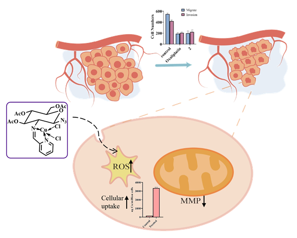
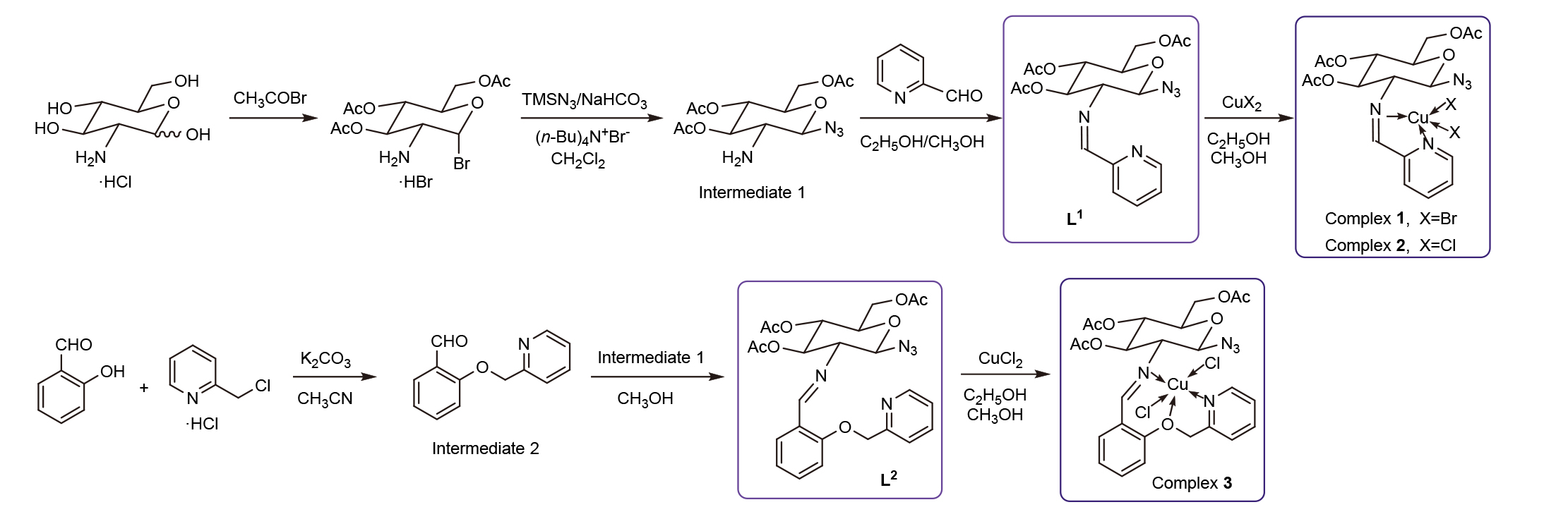
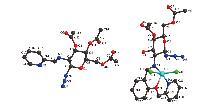
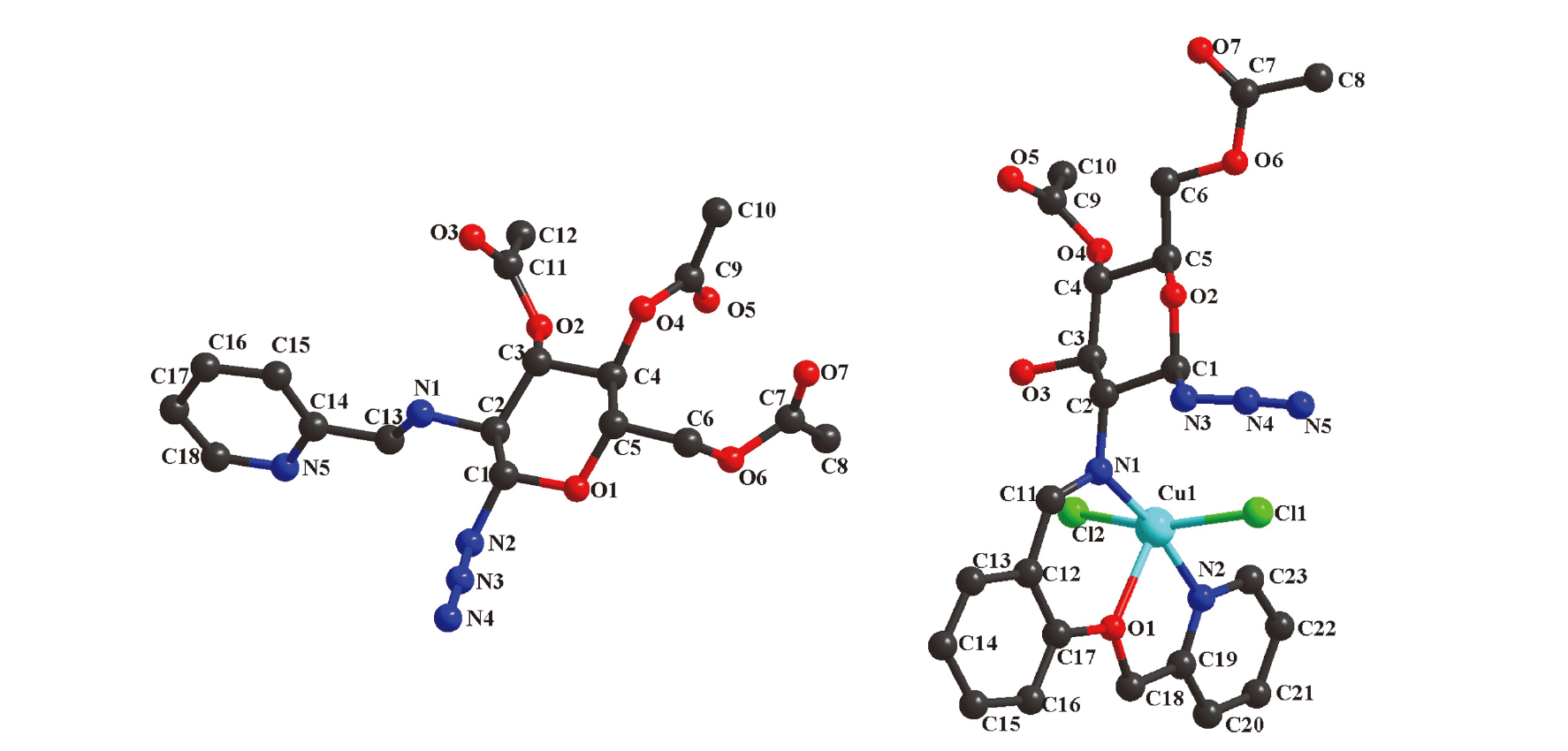
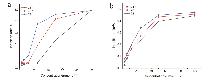
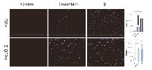
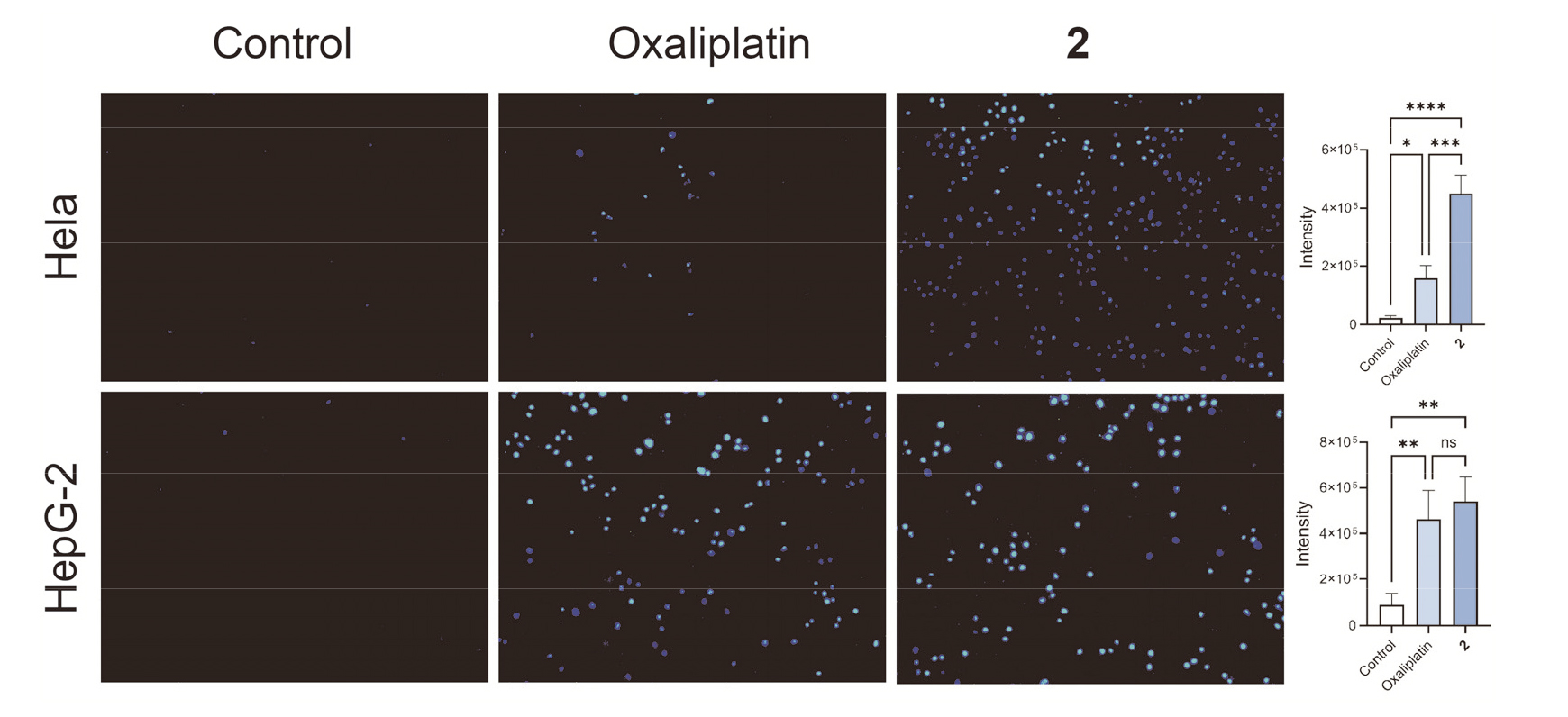
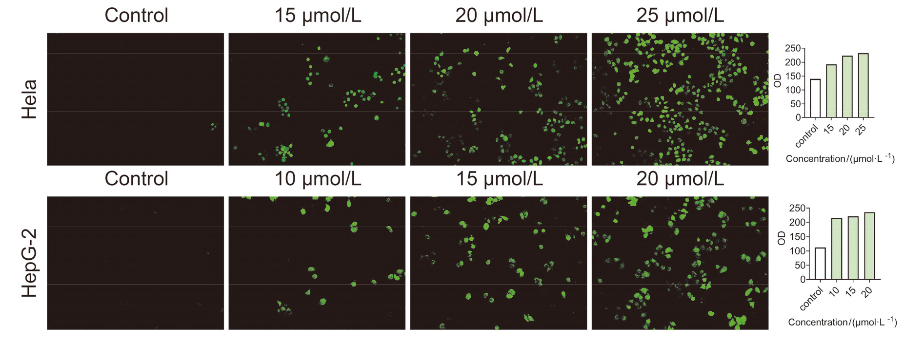
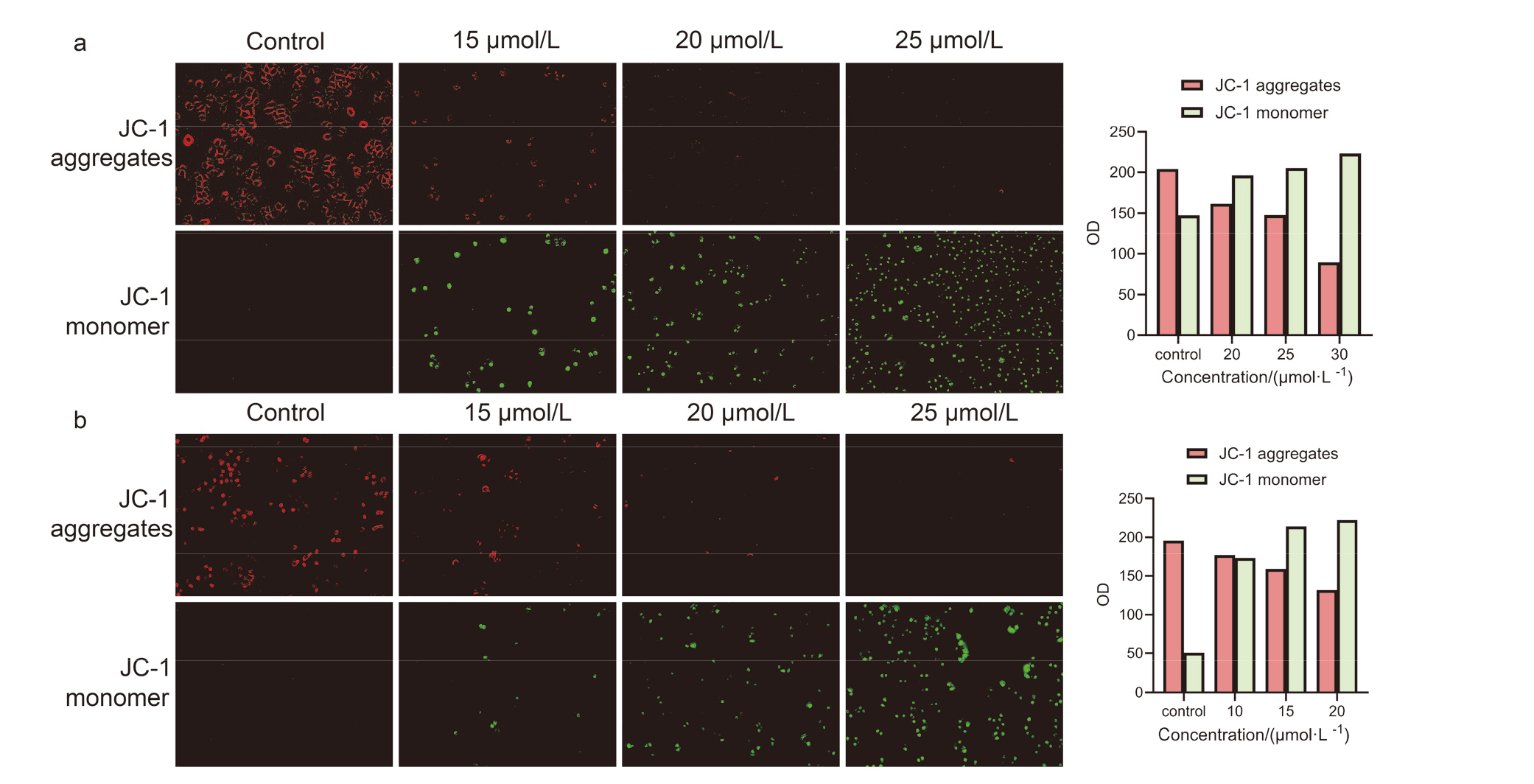
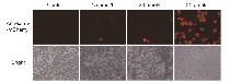
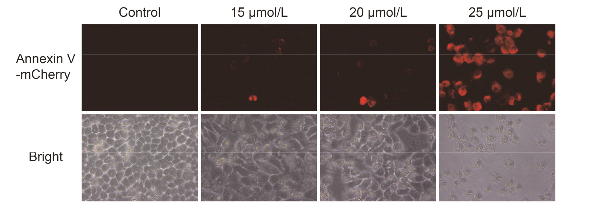
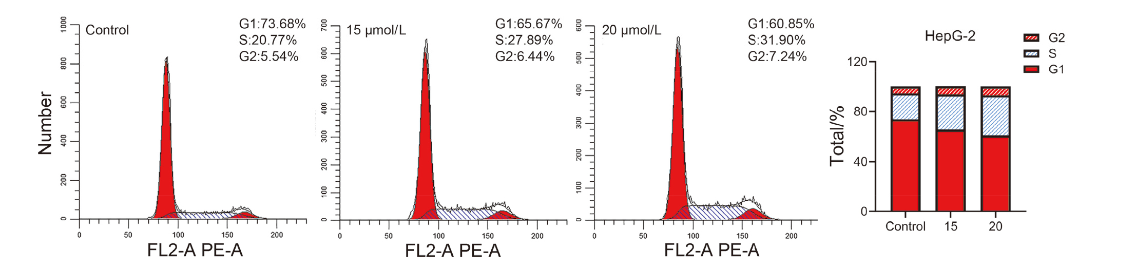
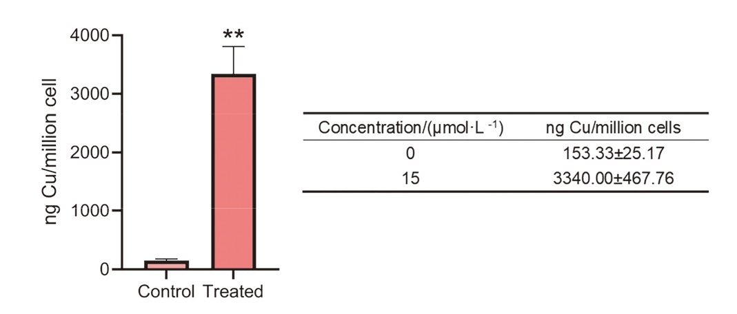
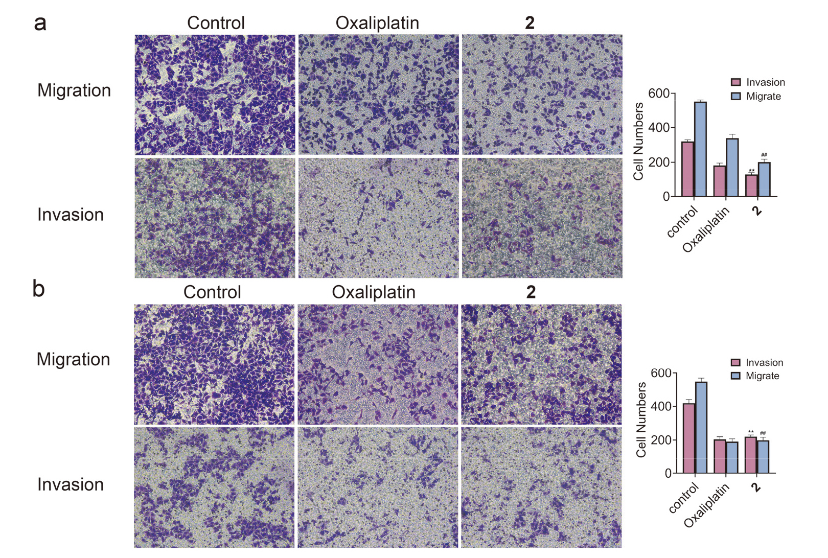
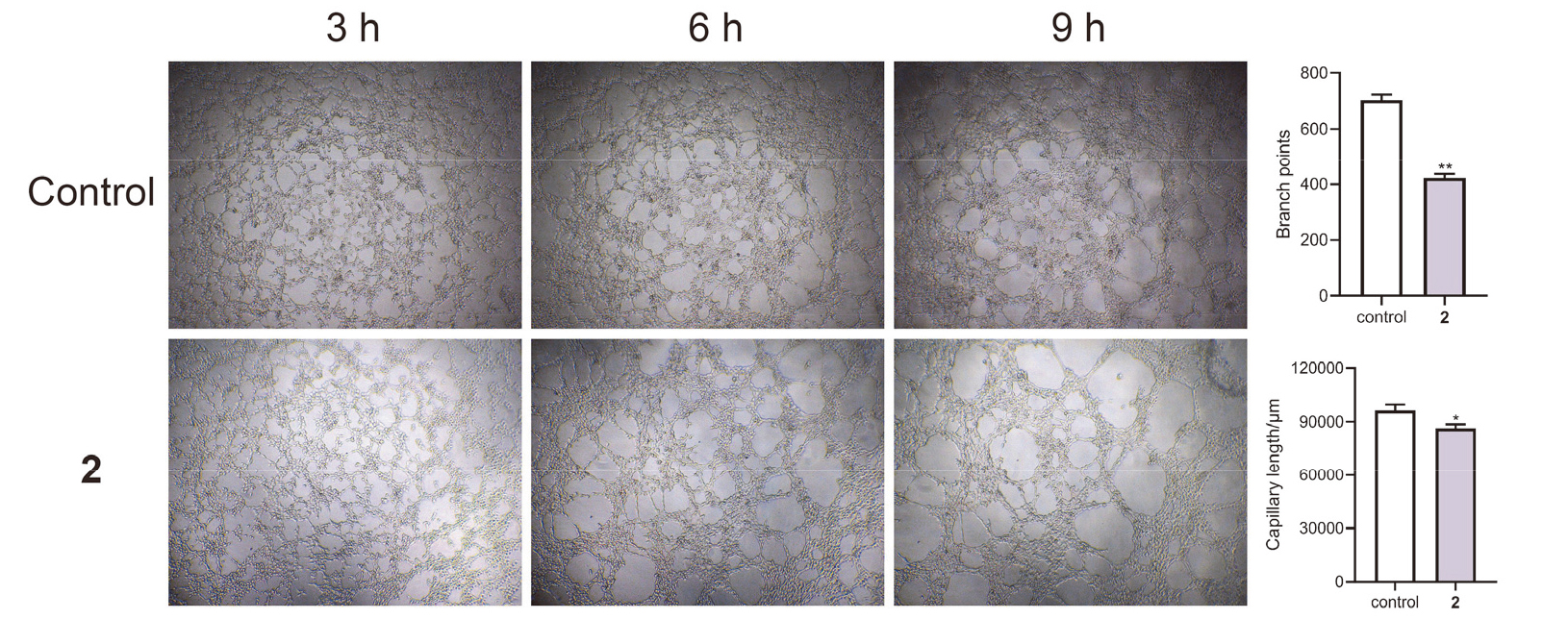
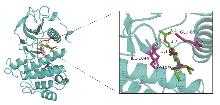
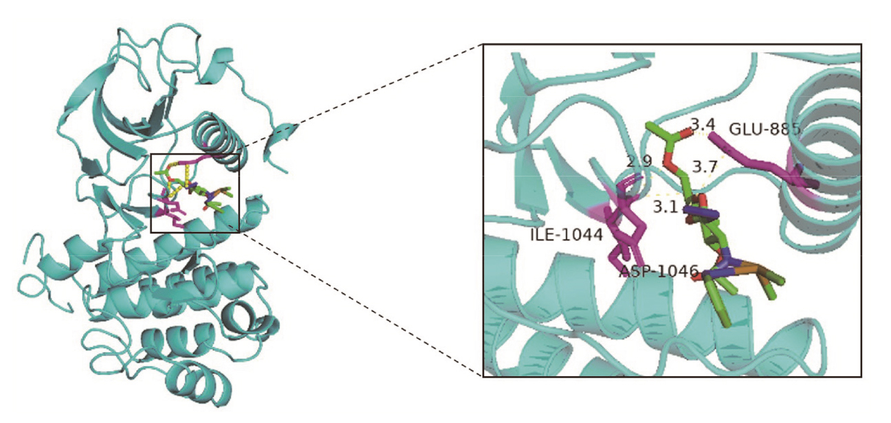
将生物活性相关的有机部分引入过渡金属配合物的配位领域已成为提高金属药物选择性和生物相容性的有效策略. 本工作设计并合成了2种具有叠氮乙酰化葡萄糖糖胺席夫碱结构的配体L1和L2, 进而与铜金属盐反应合成了3种新型铜配合物. 通过X射线单晶衍射技术解析了配体L1和配合物3的单晶结构, 通过噻唑蓝(MTT)法测定了化合物对人恶性肿瘤细胞系和正常细胞系的体外细胞毒性. 其中配合物2对肝癌细胞(HepG-2)和宫颈癌细胞(HeLa)细胞表现出高的细胞毒性, 接近或优于阳性对照的值, 但对正常细胞的毒性则远低于阳性对照. 对肿瘤细胞毒性较强的配合物 2 进行了后续抗肿瘤活性研究, 包括细胞凋亡评估、细胞内活性氧生成、线粒体膜电位、细胞周期、细胞摄取、迁移和侵袭及血管生成实验, 进一步证实了其对肿瘤细胞的抑制作用机制. 由于糖骨架在配体中具有优异的水溶性, 使得配合物2表现出良好的溶解性和稳定性. 通过细胞内活性氧(ROS)产生、赫斯特(Hoechst 33342)染色实验、线粒体膜电位实验和膜联蛋白Ⅴ-红色荧光蛋白(Annexin V-mCherry)进一步证明, 配合物2能提高癌症细胞内活性氧的水平, 通过线粒体途径诱导细胞凋亡. 细胞周期实验证明配合物2可以延迟或抑制细胞周期通过S期和G2/M期的进展. 另外, 配合物2能有效抑制癌症细胞的迁移和侵袭, 且有效抑制正常细胞系脐静脉内皮细胞(HUVEC)的血管生成, 分子对接结果显示配合物2与血管内皮细胞生长因子受体2 (VEGFR-2)具有良好的结合能力.
高春艳, 崔馨月, 王凯月, 石雪怡, 贺卿, 赵晋忠, 岳爱琴, 杜维俊, 张永坡. 新型氨基糖类席夫碱铜配合物的合成及抗肿瘤活性研究[J]. 化学学报, 2026, 84(3): 353-363.
Gao Chunyan, Cui Xinyue, Wang Kaiyue, Shi Xueyi, He Qing, Zhao Jinzhong, Yue Aiqin, Du Weijun, Zhang Yongpo. Synthesis and Antitumor Activity of Novel Amino Glucose Derivative Schiff Base Copper(II) Complexes[J]. Acta Chimica Sinica, 2026, 84(3): 353-363.
| Compound | IC50 a/(μmol•L−1) | ||||
|---|---|---|---|---|---|
| HeLa | NCI-H460 | BGC-823 | HepG-2 | HUVEC | |
| Oxaliplatin | 48.48±0.22 | 35.10±0.74 | 9.16±0.19 | 12.21±3.17 | 10.64±3.68 |
| 1 | 29.96±3.11 | 28.88±4.49 | 26.34±4.54 | 28.72±1.98 | 23.15±2.09 |
| 2 | 22.36±3.96 | 31.26±0.78 | 19.20±2.47 | 13.59±1.00 | 20.08±1.45 |
| 3 | 48.88±1.12 | 44.43±2.41 | 44.83±2.12 | 30.54±1.24 | 72.436 |
| L1 | >100 | >100 | >100 | >100 | — |
| L2 | >100 | >100 | >100 | >100 | — |
| CuBr2 | 161.5 | 106.0 | 84.7 | 117.7 | — |
| CuCl2 | 142.0 | 106.8 | 88.9 | 102.3 | — |

| [30] |
doi: 10.1016/j.arabjc.2023.105478 |
| [31] |
doi: 10.3390/ijms19071985 |
| [32] |
doi: 10.2174/1568009623666221025150239 |
| [33] |
|
|
(陈桂玲, 廖晓凤, 孙鹏涛, 岑欢, 舒盛春, 李碧晶, 黎金华, 南方医科大学学报, 2024, 44, 1109.)
doi: 10.12122/j.issn.1673-4254.2024.06.11 |
|
| [34] |
doi: 10.3390/molecules27030920 |
| [35] |
doi: 10.21037/tcr |
| [1] |
doi: 10.1016/j.ccr.2024.215698 |
| [2] |
doi: 10.1016/j.ejmech.2024.116528 |
| [3] |
doi: 10.1016/j.ccr.2024.216327 |
| [4] |
doi: 10.1016/j.ccr.2023.215231 |
| [5] |
|
| [6] |
doi: 10.1016/j.cbpa.2022.102236 |
| [7] |
doi: 10.6023/A25030093 |
|
(张一亮, 武卫龙, 许文磊, 傅玉琴, 郭辉, 卢志强, 化学学报, 2025, 83, 445.)
doi: 10.6023/A25030093 |
|
| [8] |
doi: 10.3390/cancers16162775 |
| [36] |
doi: 10.1186/1479-5876-11-276 |
| [37] |
doi: 10.1186/s12943-017-0719-3 pmid: 28893265 |
| [38] |
doi: 10.1039/D0DT00380H |
| [39] |
doi: 10.1039/C1CC15378A |
| [40] |
doi: 10.1021/jm200382r |
| [41] |
doi: 10.1002/jcc.21334 pmid: 19499576 |
| [42] |
doi: 10.3390/ijms19041264 |
| [9] |
doi: S0753-3322(17)32811-1 pmid: 28922722 |
| [10] |
doi: 10.6023/cjoc201310015 |
|
(宋沙沙, 周宏勇, 李小娜, 王丽华, 李云庆, 王家喜, 有机化学, 2014, 34, 706.)
doi: 10.6023/cjoc201310015 |
|
| [11] |
doi: 10.1016/j.ccr.2009.05.001 |
| [12] |
doi: 10.1002/ejoc.v2011.29 |
| [13] |
doi: 10.1021/acs.chemrev.0c01124 |
| [14] |
doi: 10.1039/C9BM00898E |
| [15] |
doi: 10.1002/cjoc.v40.7 |
| [16] |
doi: 10.3390/ijms232314840 |
| [17] |
|
| [18] |
doi: 10.1021/cr400135x pmid: 24102434 |
| [19] |
doi: 10.1016/j.ccr.2023.215156 |
| [20] |
doi: 10.1016/j.ccr.2025.216542 |
| [21] |
doi: 10.1016/j.molstruc.2021.130278 |
| [22] |
|
| [23] |
doi: 10.1007/s00044-011-9620-6 |
| [24] |
pmid: 16791578 |
| [25] |
doi: 10.1016/s1063-4584(03)00150-x pmid: 13129694 |
| [26] |
doi: 10.3109/1061186X.2015.1108325 |
| [27] |
doi: 10.1002/adma.v33.43 |
| [28] |
doi: 10.1016/j.molstruc.2023.136090 |
| [29] |
|
| [43] |
doi: 10.1016/j.molstruc.2021.132272 |
| [44] |
doi: 10.1016/j.tet.2013.03.075 |
| [45] |
|
| [46] |
|
| [1] | 王衍军, 王瑞欣, 杨浦曦, 陈慧芷, 郭德复, 高广鹏, 李晓芳. 新型杯呋喃大环的合成、结构及其与酸相互作用的研究[J]. 化学学报, 2025, 83(6): 588-595. |
| [2] | 赵雨晴, 梁栋, 贾吉慧, 余荣民, 卢灿忠. 具有双吸电子基团D-A型配体的Ag(I)发光配合物的合成与性能研究[J]. 化学学报, 2024, 82(5): 486-492. |
| [3] | 刘洋, 高丰琴, 马占营, 张引莉, 李午戊, 侯磊, 张小娟, 王尧宇. 一例钴基金属有机框架化合物活化过氧单硫酸盐高效降解水中亚甲基蓝研究[J]. 化学学报, 2024, 82(2): 152-159. |
| [4] | 吕天天, 马文, 詹冬笋, 邹燕敏, 李继龙, 冯美玲, 黄小荥. 两例新的镧系金属-有机框架化合物高效去除Cs+离子研究※[J]. 化学学报, 2022, 80(5): 640-646. |
| [5] | 赵锦旭, 张铭枢, 陈文发, 姜小明, 刘彬文, 郭国聪. KAg3Ga8S14: 一种高激光损伤阈值的中远红外非线性光学材料※[J]. 化学学报, 2022, 80(3): 259-264. |
| [6] | 方婧, 赵文娟, 张明浩, 方千荣. 一种新型酰胺功能化的共价有机框架用于选择性染料吸附[J]. 化学学报, 2021, 79(2): 186-191. |
| [7] | 付静茹, 贲腾. 一种新型的共价有机骨架膜的制备与气体分离性能[J]. 化学学报, 2020, 78(8): 805-814. |
| [8] | 陈光辉, 何燕萍, 张磊, 张健. 系列Ti4L6-笼基配合物的合成与结构研究[J]. 化学学报, 2020, 78(12): 1411-1417. |
| [9] | 王志涛, 李辉, 颜士臣, 方千荣. 一种沿骨架进行质子传导的二维共价有机框架的合成[J]. 化学学报, 2020, 78(1): 63-68. |
| [10] | 何燕萍, 谭衍曦, 张健. 基于尺寸识别和离子交换实现有机染料分离的一例阴离子型MOF[J]. 化学学报, 2014, 72(12): 1228-1232. |
| [11] | 韩聪, 徐喆, 刁春华, 陈鑫, 刘靖, 郭敏杰, 樊志. 2-呋喃甲硫醇修饰环糊精形成互锁式螺旋柱状超分子的自组装行为[J]. 化学学报, 2013, 71(03): 439-442. |
| [12] | 段显英, 郭利兵, 王建莉. 具有优良传导性能杂多酸复合物{[Co(H2O)8][H(H2O)3](HINO)4(PW12O40)}n的合成, 结构和性质研究[J]. 化学学报, 2013, 71(01): 107-113. |
| [13] | 郭琼, 李连之, 董建方, 刘鸿雁, 薛泽春, 许涛. 氧钒配合物[VO(o-Van-Asn)(Phen)]·1.5CH3OH的合成、晶体结构及与DNA和BSA的相互作用[J]. 化学学报, 2012, 70(15): 1617-1624. |
| [14] | 卢艳梅, 区志镔, 胡伟, 乐学义. (2-(2'-吡啶)苯并咪唑)(L-丙氨酸根)铜(II)配合物结构、抗菌活性及DNA断裂作用[J]. 化学学报, 2012, 70(08): 973-979. |
| [15] | 卢冬飞, 邸友莹, 窦建民. 四氯合镉酸正十一烷铵的合成、晶体结构及低温热容[J]. 化学学报, 2012, 70(07): 889-896 . |
| 阅读次数 | ||||||
|
全文 |
|
|||||
|
摘要 |
|
|||||
