Acta Chimica Sinica ›› 2021, Vol. 79 ›› Issue (3): 331-337.DOI: 10.6023/A20100459 Previous Articles Next Articles
Article
黄靖1, 王超1, 林敏刚1, 曾钫1,*( ), 吴水珠1,*()
投稿日期:2020-10-06
发布日期:2020-11-17
通讯作者:
曾钫, 吴水珠
作者简介:基金资助:
Jing Huang1, Chao Wang1, Mingang Lin1, Fang Zeng1,*(), Shuizhu Wu1,*()
Received:2020-10-06
Published:2020-11-17
Contact:
Fang Zeng, Shuizhu Wu
Supported by:Share
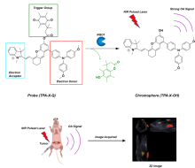
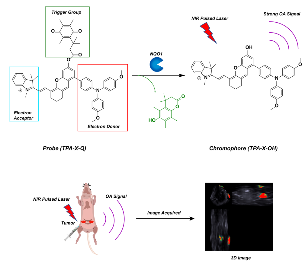
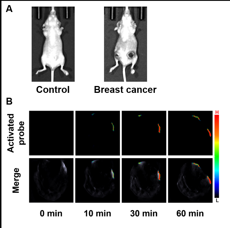
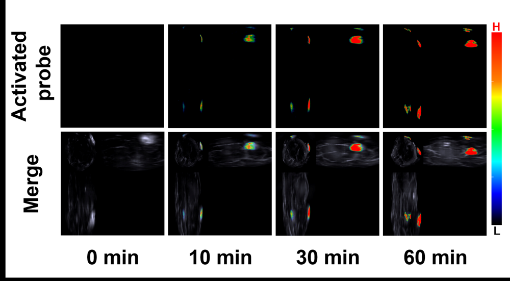
Jing Huang, Chao Wang, Mingang Lin, Fang Zeng, Shuizhu Wu. Synthesis of NQO1-activatable Optoacoustic Probe and Its Imaging of Breast Cancer[J]. Acta Chimica Sinica, 2021, 79(3): 331-337.


| [1] |
Asher, G.; Reuven, N.; Shaul, Y. BioEssays 2006, 28,844.
doi: 10.1002/bies.20447 pmid: 16927316 |
| [2] |
Clavería-Gimeno, R.; Velazquez-Campoy, A.; Pey, A. L. Arch. Biochem. Biophys. 2017, 636,17.
doi: 10.1016/j.abb.2017.10.020 pmid: 29100982 |
| [3] |
Pey, A. L. Int. J. Biol. Macromol. 2019, 126,1223.
pmid: 30615965 |
| [4] |
Zhao, Z.; Li, X.; Wu, B.; Zhou, Y. Chin. J. Chem. 2020, 38,714.
doi: 10.1002/cjoc.v38.7 |
| [5] |
Wang, N.; Zhao, J. Chin. J. Org. Chem. 2006, 26,775. (in Chinese)
doi: 10.1002/(ISSN)1614-7065 |
|
( 王乃兴, 赵嘉, 有机化学, 2006, 26,775.) 83d27848-ce6f-4042-bcec-2202ef50c36e
|
|
| [6] |
Mizumoto, A.; Ohashi, S.; Kamada, M.; Saito, T.; Nakai, Y.; Baba, K.; Hirohashi, K.; Mitani, Y.; Kikuchi, O.; Matsubara, J.; Yamada, A.; Takahashi, T.; Lee, H.; Okuno, Y.; Kanai, M.; Muto, M. J. Gastroenterol 2019, 54,687.
pmid: 30737573 |
| [7] |
Luo, S.; Lei, K.; Xiang, D.; Ye, K. Oxid. Med. Cell. Longevity 2018, 2018,1.
|
| [8] |
Sies, H.; Berndt, C.; Jones, D. P. Annu. Rev. Biochem. 2017, 86,715.
pmid: 28441057 |
| [9] |
Sabharwal, S. S.; Schumacker, P. T. Nat. Rev. Cancer 2014, 14,709.
doi: 10.1038/nrc3803 pmid: 25342630 |
| [10] |
Hayes, J. D.; Dinkova-Kostova, A. T.; Tew, K. D. Cancer Cell 2020, 38,167.
pmid: 32649885 |
| [11] |
Sharma, C.; Wang, H.-X.; Li, Q.; Knoblich, K.; Reisenbichler, E. S.; Richardson, A. L.; Hemler, M. E. Cancer Res. 2017, 77,6880.
pmid: 29055014 |
| [12] |
Danson, S.; Ward, T. H.; Butler, J.; Ranson, M. Cancer Treat. Rev. 2004, 30,437.
pmid: 15245776 |
| [13] |
Dinkova-Kostova, A. T.; Talalay, P. Arch. Biochem. Biophys. 2010, 501,116.
doi: 10.1016/j.abb.2010.03.019 pmid: 20361926 |
| [14] |
Li, Z.; Zhang, Y.; Jin, T.; Men, J.; Lin, Z.; Qi, P.; Piao, Y.; Yan, G.. BMC Cancer 2015, 15,207.
pmid: 25880877 |
| [15] |
Li, L.-S.; Reddy, S.; Lin, Z.-H.; Liu, S.; Park, H.; Chun, S. G.; Bornmann, W. G.; Thibodeaux, J.; Yan, J.; Chakrabarti, G.; Xie, X.; Sumer, B. D.; Boothman, D. A.; Yordy, J. S. Mol. Cancer Ther. 2016, 15,1757.
pmid: 27196777 |
| [16] |
Pani, G.; Giannoni, E.; Galeotti, T.; Chiarugi, P. Antioxid. Redox Signaling 2009, 11,2791.
doi: 10.1089/ars.2009.2739 |
| [17] |
Yang, Y.; Zhang, Y.; Wu, Q.; Cui, X.; Lin, Z.; Liu, S.; Chen, L. J. Exp. Clin. Cancer Res. 2014, 33,14.
doi: 10.1186/1756-9966-33-14 pmid: 24499631 |
| [18] |
Fagerholm, R.; Hofstetter, B.; Tommiska, J.; Aaltonen, K.; Vrtel, R.; Syrjäkoski, K.; Kallioniemi, A.; Kilpivaara, O.; Mannermaa, A.; Kosma, V.-M.; Uusitupa, M.; Eskelinen, M.; Kataja, V.; Aittomäki, K.; von Smitten, K.; Heikkilä, P.; Lukas, J.; Holli, K.; Bartkova, J.; Blomqvist, C.; Bartek, J.; Nevanlinna, H. Nat. Genet. 2008, 40,844.
pmid: 18511948 |
| [19] |
Jamshidi, M.; Bartkova, J.; Greco, D.; Tommiska, J.; Fagerholm, R.; Aittomäki, K.; Mattson, J.; Villman, K.; Vrtel, R.; Lukas, J.; Heikkilä, P.; Blomqvist, C.; Bartek, J.; Nevanlinna, H. Breast Cancer Res. Treat. 2012, 132,955.
doi: 10.1007/s10549-011-1629-5 pmid: 21706157 |
| [20] |
Qi, Y.; Huang, Y.; Li, B.; Zeng, F.; Wu, S. Anal. Chem. 2018, 90,1014.
doi: 10.1021/acs.analchem.7b04407 pmid: 29182316 |
| [21] |
Xiong, L.; Fan, Y.; Zhang, F. Acta Chim. Sinica 2019, 77,1239. (in Chinese)
doi: 10.6023/A19080305 pmid: 73282259-D05E-4539-8A88-A678B317D50D |
|
( 熊麟, 凡勇, 张凡, 化学学报, 2019, 77, 1239.)
pmid: 73282259-D05E-4539-8A88-A678B317D50D |
|
| [22] |
Niu, H.; Tang, J.; Zhu, X.; Li, Z.; Zhang, Y.; Ye, Y.; Zhao, Y. Chem. Commun. 2020, 56,7710.
doi: 10.1039/D0CC02668A |
| [23] |
Specht, E. A.; Braselmann, E.; Palmer, A. E. Annu. Rev. Physiol. 2017, 79,93.
pmid: 27860833 |
| [24] |
Wu, Y.; Wang, J.; Zeng, F.; Huang, S.; Huang, J.; Xie, H.; Yu, C.; Wu, S. ACS Appl. Mater. Interfaces 2016, 8,1511.
pmid: 26701212 |
| [25] |
Chang, Y.; Li, B.; Guo, M.; Cai, Y.; Xu, K. Chin. J. Org. Chem. 2019, 39,2485. (in Chinese)
doi: 10.6023/cjoc201903010 |
|
( 常永新, 李白, 郭淼, 蔡永红, 徐括喜, 有机化学, 2019, 39,2485.) ce3fcf14-0b02-4e50-a864-d4e0aa974467
doi: 10.6023/cjoc201903010 |
|
| [26] |
Christensen, P. R.; Wolf, M. O. Adv. Funct. Mater. 2016, 26,8471.
doi: 10.1002/adfm.v26.46 |
| [27] |
Wang, J.; Wu, Y.; Sun, L.; Zeng, F.; Wu, S. Acta Chim. Sinica 2016, 74,910. (in Chinese)
doi: 10.6023/A16070342 pmid: 038E5B9E-3975-4E43-99F1-BAB63AF9C904 |
|
( 王俊, 武英龙, 孙立和, 曾钫, 吴水珠, 化学学报, 2016, 74,910.) 6d05ea64-37bd-41b8-8490-f1e8f5c3f335
doi: 10.6023/A16070342 pmid: 038E5B9E-3975-4E43-99F1-BAB63AF9C904 |
|
| [28] |
Liu, P.; Xu, J.; Yan, D.; Zhang, P.; Zeng, F.; Li, B.; Wu, S. Chem. Commun. 2015, 51,9567.
doi: 10.1039/C5CC02149A |
| [29] |
Yang, Q.; Wen, Y.; Xu, J.; Shao, S. Talanta 2020, 216,120982.
doi: 10.1016/j.talanta.2020.120982 pmid: 32456908 |
| [30] |
Yuan, Z.; Xu, M.; Wu, T.; Zhang, X.; Shen, Y.; Ernest, U.; Gui, L.; Wang, F.; He, Q.; Chen, H. Talanta 2019, 198,323.
doi: 10.1016/j.talanta.2019.02.009 pmid: 30876568 |
| [31] |
Shen, Z.; Prasai, B.; Nakamura, Y.; Kobayashi, H.; Jackson, M. S.; McCarley, R. L. ACS Chem. Biol. 2017, 12,1121.
pmid: 28240865 |
| [32] |
Hettiarachchi, S. U.; Prasai, B.; McCarley, R. L. J. Am. Chem. Soc. 2014, 136,7575. e701d009-6c49-4c13-aed7-52d3b59ac8c3
doi: 10.1021/ja5030707 |
| [33] |
Pan, D.; Luo, F.; Liu, X.; Liu, W.; Chen, W.; Liu, F.; Kuang, Y.-Q.; Jiang, J.-H. Analyst 2017, 142,2624.
pmid: 28608874 |
| [34] |
Cheng, Z.; Valença, W. O.; Dias, G. G.; Scott, J.; Barth, N. D.; de Moliner, F.; Souza, G. B. P.; Mellanby, R. J.; Vendrell, M.; da Silva Júnior, E.N. Bioorg. Med. Chem. 2019, 27,3938.
doi: 10.1016/j.bmc.2019.07.017 pmid: 31327676 |
| [35] |
Ovsepian, S. V.; Olefir, I.; Westmeyer, G.; Razansky, D.; Ntziachristos, V. Neuron 2017, 96,966.
doi: S0896-6273(17)30990-X pmid: 29216459 |
| [36] |
Huang, J.; Wu, Y.; Zeng, F.; Wu, S. Theranostics 2019, 9,7313.
pmid: 31695770 |
| [37] |
Ouyang, J.; Sun, L.; Zeng, Z.; Zeng, C.; Zeng, F.; Wu, S. Angew. Chem. Int. Ed.Eng. 2020, 59,10111.
|
| [38] |
Reber, J.; Willershäuser, M.; Karlas, A.; Paul-Yuan, K.; Diot, G.; Franz, D.; Fromme, T.; Ovsepian, S. V.; Bézière, N.; Dubikovskaya, E.; Karampinos, D. C.; Holzapfel, C.; Hauner, H.; Klingenspor, M.; Ntziachristos, V. Cell Metab. 2018, 27,689.
doi: 10.1016/j.cmet.2018.02.002 pmid: 29514074 |
| [39] |
Zhan, C.; Huang, Y.; Lin, G.; Huang, S.; Zeng, F.; Wu, S. Small 2019, 15,1900309.
doi: 10.1002/smll.v15.33 |
| [40] |
Wu, Y.; Chen, J.; Sun, L.; Zeng, F.; Wu, S. Adv. Funct. Mater. 2019, 29,1807960.
|
| [41] |
Diot, G.; Metz, S.; Noske, A.; Liapis, E.; Schroeder, B.; Ovsepian, S. V.; Meier, R.; Rummeny, E.; Ntziachristos, V. Clin. Cancer Res. 2017, 23,6912.
pmid: 28899968 |
| [42] |
Ntziachristos, V.; Razansky, D. Chem. Rev. 2010, 110,2783.
doi: 10.1021/cr9002566 pmid: 20387910 |
| [43] |
Luís Dean-Ben, X.; Razansky, D. Light: Sci. Appl. 2018, 7,18004.
|
| [44] |
Gujrati, V.; Mishra, A.; Ntziachristos, V. Chem. Commun. 2017, 53,4653.
doi: 10.1039/C6CC09421J |
| [45] |
Schellenberg, M. W.; Hunt, H. K. Photoacoustics 2018, 11,14.
doi: 10.1016/j.pacs.2018.07.001 pmid: 30073147 |
| [46] |
Song, X.; Bian, H.; Wang, C.; Hu, M.; Li, N.; Xiao, Y. Org. Biomol. Chem. 2017, 15,8091.
pmid: 28905964 |
| [47] |
Kim, H.; Lee, M.; Kim, H.; Kim, S.; Yoon, J. Chem. Soc. Rev. 2008, 37,1465.
doi: 10.1039/b802497a pmid: 18648672 |
| [48] |
Han, J.; Burgess, K.; Chem. Rev. 2010, 110,2709.
pmid: 19831417 |
| [49] |
Mendoza, M. F.; Hollabaugh, N. M.; Hettiarachchi, S. U.; McCarley, R. L. Biochemistry 2012, 51,8014.
pmid: 22989153 |
| [50] |
Wu, Y.; Huang, S.; Wang, J.; Sun, L.; Zeng, F.; Wu, S. Nat. Commun. 2018, 9,3983.
pmid: 30266905 |
| [1] | Haipeng Wang, Wensheng Cai, Xueguang Shao. Antifreeze Mechanism of Antifreeze Agents by Near Infrared Spectroscopy and Molecular Simulations★ [J]. Acta Chimica Sinica, 2023, 81(9): 1167-1174. |
| [2] | Wenshan Zheng, Guanbin Gao, Hao Deng, Taolei Sun. Room Temperature Synthesis and Near-infrared Fluorescence Performance Optimization of Ag2Se@Ag2S Core-shell Quantum Dots [J]. Acta Chimica Sinica, 2023, 81(7): 763-770. |
| [3] | Xiaomeng He, Fang Yuan, Suya Zhang, Jianjian Zhang. Development of a Near-Infrared Fluorescent Probe Based on Nile Red for ONOO– and Its Imaging Applications [J]. Acta Chimica Sinica, 2023, 81(11): 1515-1521. |
| [4] | Wei Lian, Zekai Fang, Datao Tu, Jiayao Li, Siyuan Han, Renfu Li, Xiaoying Shang, Xueyuan Chen. Template-Based Controlled Synthesis and Bioapplication of AgInSe2:Zn2+ Near-Infrared Luminescent Quantum Dots※ [J]. Acta Chimica Sinica, 2022, 80(5): 625-632. |
| [5] | Ruomei Liu, Yanhui Feng, Zhuo Li, Shan Lu, Tianyong Guan, Xingjun Li, Yan Liu, Zhuo Chen, Xueyuan Chen. A Novel Near-infrared Responsive Lanthanide Upconversion Nanoplatform for Drug Delivery Based on Photocleavage of Cypate※ [J]. Acta Chimica Sinica, 2022, 80(4): 423-427. |
| [6] | Qi Wang, Hui Xia, Yanwei Xiong, Xinmin Zhang, Jie Cai, Chong Chen, Yicong Gao, Feng Lu, Quli Fan. Simple Preparation of Near-infrared-II Organic Small Molecule-based Phototheranostics by Manipulation of the Electron-donating Unit [J]. Acta Chimica Sinica, 2022, 80(11): 1485-1493. |
| [7] | Heqi Gao, Di Jiao, Hanlin Ou, Jingtian Zhang, Dan Ding. High Performance Aggregation-Induced Emission Nanoprobes for Image-Guided Cancer Surgery [J]. Acta Chimica Sinica, 2021, 79(3): 319-325. |
| [8] | Meng Yu, Zijun Zhang, Guowei Zhu, Zhenhua Gu, Yulin Duan, Liangchong Yu, Guanbin Gao, Taolei Sun. Synthesis of Ag2S Based Quantum Dots with Near-infrared-II Fluorescence Emission in Water [J]. Acta Chimica Sinica, 2021, 79(10): 1281-1285. |
| [9] | Sang Ruoyu, Xu Xingpeng, Wang Qi, Fan Quli, Huang Wei. Near-Infrared-II Fluorescence Probes Based on Organic Small Molecules [J]. Acta Chimica Sinica, 2020, 78(9): 901-915. |
| [10] | Wang Mingyuan, Cui Xiaoyu, Cai Wensheng, Shao Xueguang. Temperature-Dependent Near-Infrared Spectroscopy for Sensitive Detection of Glucose [J]. Acta Chimica Sinica, 2020, 78(2): 125-129. |
| [11] | Wang Yingmei, Zhu Daoming, Yang Yang, Zhang Ke, Zhang Xiuke, Lv Mingshan, Hu Li, Ding Shuaijie, Wang Liang. Rapid Synthesis of Bi@ZIF-8 Composite Nanomaterials for the Second Near-infrarad Window Photothermal Therapy and Controlled Drug Release [J]. Acta Chimica Sinica, 2020, 78(1): 76-81. |
| [12] | Jiao Yang, Zhang Xi. Supramolecular Free Radicals: Fabrication, Modulation and Functions [J]. Acta Chim. Sinica, 2018, 76(9): 659-665. |
| [13] | Zhu Xuewei, Cui Xiaoyu, Cai Wensheng, Shao Xueguang. Temperature Dependent Near Infrared Spectroscopy for Understanding the Hydrogen Bonding of Amines [J]. Acta Chim. Sinica, 2018, 76(4): 298-302. |
| [14] | Chen Meihua, Pan Zheng, Yin Yuefeng, Liu Jie, Liu Mengyuan, Jia Zijun, Liang Guijie. Panchromatic and High-efficient Energy Transfer Assembly Based on Type I Core-shell Quantum Dots [J]. Acta Chim. Sinica, 2016, 74(4): 330-334. |
| [15] | Gao Guanbin, Gong Dejun, Zhang Mingxi, Sun Taolei. Chiral Gold Nanoclusters: A New Near-Infrared Fluorescent Probe [J]. Acta Chim. Sinica, 2016, 74(4): 363-368. |
| Viewed | ||||||
|
Full text |
|
|||||
|
Abstract |
|
|||||
