



1 Introduction
Amino acids are not only the basic building blocks of proteins, but also metabolites necessary for the production of nucleotides, antioxidants (e.g., glutathione), and energy (e.g., the components of the tricarboxylic acid cycle).[1-5] It plays an important role in different biological fields, such as maintaining specific physiological processes, participating in neuromodulation, influencing organ function, etc. Due to their importance in chemistry and biology, their identification and detection have attracted a lot of attention from scientists. Among amino acids, L-arginine (L-Arg) is an important amino acid in the body, and L-Arg has a guanidinium group and a glycine portion.[6] L-Arg has important roles in many biological functions, and its physiological functions include participation in body metabolism, cardiovascular function, and regulation of immunity.[7-9] In addition, L-Arg can be used in food processing as a food flavor enhancer, coloring agent, and nutrient fortifier.[10-11] Thus, there is a great need to vigorously research and develop sensors for the detection of L-Arg. Many works on L-Arg recognition have been reported,[12-13] such as high-perfor- mance liquid chromatography, spectrophotometry, electrochemistry and mass spectrometry.[14-17] However, how to improve sensitivity and selectivity has been the focus of research in this field. Despite the advances in analytical methods and instruments in recent years, high sensitivity, low cost and reliable detection of chemical substances are still pursued by researchers. Fluorescence sensors have been widely used by scientists due to their good real-time performance, fast response, low detection limit, and high detection selectivity and sensitivity.[18-20] Currently, fluorescence sensors have been widely used in various applications such as the detection of metal ions,[21-23] carbohydrates,[24-27] and amino acids.[28-30]
Pillar[n]arenes were first synthesized and reported by the Ogoshi group in 2008.[30] As an important class of macrocyclic compounds, pillar[n]arenes are characterized by their ease of synthesis and modification, electron-rich cavities, and excellent host-guest assembly properties,[31] which have made it widely used in the field of supramolecular chemistry. Pillar[n]arenes have broad research potential for detecting amino acids, as their cavity structures can encapsulate specific sizes of amino acids.[32] However, the fluorescence of pillar[n]arenes is very weak, which limits their development in the field of fluorescence sensing of amino acids. Numerous efforts have shown that axial functionalization of pillar[n]arenes using π-conjugated fluorescent groups can effectively improve their luminescent proper-ties.[33] Therefore, it is of great importance to rationally design specific π-conjugated pillar[n]arenes and apply them to fluorescence detection of amino acids.
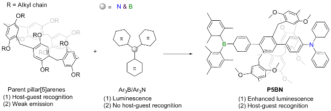
Recently, boryl-functionalized π-conjugated lumino- phors are attracting considerable attention because of their great potential in nonlinear optics,[34] organic electronics[35] and anion sensors.[36] Triarylborane (Ar3B), as a typical electron acceptor, has been widely used in luminescent materials and stimulus responsive materials.[37] Once Ar3B is coupled with electron donors through π-conjugation, they can exhibit unusual charge-transfer (CT) emission.[38] Based on this, in 2020, Chen and co-workers[39] synthesized a pillar[5]arene-based π-conjugated luminescent compound P5BN by covalently coupling fluorophores of both electron-donating triphenylamine (Ph3N) and electron-deficient Ar3B to pillar[5]arene skeleton. The axially extended π-con- jugated structure endowed compound P5BN with strong fluorescence. However, the group did not study the fluorescent recognition performance of P5BN for biological small molecules, especially amino acids. In fact, P5BN provides a good cavity structure that can encapsulate guest molecules of appropriate size. Therefore, here, we conducted a detailed study on the fluorescence recognition performance of P5BN as a fluorescent sensor for amino acids. It is found that P5BN can selectively detect L-Arg with a fluorescence color change from yellowish-brown to sky-blue, due to the effective matching L-Arg with the cavity of P5BN and significant host-guest interaction (Ka=4.88×10⁵ L•mol-1). The details are presented herein.
2 Results and discussion
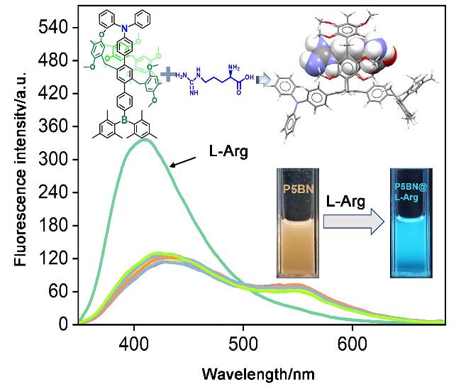
The pillar[5]arene-based π-conjugated fluorescent molecule P5BN was synthesized using a previously reported method in the literature.[39] P5BN exhibited brownish- yellow fluorescence in dimethyl sulfoxide (DMSO) solution (c=2.0×10⁻⁶ mol/L), accompanied by a significantly broadened dual emission bands at 430 and 547 nm. The former is attributed to the locally excited (LE) state radiative transition, while the latter originates from CT emission. First of all, P5BN was used as a new fluorescent sensor, and twenty natural amino acids (L-Trp, β-Ala, L-Arg, L-Cys, Gly, L-Lys, L-Pro, L-Met, L-Thr, L-Ser, L-His, L-Val, L-Tyr, L-Asn, L-Glu, L-Gln, L-Lie, L-Phe, L-Asp and L-Leu) were selected as guests to evaluate its sensing capability towards amino acid species. In the fluorescence spectra (Figure 1), when 2.0 equiv. of the above twenty amino acids were added to DMSO solution of P5BN, respectively, only L-Arg caused a significant change in the fluorescence emission band of P5BN. Quite evidently, the addition of L-Arg directly led to the decrease of emission peak at 547 nm and the increase of emission peak at 430 nm of P5BN, and meanwhile, the luminescent color changes from brownish-yellow to sky-blue. Therefore, P5BN can univocally and selectively detect L-Arg in twenty natural amino acids by fluorescence.
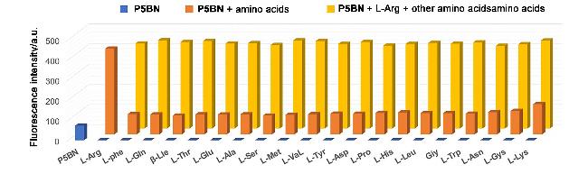
In order to further verify the selectivity of P5BN as a sensor for the actual detection of L-Arg, the competitive experiments were carried out by adding 2.0 equiv. of L-Arg to the DMSO solution of 2.0 equiv. of competitive amino acids. As shown in Figure 2, all competing amino acids did not have a significant influence on the detection of L-Arg. These results further confirmed that P5BN displayed a gratifying selectivity for the detection of L-Arg.
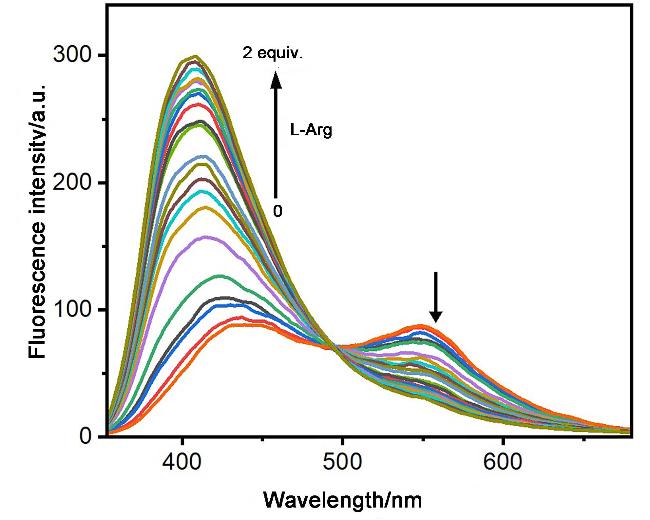
Next, the sensitivity of P5BN toward the L-Arg detection was investigated via fluorescence titration experiments (Figure 3). In the fluorescence spectrum, as the concentration of L-Arg increased in the range of 0~2.0 equiv., the fluorescence intensity at 547 nm of P5BN slowly declined, while the fluorescence intensity at 408 nm rapidly enhanced, accompanied by the formation of an isoemissive point at 495 nm. The ratio of fluorescence intensities at 408 and 547 nm of P5BN increased linearly with the equiv. of L-Arg (0~2.0 equiv.). Based on this good linear relationship, the limit of detection (LOD) calculated using the 3σ/S method is 2.21×10⁻⁸ mol/L, implying the high sensitivity of P5BN for L-Arg. Therefore, P5BN has potential appli- cation in L-Arg detection. Amino acids as guests bind to pillar[5]arene motif via 1∶1 complexation;[32,40] therefore, the binding constant (Ka) of P5BN towards L-Arg was furt-her estimated to be 4.88×10⁵ L•mol-1 using a non-linear curve fitting of the fluorescence titration results.
The recognition mechanism of P5BN for L-Arg was analyzed by the 1H NMR titration experiment. When L-Arg was gradually added to the DMSO-d6 solution of P5BN, the proton signals of Ha-d on P5BN showed a gradual upfield shift. This result indicates that host-guest recognition has occurred between P5BN and L-Arg, forming a complex P5BN@L-Arg.
To deeply understand the fluorescence response mechanism of P5BN for L-Arg, the density functional theory (DFT) was carried out based on the quantum chemical calculations at the B3LYP/6-31G(d,p) level. Electrostatic potential maps (ESP) showed that the macrocyclic skeleton of P5BN is negatively charged (Figure 4a), while L-Arg shows the most positive charge at both ends, which provides support for L-Arg penetrating into the cavity of P5BN to form P5BN@L-Arg. In P5BN, the highest occupied orbital (HOMO) is evenly distributed in the half-moiety of pillar[5]arene; however, the lowest occupied orbital (LUMO) is located on the Ar3B group. Compared with P5BN, the HOMO of P5BN@L-Arg is mainly distributed on the Ar3N unit, while the distribution of LUMO is almost unchanged. The change of HOMO distribution directly leads to the quenching of CT emission at 547 nm of P5BN. The HOMO-LUMO gap of P5BN@L-Arg is smaller than that of P5BN, indicating that the binding of L-Arg in the cavity has a certain stability.
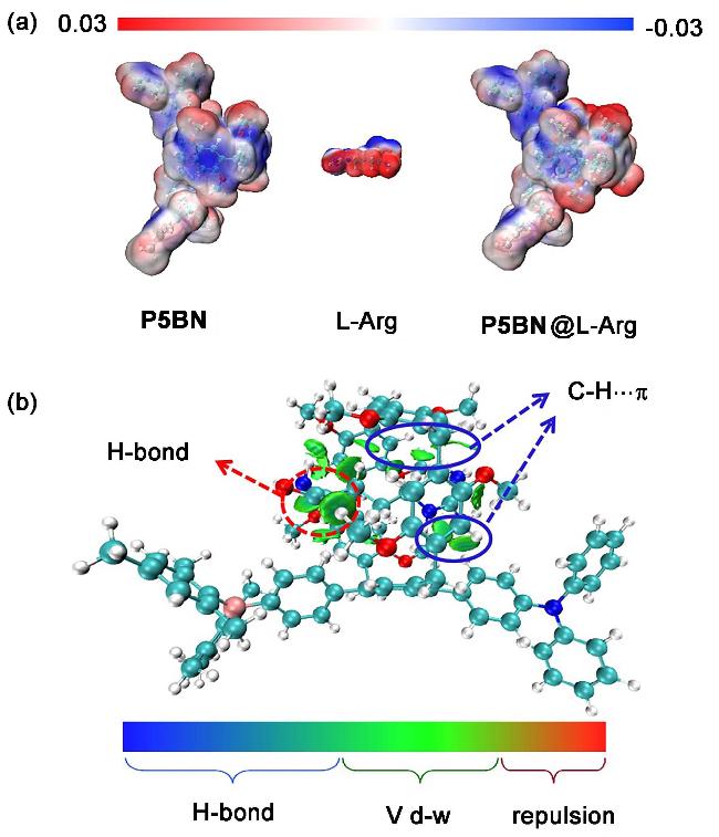


Finally, the independent gradient model was used based on Hirshfeld partition (IGMH) to analyze the weak interaction force and the binding energy of P5BN and L-Arg. It can be seen from the IGMH diagram of P5BN@L-Arg that there are significant van der Waals forces and hydrogen bonds between P5BN and L-Arg (Figure 4b). The guanidine and carboxylic acid groups of L-Arg provided strong hydrogen bonds. These multiple interactions stabilize the complexation between P5BN and L-Arg. By calculating the difference between the energy of each component of the host and guest molecule and the overall energy separately, and introducing the basis set overlap error correction (BSSE) to correct the final binding energy, a binding energy of -21.76 kJ/mol for P5BN and L-Arg was finally obtained.
3 Conclusions
In summary, we have developed a pillar[5]arene-based π-conjugated amino acid sensor P5BN, which provided a good cavity structure for encapsulating guest molecules of suitable size. L-Arg is precisely matched with its cavity to complete the host-guest recognition, resulting in enhanced fluorescence at 408 nm of P5BN. Therefore, it could sense L-Arg with excellent sensitivity by fluorescence (LOD=2.21×10⁻⁸ mol/L), with the fluorescence color changing from brownish-yellow to sky-blue. Meanwhile, the sensor exhibited high selectivity for L-Arg, and the other natural amino acids did not show any significant competitive response. The recognition mechanism was studied through experiments and theoretical calculations, indicating that P5BN and L-Arg formed a 1∶1 complex through host- guest recognition. This work not only provided an amino acid sensor for L-Arg, but also expanded the development of π-conjugated pillar[5]arene in the field of biological small molecule sensing.
4 Experimental section
4.1 General
All chemical agents and solvents, unless otherwise stated, were purchased from commercial supplies and used without further purification. 1H NMR spectra were recorded on a Bruker spectrometer. UV-visible absorption spectra were recorded on a Shimadzu UV- 2550 spectrometer. Shimadzu RF-5301PC spectrofluo- rophotometer recorded the fluorescence spectra. For titration experiments, L-Arg solutions were prepared by dissolving the desired amount of solid L-Arg in DMSO. Addition of L-Arg to the sample solution was perfor- med by a microsyringe (±0.1 μL). DFT calculations were performed with the Gaussian 16 A03 program. Geometry optimizations were calculated by means of hybrid density functional B3LYP with the basis set of 6-31G(d,p). The host-guest binding energy calculation formula is as follows:
Ecom=EAB-(EA+EB)+EBSSE.
4.2 Synthesis
4.2.1 Synthesis of P5-OTf
Compound P5-OTf was synthesized according to a previously reported procedure.[41] 1H NMR (600 MHz, CDCl3) δ: 7.37 (s, 3H), 6.84~6.81 (m, 3H), 6.81~6.81 (m, 3H), 6.81~6.79 (m, 2H), 6.72 (s,3H), 4.44 (s, 1H), 3.81 (s, 15H), 3.73~3.63 (m, 28H), 1.24~1.18 (m, 2H), 1.17~1.11 (m, 2H), 0.86 (s, 2H); 13C NMR (151 MHz, CDCl3) δ: 150.68, 150.65, 150.45, 146.13, 133.80, 129.64, 128.34, 127.90, 124.47, 124.10,119.59, 113.95, 113.78, 113.58, 113.52, 77.20, 55.69, 55.64, 55.62, 55.07, 52.81, 31.52, 30.51, 29.45, 29.20, 22.57, 14.08. ESI-HRMS calcd for C45H44- F6O14S2 [P5-OTf+H]+ 987.2149, found 987.2145.
4.2.2 Synthesis of P5N1
The mixture of P5-OTf (296 mg, 0.3 mmol), (4-(diphen-ylamino)phenyl)boronic acid (87 mg, 0.3 mmol), Pd(PPh3)4 (17 mg, 0.015 mmol), PPh3 (13 mg, 0.05mmol) and K2CO3 (276 mg, 2.0 mmol) in toluene/EtOH/H2O (3 mL/0.5 mL/ 0.5 mL) was refluxed with stirring under N2 for 12 h. The reaction mixture was then cooled to room temperature and poured into water (30 mL). After extraction with CH2Cl2, the combined organic phases were dried over Na2SO4. Purification by column chromatography on silica gel (petroleum ether/CH2Cl2/ethyl acetate, V/V/V=100/10/1) gave P5N1 as a white solid (165 mg, 51%). 1H NMR (400 MHz, CDCl3) δ: 7.35~7.27 (m, 6H), 7.23 (s, 2H), 7.19~7.14 (m, 4H), 7.12 (s, 1H), 7.11~7.03 (m, 6H), 6.78 (d, J=4.8 Hz, 4H), 6.71 (s, 3H), 5.99 (s, 1H), 3.87 (d, J=8.4 Hz, 4H), 3.81 (s, 4H), 3.73 (d, J=7.8 Hz, 6H), 3.68 (s, 3H), 3.65 (s, 10H), 3.57 (s, 7H), 3.49 (s, 3H); 13C NMR (101 MHz, CDCl3) δ: 147.63, 147.63, 130.29, 129.32, 124.53, 123.10, 122.92, 55.71, 55.71, 55.67, 53.29, 53.29. ESI-HRMS calcd for C62H59F3NO11S [P5N1+H]+ 1082.3755, found 1082.3743.
4.2.3 Synthesis of P5BN
The mixture of P5N1 (216 mg, 0.2 mmol), (4-(dimesityl- boranyl)phenyl)boronic acid[42] (74 mg, 0.2 mmol), Pd- (PPh3)4 (35 mg, 0.03 mmol), PPh3 (26 mg, 0.1 mmol) and K2CO3 (276 mg, 2.0 mmol) in toluene/EtOH/H2O (4 mL/0.5 mL/0.5 mL) was refluxed with stirring under N2 for 36 h. The reaction mixture was then cooled to room temperature and poured into water (30 mL). After extraction with CH2Cl2, the combined organic phases were dried over Na2SO4. Purification by column chromatography on silica gel (petroleum ether/CH2Cl2/ethyl acetate, V/V/V=50/1/2) gave P5BN as a white solid (98 mg, 39%). 1H NMR (400 MHz, DMSO-d6) δ: 7.36 (td, J=19.5, 17.5, 10.6 Hz, 8H), 7.10 (t, J=6.9 Hz, 8H), 6.99~6.92 (m, 4H), 6.86 (s, 4H), 6.70 (t, J=7.2 Hz, 4H), 3.71 (dd, J=60.9, 10.3 Hz, 10H), 3.55~3.37 (m, 24H), 2.28 (s, 6H), 2.00 (s, 12H); 13C NMR (101 MHz, CDCl3) δ: 150.95, 150.91, 150.89, 147.83, 140.80, 138.66, 136.50, 136.44, 136.42, 130.30, 129.41, 129.35, 128.68, 128.31, 128.24, 128.14, 124.46, 122.99, 114.40, 114.23, 114.15, 114.08, 56.02, 55.99, 55.89, 55.86, 55.62, 32.90, 32.29, 30.21, 30.11, 29.63, 26.96, 23.54, 21.27. ESI-HRMS calcd for C85H85BNO8 [P5BN+H]+ 1258.6363, found 1258.6387.
Supporting Information Synthetic procedure of P5BN, photophysical measurements, host-guest recognition, partial 1H NMR (400 MHz, DMSO-d6) titration spectrums of P5BN with adding L-Arg, theoretical calculation, and references. The Supporting Information is available free of charge via the Internet at http://sioc-journal.cn.
(Lu, Y.)