


1 Introduction
Medicinal plants of the genus Litsea are predominantly distributed in the provinces of Guizhou, Chongqing, Sichuan, and Anhui in China. Litsea coreana Lévl. var. lanuginosa (Migo), a medicinal plant from the Lauraceae family known for its nutritional properties, is referred to as "hawk tea" or "white tea" in Chinese.[1-3] Traditionally, both its bark and leaves are used to prepare tea. It is commonly used as a traditional remedy for stomach ailments, hepatitis, and inflammation. Recently, hawk tea has garnered significant interest due to its diverse bioactive properties, including anticancer, antiviral, anti-inflammatory, antibacterial, and hepatoprotective activities.[4-9] Previous phytochemical investigations of hawk tea have demonstrated that this plant contains flavonoids, polyphenols, biphenylpropanoids, coumarins, lignans, butanolides, and polysaccharides.[10-16] Previous phytochemical research on hawk tea has primarily concentrated on the leaves, while there has been comparatively less investigation into the bark. To further explore biologically active compounds from medicinal plants in southwestern China, an investigation into the chemical composition of hawk tea was conducted.[17,18] A rare sesquiterpene, hawkteasesquioid A (1, Figure 1), featuring a novel 5/6/5 tricyclic system containing a 6,5-spiroketal motif, was obtained from the bark of hawk tea. The cytotoxicity of this novel compound was evaluated against A549, HT-29, and SW1990 cells.
2 Results and discussion
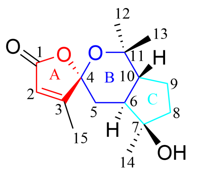
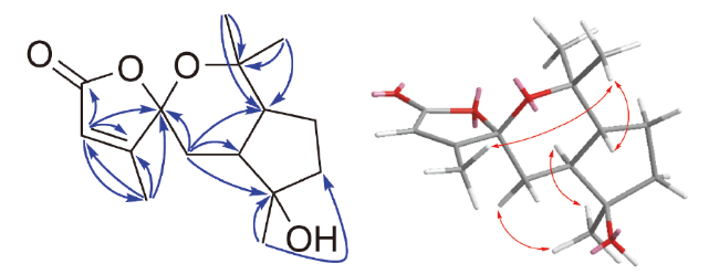
Compound 1 was obtained as a colorless needle-like crystal, and its molecular formula was determined to be C15H22O4 based on the HRESIMS ([M-H]- at m/z 265.1471, calcd for C15H21O4 m/z 265.1445), which required five degrees of unsaturation. 1H NMR (Table 1) and HSQC spectra exhibited three singlet methyls at δH 1.26, 1.28, and 1.33 (s, each 3H); one doublet methyl at δH 2.01 (d, J=1.5 Hz, 3H); three methylene groups at δH 1.63~1365 and 1.8~1.80 (m, each 1H), δH 1.34~1.36 and 1.88~1.91 (m, each 1H), and δH 1.75 and 1.85 (overlapped, each 1H); two methine groups at δH 1.75 and 1.85 (overlapped, each 1H); and one olefinic proton at δH 5.77 (q, J=1.5 Hz, 1H). The 13C NMR and DEPT spectra exhibited 15 carbon resonances, including four methyl carbons at δC 12.9, 22.1, 27.0, and 30.6; three methylene carbons at δC 24.8, 31.8, and 40.4; two methane carbons at δC 43.5 and 49.0; three oxygenated quaternary carbons at δC 79.5, 80.6, and 108.8; and three sp2 hybridized carbons at δC 117.9, 168.4, and 171.1. The 13C NMR data indicated a total of 15 carbon atoms, while the 1H NMR data revealed the presence of multiple methyl groups suggested that the skeleton of compound 1 should be sesquiterpene.[19] The presence of an unsaturated five-membered lactone moiety[20] (subunit A in Figure 1) was confirmed by 1H-1H correlated spectroscopy (COSY) correlations of H-2 (δH 5.77) with 15-CH3 (δH 2.01), together with the heteronuclear multiple bond correlation (HMBC) correlations from H-2 to C-1 (δC 171.1), C-3 (δC 168.4), and C-4 (δC 108.8), from 15-CH3 to C-2 (δC 117.9), C-3, and C-4 (Figure 2). Furthermore, the HMBC correlations from H-5 (δH 1.64) to C-4 (δC 108.8), C-6 (δC 43.5), and C-10 (δC 49.0), from 12-CH3 (δH 1.28) and 13-CH3 (δH 1.26) to C-11 (δC 80.6) and C-10, in conjunction with the nuclear Overhauser effect spectroscopy (NOESY) interactions (Figure 2) of 13-CH3 and 15-CH3, demonstrated that the six-membered spiro-heterocycle (subunit B in Figure 1) was connected to C-4. The C-7 signal (δC 79.5), together with HMBC correlations from 14-CH3 (δH 1.33) to C-6, C-7, and C-8 (δC 40.4), suggested that C-7 was an oxygenated carbon, and 14-CH3 connected to C-7. These data accounted for only four degrees of unsaturation, necessitating the addition of one more ring in the molecule. Further analysis of the 13C NMR spectrum of compound 1 implied the presence of a cyclopentane moiety (subunit C in Figure 1). Thus, compound 1 was characterized as a rare sesquiterpene featuring a unique spiro-heterocyclic skeleton. The NOESY correlations of 15-CH3 with 13-CH3, 15-CH3 with H-10, 12-CH3 with δH 1.64 (H-5α), H-5α with 14-CH3, and 14-CH3 with H-6, indicated that 15-CH3, 13-CH3 and H-10 were in the β-orientation, while 12-CH3, 14-CH3 and H-6 were in the α-orientation. The relative configuration of compound 1, (4R*,6S*,7R*,10R*), was established using single crystal X-ray diffraction (Figure 3) (CCDC No. 2403747). The CD spectrum of the compound displayed a straight line, leading to the presumption that the compound was a racemate. Therefore, the structure of compound 1 was determined and named hawkteasesquioid A.
Table 1 NMR spectroscopic data of compound 1 (400/100 MHz, in CDCl3, J in Hz) |
| No. | δH | δC | No. | δH | δC |
|---|---|---|---|---|---|
| 1 | 171.1 | 9 | 1.88~1.91 m; 1.34~1.36, m | 24.8 | |
| 2 | 5.77, q, 1.5 | 117.9 | 10 | 1.85, overlapped | 49.0 |
| 3 | 168.4 | 11 | 80.6 | ||
| 4 | 108.8 | 12 | 1.28, s | 22.1 | |
| 5 | 1.63~1.65, m; 1.78~1.80, m | 31.8 | 13 | 1.26, s | 30.6 |
| 6 | 1.75, overlapped | 43.5 | 14 | 1.33, s | 27.0 |
| 7 | 79.5 | 15 | 2.01, d, 1.5 | 12.9 | |
| 8 | 1.85, overlapped; 1.75, overlapped | 40.4 |
 ) and NOESY (
 ) correlations of compound 1
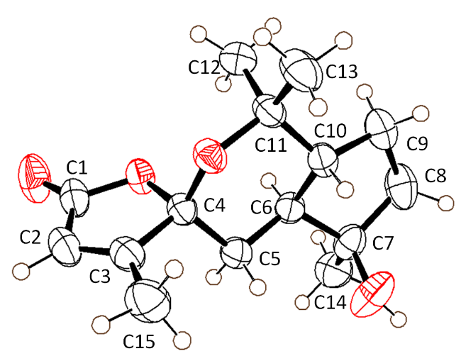
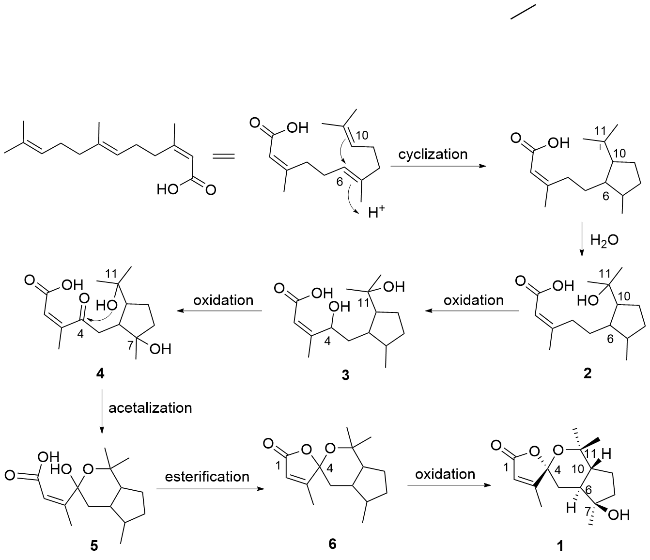
The biogenic synthesis pathway of compound 1 was proposed based on literature references and plausible chemical reactions, as illustrated in Figure 4. Compound 1 was derived from 3,7,11-trimethyldodeca-2,6,10-trienoic acid, which subsequently underwent cyclization and hydration to form compound 2, a five-membered ring derivative. Compound 2 was then oxidized at the α-carbon of the olefinic bond, and the resulting intermediate was further oxidized to yield compound 4. Subsequent acetalization of compound 4 produced compound 5, followed by esterification to form compound 6. Finally, compound 1 was obtained through the oxidation of compound 6.[21] The anti-proliferative activities of these compounds against three human tumor cell lines (pancreatic cancer cells SW1990, non-small cell lung cancer cells A549, and colon cancer cells HT-29) were evaluated using the 3-(4,5-dimethyl-thiazol-2-yl)-2,5-diphenyltetrazolium bromide (MTT) assay, with doxorubicin as the positive control. The results demonstrated that compound 1 showed no significant inhibitory effect on the tested cells (IC50>40 μmol/L).
3 Conclusions
To further investigate the active components of hawk tea, a rare sesquiterpenoid compound was isolated from the bark of the plant. This compound features a unique 5/6/5 tricyclic spiro skeleton, and a plausible biosynthetic pathway has been proposed for its formation. However, compound 1 did not exhibit significant cytotoxicity against the A549, HT- 29, and SW1990 cell lines, suggesting that further studies are needed to explore its potential biological activities.
4 Experimental section
4.1 General experimental procedures
The X-ray data were collected on Rigaku oxford diffraction (XtaLABSyne). Optical rotations were determined on a SGWR-531 polarimeter (Shanghai INESA Physico-optical instrument Co. Ltd, China). 1D and 2D-NMR spectra were recorded on an Agilent DD2400-MR instrument (Agilent Technologies, Santa Clara, USA) using tetramethylsilane (TMS) as the internal reference at room temperature. The chemical shift value was presented in δ (ppm), based on the CDCl3 (δC 77.16; δH 7.26) resonances as reference, while the coupling constants were expressed as J in Hz. HRESI- MS spectra were measured using a Thermo Scientific LTQ Orbitrap XL high-resolution mass spectrometer (Thermo Fisher Scientific, MA, U. S. A.). The UV spectra were obtained using a N4S UV-Vis spectrophotometer. Infrared (IR) spectra were measured on a PerkinElmer one FT-IR spectrometer (KBr) (PerkinElmer Inc., Waltham, Massachusetts, USA). Column chromatography (CC) was performed using silica gel (thin-layer and 200~300, 300~400 mesh from Qingdao Marine Chemical Inc., Qingdao, China). Semi-preparative HPLC was performed on an LC3000 system (Beijing Chuangxintongheng Science & Technolo- gy Co., Ltd., Beijing, China) equipped with YMC-Pack ODS-A column (10 mm×250 mm, 5 μm).
4.2 Plant material
The bark of L. coreana Lévl. var. lanuginosa (hawk tea) was collected from Meitan County, Guizhou Province, in 2020 and authenticated by Professor Fa-Ming Wu from Zunyi Medical University. A voucher specimen (No. 20201215) was deposited at the herbarium of the School of Pharmacy, Zunyi Medical University.
4.3 Extraction and isolation
The dried bark of L. coreana Lévl. var. lanuginosa (8 kg) was cut, crushed, soaked, and extracted three times with ethanol for five days each time (10 L). The ethanol extract (438 g) was obtained after concentration under reduced pressure. The extract was suspended in water and successively partitioned with petroleum ether, ethyl acetate, and n-butanol (3 times each) to obtain the corresponding petroleum ether extract, ethyl acetate extract, and n-butanol extract. The ethyl acetate fraction (226 g) was subjected to medium-pressure column chromatography over silica gel (100 mm×920 mm, petroleum ether-ethyl acetate V:V= 9:1→1:10) to yield seven fractions (Fr.1~Fr.7). Fr.4 (3.4 g) was subjected to column chromatography over silica gel (70 mm×460 mm, dichloromethane-methanol V:V= 9:1) to yield four subfractions (Fr.4.1~Fr.4.4). Fr.4.2 was separated by semi-preparative HPLC (10 mm×250 mm, 5 µm, MeCN-H2O V:V=42:58, 4 mL/min) to yield compound 1 (10 mg, tR=3.4 min).
Hawkteasesquioid A (1): Colorless needle-like crystals (MeOH), m.p. 149.5~151.4 ℃; [α]20 D±0.01 (c 0.03, MeOH); UV (MeOH) λmax [log ε/(L•mol-1•cm-1)]: 234 (1.32) nm; 1H NMR and 13C NMR data see Table 1; HRESIMS calcd for C15H21O4 [M-H]- 265.1445, found 265.1471. X-ray crystallographic data: C15H22O4, Mr=266.32, monoclinic, space group I2/a, a=1.84982(15) nm, b=0.75883(5) nm, c=2.2573(2) nm, α=90°, β=109.799°, γ=90°, V=2.9813(4) nm3, Z=8, T=240 K, μ(Cu Kα)=0.692 mm-1, ρcalc=1.187 g/cm3, F(000)=1152.0, 4552 reflections collected (8.326°≤2θ≤133.166°), 2596 independent reflections (Rint=0.0180, Rsigma=0.0234). The final R1 was 0.0407 (I>2sigma(I)) and wR2 was 0.1075 (all data). Crystallographic data for compound 1 has been deposited at the Cambridge Crystallographic Data Center (deposition number: CCDC Number: 2403747).
4.4 X-ray diffraction analysis
Crystallographic data were collected at 240 K using a Rigaku Oxford diffraction configured with a HyPix-Bantam solid-state pixel array X-ray detector and Cu Kα radiation (λ=0.154184 nm). Data collection and data process for single crystal analysis were completed on CrysAlisPro software. Compound 1 was recrystallized from ethyl acetate/CH3OH at room temperature to give colorless needle-like crystals.
4.5 Cell cytotoxicity assay
The human cancer cell lines used in this study (A549 (non-small cell lung cancer), HT-29 (colon cancer), and SW1990 (pancreatic cancer)/doxorubicin) were purchased from the Cell Bank of the Shanghai Chinese Academy of Sciences in Shanghai, China. These cell lines were cultivated in Dulbecco’s Modified Eagle Medium (DMEM) supplemented with 10% heat-inactivated fetal bovine serum (FBS), 100 units/mL penicillin, and 100 μg/mL streptomycin, and maintained at 37 ℃ in a humidified incubator with 5% CO₂ and 90% relative humidity.
Cells in the logarithmic growth phase were seeded in 96-well plates at a density of 4×10³ cells per well. After 12 h, the cells were exposed to 40 μmol/L the test compound 1 for 24 h. Each experiment was performed in at least quintuplicate. The calculation formula is as follows:
Cell viability (%)=OD value of the treated group/ mean OD value of the control group×100%
Supporting Information 1D NMR, 2D NMR, HR-ESI- MS, HSQC, HMBC, NOESY, HR-ESIMS and CD spectra of compound 1. The Supporting Information is available free of charge via the Internet at http://sioc-journal.cn.
(Lu, Y.)