

1 Introduction
The Penicillium genus has the potential to produce a large quantity of secondary metabolites with appealing bioactivities, such as cytotoxic,[1] antimicrobial[2] and antileukemia[3] activities. Penicillin, a well-known broad-spectrum antibiotic, was isolated from the genus Penicillium, inspiring researchers to investigate other potent natural products from this genus of fungi. Among them, P. janthinellum is a potential producer of secondary metabolites whose natural products have been noticed due to their various chemical structures and biological activities.[4] It has a wide distribution in terrestrial environments, such as ginseng plants in China,[5] the fruits of Melia azedarach in Brazil,[6] gold mine tailings in South Africa[7] and the soils of the Truelove Lowland in Canada,[8] as well as many marine areas, such as the Bohai Sea,[9] South China Sea[10] and Amursky Bay,[11] which may be attributed to its capacity to generate numerous secondary metabolites. Structurally diverse secondary metabolites have been found in the fungi of P. janthinellum, including polyketides, alkaloids and terpenoids, which displayed attractive biological activities, such as cytotoxic, antibacterial, antifungal and antiviral activities. Novel acorane-type sesquiterpene penijanacorane C, isolated and characterized from the deep-sea-derived fungus P. janthinellum SH0301, exhibited significant inhibitory activity against lipopolysaccharide (LPS) induced NO production in RAW264.7 macrophages with an IC50 value of 6.23 μmol/L, which was more potent than that of positive control dexamethasone (IC50=11.49 μmol/L)[12]. Brefeldin A was isolated from the fungus P. janthinellum, and showed significant cytotoxicity against HL-60, U87MG, MDA-MB- 231, A549, HEP-3B, SW480 and NCM460 with IC50 values from (0.01±0.00) to (0.11±0.02) μmol/L.[13] Three new epipolythiodioxopiperazine alkaloids, penicisulfuranols A~C, were isolated from the mangrove endophytic fungus P. janthinellum HDN13-309, and showed cytotoxicity with IC50 values ranging from 0.1 to 3.9 μmol/L.[14] These results indicate that P. janthinellum is a potential fungus for producing bioactive secondary metabolites which can be used as precursors for new drugs.[15-21]
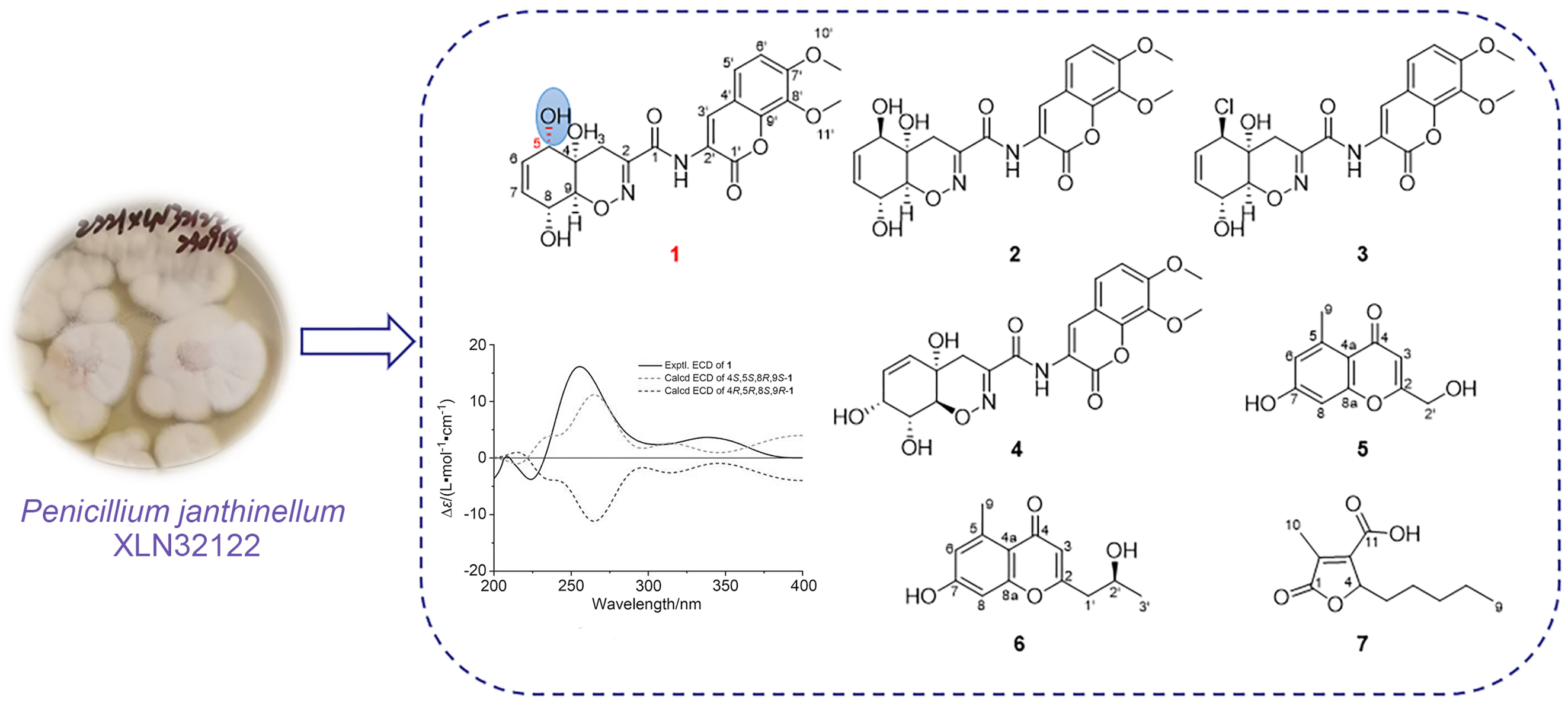
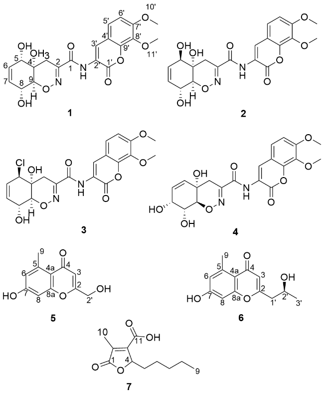
Mangroves grow in tropical and subtropical intertidal areas that feature high salinity, low oxygen content, strong UV radiation, and frequent tidal changes. These unique ecological conditions allow the microorganisms in mangroves to produce metabolites possessing distinctive chemical structures and bioactivities. Among them, the fungus genus Penicillium sp. is a significant source of pharmaceutical lead compounds. During our ongoing investigations of the fungi inhabiting mangrove environments,[22-27] the fermentation of the fungus P. janthinellum XLN32122, isolated from the mangrove Acanthus ebracteatus Vahl, displayed significant anti-inflammatory activity, with an IC50 value of (3.67±0.154) μg/mL. The subsequent purification has led to the discovery of seven compounds, including a new heterocyclic dipeptide tricho- dermamide H (1), and six known compounds trichodermamides A (2), B (3),[28] trichodermamide D (4),[29] 7-hydroxy-2-hydroxymethyl-5-methyl-4H-chromen-4-one (5),[30] 7-hydroxy-2-(2-hydroxypropyl)-5-methylchromone (6),[31] and striatisporolide A (7)[32] (Figure 1). Herein, we report the isolation, structure elucidation, and the bioactivity of these compounds.
2 Results and discussion
2.1 Structure elucidation
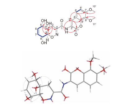
Trichodermamide H (1) was isolated as a yellow amorphous powder. The molecular formula of compound 1 was established as C20H20N2O9 based on HR-ESI-MS [M+ Na]+ ion peak (calcd for C20H20N2O9Na 455.1066, found 455.1056), indicating 12 degrees of unsaturation. The 1H NMR data (Table 1), combined with the heteronuclear multiple quantum coherence (HMQC) spectrum of 1 in DMSO-d6, showed an exchangeable hydrogen signal at δH 9.32 (s, 1H, NH), three hydroxyl groups at δH 5.46 (d, J=6.4 Hz,1H, 8-OH), 5.31 (d, J=4.8 Hz, 1H, 5-OH), and 5.09 (s, 1H, 4-OH), two aromatic protons at δH 7.49 (d, J=8.8 Hz, 1H, H-5') and 7.13 (d, J=8.8 Hz, 1H, H-6'), three olefinic protons at 5.69 (ddd, J=10.4, 4.8, 2.4 Hz, 1H, H-6) and 5.48 (dd, J=10.4, 2.4 Hz, 1H, H-7), three methine groups at δH 3.82 (br d, J=4.8 Hz, 1H, H-5), 3.97 (t, J=7.6 Hz, 1H, H-8) and 4.11 (dd, J=7.6, 2.0 Hz, 1H, H-9), two methoxy groups at δH 3.89 (1H, s, H-10') and 3.83 (1H, s, H-11'), and one methylene group at δH 2.07 (d, J=18.8 Hz, 1H, H-3α) and 2.28 (dd, J=18.8, 2.4 Hz, 1H, H-3β). The 13C NMR data (Table 1), combined with distortionless enhancement by polarization transfer (DEPT)-135 spectrum of 1, showed the presence of two carbonyl carbons at δC 160.9 (C-1) and 157.9 (C-1'), 11 aromatic and olefinic sp2-carbons at δC 149.9 (C-2), 127.4 (C-6), 130.5 (C-7), 121.0 (C-2'), 123.9 (C-3'), 1113.7 (C-4'), 123.1 (C-5'), 110.2 (C-6'), 53.9 (C-7'), 135.3 (C-8') and 143.6 (C-9'), three oxygenated methine groups at δC 69.4 (C-5), 66.4 (C-8) and 63.9 (C-4), two methoxy carbons at δC 56.5 (C-10') and 60.9 (C-11'), one oxygenated quaternary sp3-carbon at δC 81.7 (C-9), and one methylene carbon at δC 26.3 (C-3). The 1H NMR and 13C NMR data of compound 1 were similar to those of trichodermamide A (2),[28] and had the same molecular formula as 2 on the basis of the HR-ESI-MS data. The major differences were the chemical shifts of C-5 at δH 3.82 (br d, J=4.8 Hz, 1H) and δC 69.4 (CH) in 1, instead of at δH 4.24 (br d, J=2.4 Hz, 1H) and δC 73.0 (CH) in 2, and the chemical shifts of C-9 at δH 4.11 (dd, J=7.6, 2.0 Hz, 1H) and δC 81.7 (CH) in 1, in contract to the chemical shifts at δH 3.99 (br s, 1H) and δC 83.8 (CH) in 2. The above results indicated that compounds 1 and 2 had the same plane structure, but differ in their configurations at the cyclohexane ring. The relative configuration of 1 was determined on the basis of nuclear overhauser effect spectroscopy (NOESY) and coupling constants. The NOESY correlations of H-5 to H-8 indicated that H-5 and H-8 were on the same side of the cyclohexane ring, and the correlations of 4-OH to 5-OH/8-OH, and 5-OH to 8-OH suggested that 4-OH, 5-OH and 8-OH oriented to the other side of the cyclohexane ring (Figure 2). In addition, the large coupling constants between H-8 (7.6 Hz) and H-9 (7.6, 2.0 Hz), consistent with literature data,[14,28] further confirmed their trans-diaxial orientation. The absolute configuration of 1 was determined on quantum chemical electronic circular dichroism (ECD) calculations. The theoretical ECD spectra of two possible stereoisomers of 4S,5S,8R,9S and 4R,5R,8S,9R were created by the time-dependent density functional theory (TDDFT) calculations, and the calculated ECD spectrum of the isomer 4S,5S,8R,9S matched well with the experimental ECD curve of 1 (Figure 3). Thus the structure of 1 was determined, and named as trichodermamide H (1).
Table 1 1H (400 MHz) and 13C (100 MHz) NMR data of compound 1 in DMSO-d6 |
| Position | δC | δH (J in Hz) |
|---|---|---|
| 1 | 160.9 (C) | — |
| 2 | 149.9 (C) | — |
| 3 | 26.3 (CH2) | 2.07 (d, 18.8), 2.28 (dd, 18.8, 2.4) |
| 4 | 63.9 (C) | — |
| 4-OH | — | 5.09 (s) |
| 5 | 69.4 (CH) | 3.82 (br d, 4.8) |
| 5-OH | — | 5.31 (d, 4.8) |
| 6 | 127.4 (CH) | 5.69 (ddd, 10.4, 4.8, 2.4) |
| 7 | 130.5 (CH) | 5.48 (dd, 10.4, 2.4) |
| 8 | 66.4 (CH) | 3.97 (t, 7.6) |
| 8-OH | — | 5.46 (d, 6.4) |
| 9 | 81.7 (CH) | 4.11 (dd, 7.6, 2.0) |
| 1' | 157.9 (C) | — |
| 2' | 121.0 (C) | — |
| 3' | 123.9 (CH) | 8.53 (s) |
| 4' | 113.7 (C) | — |
| 5' | 123.1 (CH) | 7.49 (d, 8.8) |
| 6' | 110.2 (CH) | 7.13 (d, 8.8) |
| 7' | 153.9 (C) | — |
| 8' | 135.3 (C) | — |
| 9' | 143.6 (C) | — |
| 10' | 56.5 (OMe) | 3.89 (s) |
| 11' | 60.9 (OMe) | 3.83 (s) |
| NH | — | 9.32 (s) |
 ), HMBC (
 ) and NOESY (
 ) correlations of compound 1
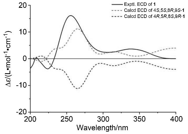
2.2 Biological activity
Compounds 1~7 were evaluated for their inhibitory activities against LPS-induced nitric oxide (NO) production in RAW 264.7 mouse macrophages, and dexamethasone was used as a positive control. Compounds 1~2 and 4~7 exhibited less than 50% inhibition at a concentration of 100 μmol/L. Only compound 3 demonstrated potent inhibition of NO accumulation induced by LPS on RAW 264.7 with the IC50 value of (13.13±0.005) μmol/L, which was stron- ger than the positive control dexamethasone [IC50=(136.84±1.33) μmol/L]. Furthermore, published studies have demonstrated that compound 3 exhibited potent antiproliferative activity against HCT-116 human colorectal carcinoma cell lines (IC50=0.32 μg/mL), HeLa cell lines [IC50=(3.1±0.5) μmol/L], and multiple human cancer cell lines.[28,33-34]
Compounds 1~7 were evaluated for their antibacterial activity against MRSA, while vancomycin was used as the positive control. Among them, only compound 3 displayed inhibitory activity against MRSA with an IC50 value of 12.5 μg/mL, while the positive control vancomycin showed an IC50 value of 1.563 μg/mL. These results indicate that chlo- rine atoms may play a crucial role in the bioactivity of these compounds.
3 Conclusions
In summary, a new heterocyclic alkaloid, and six known compounds were obtained from cultures of P. janthinellum XLN32122. Their structures were determined by extensive spectroscopic analyses and ECD calculations. Compound 3 demonstrated potent anti-inflammatory effects with an IC50 value of (13.13±0.005) μmol/L, and showed antibacterial activity against MRSA with the IC50 value of 12.5 μg/mL. This study provides a theoretical basis for the medicinal research on mangrove-derived Penicillium fungi and holds significant reference significance and value.
4 Experimental section
4.1 General experimental procedures
Modular Circular Polarimeter 500 (Anton Paar, Austria) polarimeter was used to detect optical rotation. JASCO J-715 spectrophotometer (JASCO, Japan) was used to measure ECD spectra. The 1D (1H and 13C) and 2D (HSQC, HMBC, COSY and NOESY) NMR spectra were measured on an NMR spectrometer (JEOL, 600 MHz, Japan) and a Bruker AV-400 (Bruker Corporation, Switzerland) instrument with TMS as the internal standard. Chemical shifts (δ) were referenced using the residual solvent signals of DMSO-d6 (δH 2.50, δC 39.5), CD3OD-d4 (δH 3.31, δC 49.0), and CDCl3 (δH 7.26, δC 77.2). ESI-MS and HR-ESI-MS spectra were obtained on a Bruker Daltonics Apex-Ultra 7.0 T (Bruker Corporation, Billerica, MA, USA) and a Q-TOF Ultima Global GAA076 LC mass spectrometer. For semipreparative HPLC, an Agilent 1100 prep-HPLC system with a Waters C18 semipreparative column (9.4 mm×250 mm, 7 μm) was used. Sephadex LH-20 (Pharmacia Co. Ltd., Sandwich, UK) and silica gel (200~300 and 300~400 mesh, Qingdao Marine Chemical Factory, Qingdao, China) were used for column chromatography (CC). Silica gel (GF254) for TLC was supplied by the Qingdao Marine Chemical Factory in China. All solvents used were of analytical grade (Guangzhou, China).
4.2 Fungal material
The fungal strain XLN32122 was isolated from the mangrove A. ebracteatus Vahl, collected in the Bamen Bay Mangrove Nature Reserve, Wenchang, Hainan Province, China in December 2022. It was stored in the Key Laboratory of Tropical Medicinal Resource Chemistry of Ministry of Education, College of Chemistry and Chemical Engineering, Hainan Normal University, Haikou, China. The strain was designated Penicillium janthinellum XLN32122 was based on BLAST analysis of the ITS sequence. Finally, the sequence was deposited in GenBank with the accession number PV688663.
4.3 Extraction and isolation
The fungal strain XLN32122 was grown on solid rice cultures in 1 L Erlenmeyer flasks (50 flasks; 50 mL of rice and 50 mL of water, and 0.5 g of sea salt per Erlenmeyer flask, autoclave sterilization) at 28 ℃ for 30 d. The fermentation was extracted three times with ethyl acetate (EtOAc, 25 L×3), and the combined extracts were concentrated under reduced pressure to afford a 30.0 g EtOAc extract.
The crude extract (30.0 g) was separated in ten fractions (Fr.1~Fr.10) by silica gel column chromatography (CC) eluting with a petroleum ether (PE)/EtOAc gradient system from 0∶1 to 1∶0. Fr.6 (2.5 g) was subjected to CC over octadecylsilyl (ODS) eluting with a MeOH/H2O gradient (10%~100%) to yield eight subfractions (Fr.6-1~ Fr.6-8). Fr.6-6 was purified by semipreparative HPLC (MeOH/H2O, V∶V=55∶45, 2 mL/min) to give compounds 1 (3.0 mg), 2 (2.5 mg), and 4 (3.6 mg). Fr.6-2 was purified by semipreparative HPLC (MeOH/H2O, V∶V=20∶80, 2 mL/ min) to give 3 (2.0 mg). Fr.4 (2.0 g) was subjected to CC over ODS eluting with a MeOH/H2O gradient (10%~100%) to yield eight subfractions (Fr.4-1~Fr.4-8). Fr.4-1 was purified by semipreparative HPLC (MeOH/H2O, V∶ V=20∶80, 2 mL/min) to give compound 5 (3.5 mg). Fr.4-3 was purified by semipreparative HPLC (MeOH/H2O, V∶V=45∶55, 2 mL/min) to give compounds 6 (3.5 mg) and 7 (4.2 mg).
Trichodermamide H (1): Yellow amorphous powder. m.p. 158.3~159.6 ℃; $[a]_{\mathrm{D}}^{25}$+23.3 (c 0.5, CH3OH); UV (MeOH) λmax [log ε/(L•mol-1•cm-1)]: 251, 342 nm; IR (KBr) νmax: 3430, 1628, 1386, 1086, 475 cm-1; ECD (2.98 mmol/L, MeOH) λmax [∆ε/(L•mol-1•cm-1)]: 212 (-8.45), 250 (+12.95), 305 (+1.63), 354 (+2.73) nm; 1H and 13C NMR data see Table 1; HR-ESI-MS calcd for C20H20N2- O9Na [M+Na]+ 455.1066, found 455.1056.
Trichodermamide A (2): White needles. m.p. 224.0~226.0 ℃ (lit.[28] 224.0~226.0 ℃); 1H NMR (600 MHz, DMSO-d6) δ: 9.33 (s, 1H, NH), 8.55 (s, 1H, H-3'), 7.52 (d, J = 9.0 Hz, 1H, H-5'), 7.14 (d, J = 9.0 Hz, 1H, H-6'), 5.44~5.46 (m, 1H, H-6), 5.38~5.41 (m, 1H, H-7), 5.39~5.44 (overlapped, 1H, 8-OH), 5.46~5.55 (overlapped, 2H, 4-OH, 5-OH), 4.24 (br d, J = 2.4, 1H, H-5), 3.99 (br s, 2H, H-8, H-9), 3.90 (s, 3H, 10'-OMe), 3.84 (s, 3H, 11'-OMe), 2.47 (br s, 1H, H-3a), 2.03 (d, J = 19.2 Hz, 1H, H-3b); 13C NMR (150 MHz, DMSO-d6) δ: 161.1 (C-1), 157.9 (C-1'), 153.8 (C-7'),150.2 (C-2), 143.6 (C-9'), 135.3 (C-8′), 130.0 (C-6), 128.0 (C-7), 123.7 (C-3'), 123.1 (C-5'), 121.0 (C-2'), 113.7 (C-4'), 110.1 (C-6'), 84.0 (C-9), 73.2 (C-5), 67.4 (C-4), 66.2 (C-8), 60.9 (C-11'), 56.4 (C-10'), 23.2 (C-3); HR-ESI-MS calcd for C20H20N2O9Na [M+Na]+ 455.1061, found 455.1052.
Trichodermamide B (3):[28] Colorless oil. 1H NMR (400 MHz, MeOD) δ: 8.62 (s, 1H, H-3'), 7.33 (d, J = 8.8 Hz, 1H, H-5'), 7.10 (d, J = 8.8 Hz, 1H, H-6'), 5.60~5.73 (m, 2H, H-6, H-7), 4.90 (overlapped, 1H, H-5), 4.17~4.24 (overlapped, 2H, H-8, H-9), 3.95 (s, 3H, 10'-OMe), 3.93 (s, 3H, 11'-OMe), 2.83 (dd, J = 2.2, 19.4 Hz, 1H, H-3a), 2.32 (d, J = 19.4 Hz, 1H, H-3b); 13C NMR (100 MHz, MeOD) δ: 162.5 (C-1), 159.8 (C-1'),155.8 (C-7'), 151.1 (C-2), 145.4 (C-9'), 137.3 (C-8'), 130.4 (C-6), 128.9 (C-7),125.6 (C-3'), 124.0 (C-5'), 122.4 (C-2'), 115.4 (C-4'), 111.1 (C-6'), 85.5 (C-9), 68.9 (C-4), 67.6 (C-5), 66.4 (C-8), 61.8 (C-11'), 57.0 (C-10'), 25.8 (C-3); HR-ESI-MS calcd for C20H19ClN2O8Na [M+Na]+ 473.0723, found 473.0720.
Trichodermamide D (4):[29] Pale yellow powder. m.p. 209.3~210.7 ℃; 1H NMR (600 MHz, DMSO-d6) δ: 9.33 (s, 1H, NH), 8.53 (s, 1H, H-3'), 7.50 (d, J = 9.0 Hz, 1H, H-5'), 7.14 (d, J = 9.0 Hz, 1H, H-6'), 5.81 (d, J = 10.2 Hz, 1H, H-5), 5.69 (dd, J = 10.2, 4.8 Hz, 1H, H-6), 5.17~5.48 (overlapped, 1H, 4-OH), 4.24 (dd, J = 10.8, 1.8 Hz, 1H, H-9), 4.02 (t, J = 4.2 Hz, 1H, H-7), 3.90 (s, 3H, 10'-OMe), 3.84 (s, 3H, 11'-OMe), 2.61 (dd, J = 19.2, 2.4 Hz, 1H, H-3a), 2.17 (d, J = 19.2 Hz, 1H, H-3b); 13C NMR (150 MHz, DMSO-d6) δ: 160.8 (C-1), 157.9 (C-1'), 153.9 (C-7'), 147.7 (C-2), 143.6 (C-9'), 135.5 (C-5), 135.3 (C-8'), 126.8 (C-6), 124.0 (C-3'), 123.0 (C-5'), 120.9 (C-2'), 113.7 (C-4'), 110.1 (C-6'), 79.4 (C-9), 67.8 (C-8), 66.6 (C-7), 64.1 (C-4), 60.9 (C-11'), 56.4 (C-10'), 28.9 (C-3); HR-ESI-MS calcd for C20H20N2O9Na [M+Na]+ 455.1061, found 455.1057.
7-Hydroxy-2-hydroxymethyl-5-methyl-4H-chromen-4-one (5): White amorphous powder. m.p. 231.0~234.0 ℃ (lit.[30] 231.0~234.0 ℃); 1H NMR (400 MHz, DMSO-d6) δ: 6.63 (d, J = 2.4 Hz, 1H, H-8), 6.60~6.62 (m, 1H, H-6), 6.08 (t, J = 1.2 Hz, 1H, H-3), 5.69 (t, J = 6.4 Hz, 1H, 2'-OH), 4.33 (d, J = 5.2 Hz, 2H, H-2'), 2.65 (s, 3H, H-9); 13C NMR (100 MHz, DMSO-d6) δ: 178.3 (C-4), 166.8 (C-2), 161.1 (C-7), 158.9 (C-8a), 141.6 (C-5), 116.6 (C-6), 114.7 (C-4a), 108.3 (C-3), 100.6 (C-8), 59.5 (C-2'), 22.5 (C-9); HR-ESI-MS calcd for C11H9O4 [M-H]- 205.0506, found 205.0500.
7-Hydroxy-2-(2-hydroxypropyl)-5-methylchromone (6):[31] White amorphous powder. m.p. 205.4~207.9 ℃; 1H NMR (400 MHz, DMSO-d6) δ: 6.62 (d, J = 2.4 Hz, 1H, H-8), 6.58~6.60 (m, 1H, H-6), 5.96 (s, 1H, H-3), 4.84 (br s, 1H, 2'-OH), 3.98~4.07 (m, 1H, H-2'), 2.65 (s, 3H, H-3'), 2.52~2.62 (m, 2H, H-1'), 1.14 (d, J = 6.4 Hz, 3H, H-9); 13C NMR (100 MHz, DMSO-d6) δ: 178.3 (C-4), 164.8 (C-2), 160.9 (C-7), 159.2 (C-8a), 141.4 (C-5), 116.5 (C-6), 114.5 (C-4a), 111.6 (C-3), 100.6 (C-8), 61.4 (C-2'), 42.9 (C-1'), 23.5 (C-3'), 22.4 (C-9). HR-ESI-MS calcd for C13H13O4 [M-H]- 211.0975, found 211.0971.
Striatisporolide A (7):[32] Pale yellow amorphous solid. m.p. 116.2~117.8 ℃; 1H NMR (400 MHz, DMSO-d6) δ: 5.11~5.17 (m, 1H, H-4), 2.03 (d, J = 2.0 Hz, 3H, H-10), 1.94~2.02 (m, 1H, H-5a), 1.49~1.59 (m, 1H, H-5b), 1.20~1.31 (m, 6H, H-6, H-7, H-8), 0.85 (t, J = 6.4 Hz, 3H, H-9); 13C NMR (100 MHz, DMSO-d6) δ: 172.9 (C-1), 163.6 (C-11), 150.1 (C-3), 134.3 (C-2), 81.0 (C-4), 31.8(C-5), 30.9 (C-7), 23.8 (C-6), 21.9 (C-8), 13.8 (C-9), 10.3 (C-10); HR-ESI-MS calcd for C11H15O4 [M-H]- 233.0819, found 233.0817.
4.4 ECD computation section
The TDDFT ECD calculations for compound 1 was performed with the Gaussian 09 program.[35] The conformational searching was conducted by the Spartan 14 software using the MMFF force field within an energy window of 20.9 kJ/mol. The conformers with the Boltzmann population above 5% were re-optimized at the B3LYP/631G(d) level in vacuo or M06-2X/6-311G(d,p). TDDFT ECD calculations were run at the CAM-B3LYP/TZVP or M06-2X/ TZVP level with the SMD solvent model for methanol.[36] ECD spectra were generated using the SpecDis Version 1.71 with 0.3 sigma/gamma (eV) after UV correction.[37]
4.5 Assay of anti-inflammatory activity
The anti-inflammatory activity was evaluated by determination of NO production in RAW264.7 cell culture supernatants induced by LPS with Griess reagent.[38] The RAW264.7 cells were inoculated in 96-well plates with a density of 1×10⁵ cells per well, and treated with samples and positive control (dexamethasone) after 12 h. One hour after this treatment, the cells were exposed to 1 μg/mL LPS and cultured at 37 ℃ and 5% (ϕ) CO2 for 24 h. Next, 50 μL of culture supernatant and 100 μL of Griess reagent were mixed. The absorbance values at a wavelength of 540 nm were recorded by a microplate reader.
4.7 Assay of anti-MRSA activity
Compounds 1~7 were tested for antibacterial activities against methicillin-resistant Staphylococcus aureus ATCC- 43300, and were evaluated in 96-well plates using a modified broth microdilution method.[39]
Supporting Information HR-ESI-MS, 1D NMR, 2D NMR, IR and UV spectra of compound 1. The Supporting Information is available free of charge via the Internet at http://sioc-journal.cn.
(Zhao, C.)