


1 Introduction
Microbial secondary metabolites (MSMs), characterized by their diverse chemical structures and potent bioactivities, have become a significant source for the discovery of bioactive natural products and drug-leads.[1] Marine-derived microorganisms, shaped by the unique conditions of the marine environment, hold great potential for the discovery of novel secondary metabolites. This has drawn sustained interest from chemists and biologists in recent years, particularly in the field of actinobacteria research.[2-7] For over two decades, our group has been dedicated to studying bioactive MSMs from marine-derived microorganisms. During this time, we have accumulated an extensive collection of microorganisms and metabolite resources, leading to notable achievements that provide valuable support for drug discovery research.[8-9]
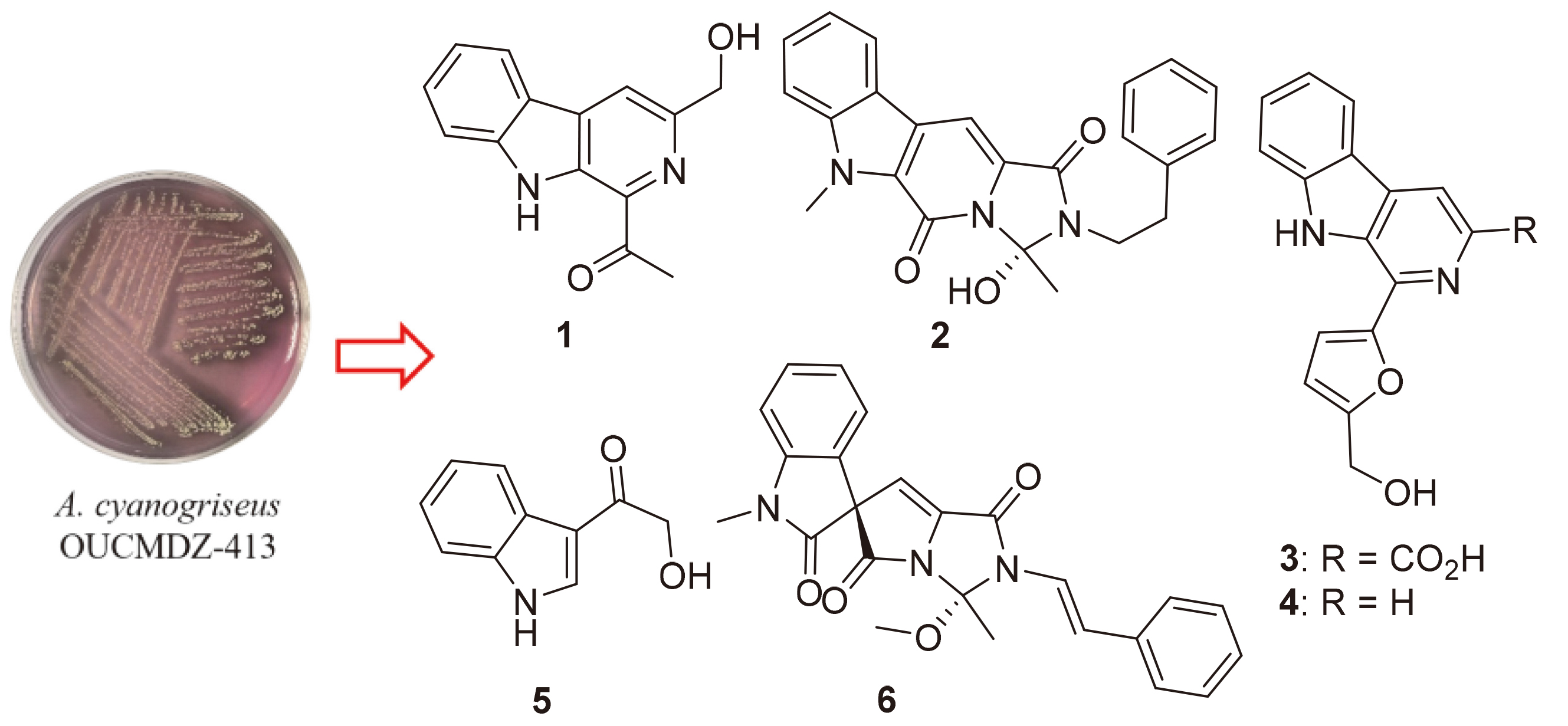
Actinoalloteichus cyanogriseus WH1-2216-6 (now designated as OUCMDZ-413) was a high-yielding strain isolated from marine sediments.[10] This strain is known to produce a variety of secondary metabolites, including cae- rulomycins,[10-14] 5,5,6-polycyclic tetramate macrolactams[15] and cyanogramide,[16-17] all of which feature novel structures and bioactivities. Notably, cyanogramide (6) is distinguished by its unique spirocyclic pyrrolo[1,2-c]imi- dazole skeleton. To obtain analogues of cyanogramide for further study, various methods have been employed, including the isolation of trace components through large- scale fermentation, solid and liquid fermentation techniques, as well as putative precursor-feeding experiments.
Fortunately, during our ongoing exploration of trace components from the large-scale fermentation of A. cyanogriseus OUCMDZ-413, cyanogramide (6) was successfully re-isolated alongside the key intermediate (S)- cyanogramide B (2), which was first identified in the fermentation broth of the wild A. cyanogriseus OUCMDZ- 413.[16-17] In addition, a new β-carboline analogue (1) was discovered, along with three know analogues (3~5) were also identified (Figure 1). The structures of these compounds were fully elucidated through comprehensive spectroscopic analysis.
2 Results and discussion
2.1 Structure identification
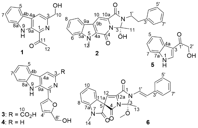
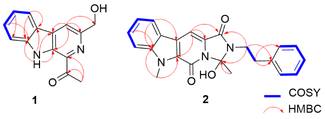
Compound 1 was obtained as a yellow powder with its molecular formula determined to be C14H12N2O2, based on its HRESIMS peaks at 241.0978 [M+H]+ and 263.0786 [M+Na]+. The 13C NMR resolved 14 carbon signals, which were classified using distortionless enhancement by polarization transfer (DEPT) and heteronuclear single quantum coherence (HSQC) spectra as seven quaternary carbons, five olefinic methines, one oxidized methylene (hydroxy- methyl) and one methyl carbon (Table 1). The coupling aromatic protons at H-5 (δH 8.29, d, J=7.8 Hz), H-6 (δH 7.28, t, J=7.8 Hz), H-7 (δH 7.57, dt, J=7.8, 0.8 Hz) and H-8 (δH 7.77, dd, J=7.8, 0.8 Hz) showed continuous 1H-1H correlation spectroscopy (COSY) and key heteronuclear multiple bond correlation spectroscopy (HMBC) from H-5 to C-8a (δC 142.3) and C-4b (δC 132.0), confirming the presence of a 1,2-disubstituted phenyl fragment (Figure 1). An exchangeable proton at H-9 (δH 11.78, s) exhibited HMBC correlations with four quaternary carbons: C-4a (δC 120.0), C-4b, C-8a and C-9a (δC 133.3), indicating the presence of an NH group. This finding further linked the 1,2-disubstituted phenyl fragment to form a 2,3-disub- stituted indole nucleus. Additionally, the aromatic proton at H-4 (δH 8.48, s) showed HMBC correlations with C-3 (δC 149.9), C-4a and C-9a, revealing the presence of β-car- boline nucleus. The methyl signals at δH/C 2.77/25.8 and the carbonyl carbon at δC 201.3 were assigned to an acetyl group from the HMBC correlation of H-12 (δH 2.77, s) to C-11 (δC 149.9). The 1H NMR closely resemble those of 1-formyl-3-hydroxymethyl-β-carboline,[18] with the primary difference being the replacement of the formyl group by an acetyl group. Key HMBC correlations from H-12 to C-1 (δC 134.5) and from H-4 to C-10 (δC 64.4) (Figure 2) located the positions of acetyl and hydroxymethyl groups at C-1 and C-3, respectively. Compound 1 was thus identified as 1-acetyl-3-hydroxymethyl-β-carboline.
Table 1 1H NMR and 13C NMR data for compounds 1~3 (500/125 MHz) and 4 (400/125 MHz) in DMSO-d6 |
| No. | 1 | 2 | 3 | 4 | ||||||||
|---|---|---|---|---|---|---|---|---|---|---|---|---|
| δC | δH | δC | δH | δC | δH | δC | δH | |||||
| 1 | 134.5 (C) | 159.2 (C) | 132.5 (C) | 133.2 (C) | ||||||||
| 3 | 149.9 (C) | 99.2 (C) | 137.1 (C) | 8.36 (d, J=5.3 Hz) | 138.2 (CH) | 8.36 (d, J=5.3 Hz) | ||||||
| 4 | 116.4 (CH) | 8.48 (s) | 152.8 (C) | 115.8 (CH) | 8.06 (d, J=5.3 Hz) | 113.7 (CH) | 8.06 (d, J=5.3 Hz) | |||||
| 5 | 121.8 (CH) | 8.29 (t, J=7.8 Hz) | 122.1 (CH) | 8.26 (d, J=8.0 Hz) | 121.7 (CH) | 8.26 (d, J=8.0 Hz) | ||||||
| 6 | 120.2 (CH) | 7.28 (t, J=7.8 Hz) | 111.0 (CH) | 7.70 (dt, J=8.4, 1.0 Hz) | 120.6 (CH) | 7.28 (t, J=7.5 Hz) | 119.8 (CH) | 7.28 (t, J=7.5 Hz) | ||||
| 7 | 128.9 (CH) | 7.57 (dt, J=7.8, 0.8 Hz) | 127.1 (CH) | 7.56 (ddd, J=8.4, 7.0, 1.0 Hz) | 129.0 (CH) | 7.59 (t, J=7.5 Hz) | 128.5 (CH) | 7.59 (t, J=7.5 Hz) | ||||
| 8 | 113.1 (CH) | 7.77 (dd, J=7.8, 0.8 Hz) | 120.9 (CH) | 7.30 (ddd, J=7.9, 7.0, 1.0 Hz) | 112.9 (CH) | 7.78 (d, J=8.2 Hz) | 112.5 (CH) | 7.78 (d, J=8.2 Hz) | ||||
| 9 | 11.78 (s) | 121.8 (CH) | 8.25 (dt, J=7.9, 1.0 Hz) | 11.29 (s) | 11.29 (s) | |||||||
| 10 | 64.6 (CH2) | 4.80 (s) | 97.6 (CH) | 7.72 (s) | 166.5 (C) | |||||||
| 11 | 201.3 (C) | 22.5 (CH3) | 2.03 (s) | |||||||||
| 12 | 25.8 (CH3) | 2.77 (s) | 31.4 (CH3) | 4.30 (s) | ||||||||
| 4a | 120.0 (C) | 128.9 (C) | 129.9 (C) | 129.5 (C) | ||||||||
| 4b(9b) | 132.0 (C) | 123.2 (C) | 121.0 (C) | 120.7 (C) | ||||||||
| 5a | 140.8 (C) | |||||||||||
| 8a(10a) | 142.3 (C) | 127.5 (C) | 141.4 (C) | 141.0 (C) | ||||||||
| 9a | 133.3 (C) | 121.5 (C) | 131.9 (C) | 130.5 (C) | ||||||||
| 1' | 40.5 (CH2) | 3.59 (t, J=8.1 Hz) | ||||||||||
| 2' | 34.4 (CH2) | 3.01 (t, J=8.1 Hz) | 151.3 (C) | 152.2 (C) | ||||||||
| 3' | 138.9 (C) | 111.1 (CH) | 7.20 (d, J=3.2 Hz) | 109.7 (CH) | 7.20 (d, J=3.2 Hz) | |||||||
| 4'(8') | 128.5 (CH) | 7.30 (m) | 109.3 (CH) | 6.58 (d, J=3.2 Hz) | 109.2 (CH) | 6.58 (d, J=3.2 Hz) | ||||||
| 5'(7') | 128.8 (CH) | 7.31 (m) | 157.3 (C) | 156.8 (C) | ||||||||
| 6' | 126.4 (CH) | 7.24 (tt, J=6.8, 1.9 Hz) | 56.0 (CH2) | 4.66 (s) | 56.0 (CH2) | 4.66 (s) | ||||||
| 6'-OH | 5.57 (brs) | 5.57 (brs) | ||||||||||
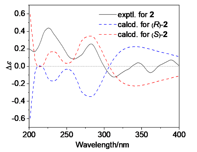
Cyanogramide B (2) was originally isolated as a racemic mixture from a Streptomyces coelicolor YF11 strain heterologously expressing the biosynthetic gene cluster of cyanogramide (6) derived from the wild A. cyanogriseus WH1-2216-6.[17] Subsequently, enantiomerically pure (S)- cyanogramide B was isolated and characterized from Actinoalloteichus sp. ZZ1866.[19] In this study, enantiomerically pure (S)-cyanogramide B (2) was isolated for the first time from the fermentation broth of the wild A. cyanogriseus OUCMDZ-413. This compound was identified by comparing its NMR data (Table 1) with previously reported values.[17,19] Its absolute configuration was determined by comparing the calculated electronic circular dichroism (ECD) spectrum with the experimentally measured one (Figure 3).


In addition, other four known compounds were identified as flazin (3),[20] perlolyrine (4),[20] 2-hydroxy-1-(1H-indol- 3-yl)ethan-1-one (5),[20-21] and cyanogramide (6),[16] respectively. Their identification was accomplished through MS analysis and by comparing their 1D NMR data (Table 1) and specific rotation measurements with previously reported literature. The re-isolation of cyanogramide (6) further confirmed that fermentation optimization is an efficient strategy to explore the metabolic potential of microbial strains and provides a valuable reference for further in- depth studies. Cyanogramide (6), which features a novel spirocyclic pyrrolo[1,2-c]imidazole lactone skeleton, has drawn significant attention due to its unique structure. Its biosynthetic pathway has been a subject of interest and has led to important progress.[17] Moreover, the isolation of (S)- cyanogramide B (2) in this study confirmed its role as a key intermediate in the biosynthesis of cyanogramide (6), which is derived from L-tryptophan and L-phenylalanine during the early stages of its biosynthetic pathway.[17]
2.2 Bioactivities of compounds
Compounds 1~6 were evaluated for their antimicrobial efficacy against a range of pathogenic bacteria, including Staphylococcus aureus ATCC 6538, methicillin-resistant S. aureus ATCC 43300, Pseudomonas aeruginosa ATCC- 10145 and Bacillus subtilis CGMCC1.3376, as well as fungi such as Candida albicans ATCC10231 and Candida glabrata ATCC2001.[22] Unfortunately, none of these compounds exhibited observable antimicrobial activity with minimum inhibitory concentration (MIC) values exceeding 64 μg/mL. Among the isolated compounds, cyanogramide B (2) also showed no notable activities against human glioma U87MG and U251 cells.[19] In contrast, cyanogramide (6) demonstrated significant activity in reversing multidrug resistance in K562/A02, MCF-7/Adr, and the vincristine-induced resistant KB/VCR cells at a concentration of 5 μmol/L with reversal fold values of 15.5, 41.5, and 9.7, respectively.[16] Additionally, perlolyrine or 1-(5-hydr- oxymethylfuran-2-yl)-β-carboline (4) was found to be active against the influenza A virus subtype H1N1 with an IC50 value of 38.3 μg/mL and a selectivity index (SI) of 3.0. For comparison, the positive control, ribavirin, exhibited IC50 and SI values of 23.1 μg/mL and 32.2, respectively.[20] However, flazin or 1-(5-hydroxymethylfuran-2-yl)-β-car- boline-3-carboxylic acid (3) and 2-hydroxy-1-(1H-indol-3- yl)ethan-1-one (5) were inactive against H1N1 virus.
3 Conclusion
In conclusion, microbial secondary metabolites play a crucial role in various fields, including medicine, industrial applications, sustainable agriculture, chemical biology, and biochemistry research.[1,23-26] In this study, a new β-carbo- line derivative (1) was identified, along with two known analogues (3 and 4), as well as the cyanogramide (6) and its key intermediate cyanogramide B (2), from the marine- derived A. cyanogriseus OUCMDZ-413. The isolation of (S)-cyanogramide B (2) and cyanogramide (6) further confirmed the biosynthetic pathway of cyanogramides in the A. cyanogriseus OUCMDZ-413.[17]
4 Experimental section
4.1 General experimental procedures
UV spectra were recorded on a HITACHI UH5300 spec- trophotometer using continuous wavelength scanning from 200 nm to 700 nm. IR spectra were measured on a Nicolet Nexus 470 spectrophotometer using KBr discs. Optical rotations and electronic circular dichroism (ECD) data were obtained with a JASCO P-1020 digital polarimeter and a JASCO J-815 spectropolarimeter, respectively. High- resolution electrospray ionization mass spectrometry (HRESIMS) data were acquired using a Q-TOF ULTIMA GLOBAL GAA076 LC mass spectrometer. Nuclear magnetic resonance (NMR) spectra for compounds 1~6 were collected on a Bruker Avance NEO 400, an Agilent DD2 500, or a JEOL JNM-ECZ600R/S1 600 MHz spectrometers in DMSO-d6 solution. Chemical shifts (δ) were referenced to the residual solvent signals (δH/C 2.50/39.5 for DMSO-d6) using tetramethylsilane (TMS) as an internal standard. Vacuum-liquid chromatography (VLC) and flash column chromatography (FCC) were performed using silica gel H (200~300 mesh, Qingdao Marine Chemical Factory). Gel column chromatography was conducted using Sephadex LH-20 (Amersham Biosciences) instruments. Semi-pre- parative HPLC was carried out on an ODS column (YMC- pack ODS-A, 10 mm×250 mm, 5 μm) with a flow rate of 4.0 mL/min.
4.2 Actinobacterial strain
The strain OUCMDZ-413 was isolated from marine sediments collected at the Weihai seashore and identified as Actinoalloteichus cyanogriseus based on 16S rRNA gene sequences and morphological characteristics. The strain has been preserved in our laboratory (Zhu’s Lab) as well as in the China Center for Type Culture Collection (CCTCC) under the patent depository number CCTCCM 209277[10].
4.3 Fermentation and extraction of the strain
Colonies of the strain A. cyanogriseus OUCMDZ-413 growing on Gauze’s agar medium were transferred into 500 mL flasks, each containing 150 mL of liquid culture medium 1# (Table 2). The flasks were incubated at 28 ℃ for 5 d on a rotary shaker set at 180 r/min to produce seed cultures. The seed cultures were then inoculated into a 500 mL flask containing 150 mL of a different liquid culture medium 2# (Table 2). A total of 100 flasks were applied to culture the strain OUCMDZ-413. The cultures were incubated at 28 ℃ on a rotary shaker at 180 r/min for 25 d. Following fermentation, the culture broth in each flask was extracted three times with ethyl acetate (EtOAc, 150 mL). The combined EtOAc solution was concentrated under reduced pressure to yield a crude extract weighing 10.1 g.
Table 2 List of culture medium formulations |
| Medium name | Medium component |
|---|---|
| LB | Tryptone 10 g, NaCl 5 g, yeast extract 5 g, sea water 1 L |
| YPD | Yeast extract 10 g, peptone 20 g, glucose 20 g, sea water 1 L |
| Medium 1# | Peptone 20 g, soluble starch 20 g, glycerol 20 g, CaCO3 2 g, sea water 1 L |
| Medium 2# | Peptone 20 g, soluble starch 20 g, glycerol 20 g, CaCO3 2 g, 2-pyridinecarboxylic acid 0.4 g, XAD-16 macroporous adsorption resin 50 g, sea water 1 L |
4.4 Isolation and purification of compounds
The extract (10.1 g) was fractionated using VLC over a silica gel column (7.5 cm×25 cm) with stepwise gradient elution of CH2Cl2-MeOH (V∶V=1∶0 to 0∶1), yielding 18 fractions (Fr.n1~Fr.n18). Fr.n3 (1.5 g) and Fr.n4 (1.9 g) were combined and further subjected to VLC separation on a silica gel column (4.5 cm×18 cm) using a stepwise gradient of petroleum ether (PE)-EtOAc (V∶V=10∶1 to 3∶1), producing 18 subfractions (Fr.n3-1~Fr.n3-18). From these subfractions, Fr.n3-14 (40.0 mg) was separated using a Sephadex LH-20 column (2.5 cm×100 cm) eluted with MeOH, followed by purification through semipreparative HPLC (YMC-pack ODS-A, 10 mm×250 mm, 4.0 mL/min) with a MeCN-H2O (V∶V=1∶1) eluent, yielding compound 2 (2.1 mg, tR=16.2 min). Subfraction Fr.n3-15 was further separated into 25 subfractions (Fr.n3-15-1~Fr.n3- 15-25) using a Sephadex LH-20 column (2.5 cm×100 cm) eluted with a mixture of MeOH and CH2Cl2 (V∶V=1∶1). Compound 6 (0.8 mg, tR=11.8 min) was purified from Fr.n3-15-13 (23.5 mg) via semipreparative HPLC (YMC- pack ODS-A, 10 mm×250 mm, 4.0 mL/ min) using MeCN-H2O (V∶V=3∶2) as eluent, and compound 1 (2.5 mg, tR=9.3 min) was purified from Fr.n3-15-18 (13.5 mg) via semipreparative HPLC (YMC- pack ODS-A, 10 mm×250 mm, 4.0 mL/min) using MeCN- H2O (V∶V=9∶11) as eluent. Meanwhile, fraction Fr.n5 (1.3 g) was subjected to VLC over a silica gel column (2.5 cm×10 cm) with a stepwise gradient of PE-EtOAc (V∶ V=5∶1 to 2∶1), resulting in 13 subfractions (Fr.n5-1~Fr.n5-13). Among these, Fr.n5-5 was separated into 13 subfractions (Fr.n5-5-1~Fr.n5-5-13) employing a Sephadex LH-20 column (2.5 cm×100 cm) eluted with CH2Cl2-MeOH (V∶V=1∶1). Fr.n5-5-12 was further purified by semipreparative HPLC (YMC-pack ODS-A, 10 mm×250 mm, 4.0 mL/min) with MeCN-H2O (V∶V=3∶7) as eluent, yielding compounds 4 (1.5 mg, tR=7.5 min) and 3 (0.8 mg, tR=16.5 min). Additionally, compound 5 (2.0 mg, tR=8.7 min) was isolated by semipreparative HPLC (YMC-pack ODS-A, 10 mm×250 mm, 4.0 mL/min) using MeCN-H2O (V∶V=9∶11) as eluent.
1-Acetyl-3-hydroxymethyl-β-carboline (1): Yellow amorphous powder. UV (MeOH) λmax [log ε/(L•mol-1• cm-1)]: 216 (3.98), 285 (3.58), 384 (3.16) nm; 1H NMR (500 MHz, DMSO-d6) and 13C NMR (125 MHz, DMSO-d6) data see Table 1; IR (KBr disk) νmax: 3863, 3749, 3648, 3544, 3523, 2943, 1744, 1602, 1584, 1463, 1384, 1365, 1339, 1225, 1198, 1049, 796 cm-1; HRESIMS calcd for C14H13N2O2 (M+H+) 241.0972, found 241.0978.
(S)-Cyanogramide B (2): Yellow oil. $[\alpha]_{\mathrm{D}}^{20}$+36 (c 0.15, CHCl3); UV (MeOH) λmax [log ε/(L•mol-1•cm-1)]: 242 (3.75), 278 (3.67), 286 (3.69), 332 (3.41), 346 (3.45), 364 (3.46) nm; ECD (1.29 mmol/L, MeOH) λmax [∆ε/(L• mol-1•cm-1)]: 390 (+0.17), 362 (-0.17), 346 (+0.10), 314 (-0.29), 283 (+0.60), 260 (+0.20), 226 (+1.03), 207 (+0.39) nm; 1H (500 MHz, DMSO-d6) and 13C (125 MHz, DMSO-d6) NMR data see Table 1; HRESIMS calcd for C23H22N3O3 (M+H+) 388.1656, found 388.1662.
Flazin or 1-(5-hydroxymethylfuran-2-yl)-β-carboline-3- carboxylic acid (3): Yellow amorphous powder. 1H (500 MHz, DMSO-d6) and 13C (125 MHz, DMSO-d6) NMR data see Table 1; ESI-MS m/z 309.36 [M+H]+.
Perlolyrine or 1-(5-hydroxymethylfuran-2-yl)-β-carbo- line (4): Yellow amorphous powder. 1H NMR (400 MHz, DMSO-d6) and 13C NMR (125 MHz, DMSO-d6) data see Table 1; ESI-MS m/z: 265.40 [M+H]+.
2-Hydroxy-1-(1H-indol-3-yl)ethan-1-one (5): Yellow oil. 1H NMR (500 MHz, DMSO-d6) δ: 8.34 (s, 1H, H-2), 8.16 (d, J=7.2 Hz, 1H, H-4), 7.17 (td, J=7.2, 1.2 Hz, 1H, H-5), 7.23 (td, J=7.2, 1.2 Hz, 1H, H-6), 7.47 (d, J=7.2 Hz, 1H, H-7), 4.58 (s, 2H, H-9); 13C NMR (125 MHz, DMSO- d6) δ: 133.4 (CH, C-2), 113.2 (C, C-3), 125.5 (C, C-4a), 121.2 (CH, C-4), 121.8 (CH, C-5), 122.8 (CH, C-6), 122.3 (CH, C-7), 136.4 (C, C-7a), 194.4 (C, C-1'), 65.3 (CH2, C-2'); ESI-MS m/z: 176.16 [M+H]+.
Cyanogramide (6): Yellow oil. $[\alpha]_{\mathrm{D}}^{18}$-75 (c 0.1, MeOH); UV (MeOH) λmax [log ε/(L•mol-1•cm-1)]: 206 (3.92), 253 (3.55), 318 (3.60) nm; 1H NMR (600 MHz, DMSO-d6) δ: 7.16 (d, J=8.3 Hz, 1H, H-8), 7.44 (t, J=8.3 Hz, 1H, H-9), 7.12 (t, J=7.4 Hz, 1H, H-10), 7.35 (d, J=7.4 Hz, 1H, H-11), 6.28 (s, 1H, H-12), 2.08 (s, 3H, H-13), 3.23 (s, 3H, H-14), 7.18 (d, J=15.2 Hz, 1H, H-1'), 7.04 (d, J=15.2 Hz, 1H, H-2'), 7.54 (d, J=7.7 Hz, 2H, H-4'/8'), 7.36 (d, J=7.7 Hz, 2H, H-5'/7'), 7.27 (t, J=7.4 Hz, 1H, H-6'), 3.21 (s, 3H, 3-OCH3); 13C NMR (150 MHz, DMSO-d6) δ: 154.6 (C, C-1), 98.7 (C, C-3), 167.4 (C, C-4), 69.8 (C, C-5), 169.5 (C, C-6), 144.8 (C, C-7a), 109.6 (CH, C-8), 130.1 (CH, C-9), 123.1 (CH, C-10), 124.5 (CH, C-11), 124.6 (C-11a, C), 107.2 (CH, C-12), 138.6 (C, C-12a), 21.8 (CH3, C-13), 26.9 (CH3, C-14), 118.9 (CH, C-1'), 118.0 (CH, C-2'), 135.5 (C, C-3'), 126.1×2 (CH, C-4'/8'), 128.8×2 (CH, C-5'/7'), 127.6 (CH, C-6'), 49.4 (CH3, 3-OCH3); HRESIMS calcd for C24H21N3O4Na (M+Na+) 438.3348, found 438.3351.
4.5 Antibacterial and antifungal assays
4.5.1 Preparation of bacterial suspension
Four strains of human pathogenic bacteria, namely Staphylococcus aureus ATCC 6538, methicillin-resistant S. aureus ATCC 43300, Pseudomonas aeruginosa ATCC- 10145 and Bacillus subtilis CGMCC1.3376, as well as fungi such as Candida albicans ATCC10231 and Candida glabrata ATCC2001 were inoculated into LB and YPD medium, respectively. The volume of the medium for each strain was 20/50 mL (Table 2 for the medium component). The cultures were incubated on a shaker at 28 ℃ and 180 r/min for 12 h until the culture medium became visibly turbid. The medium was filtered through a 0.22 μm sterile microporous membrane. The cultured medium was diluted 1000 times with the corresponding medium filtered through a 0.22 μm sterile microporous membrane for later use.
4.5.2 Sample preparation
The test compounds and positive controls (ciprofloxacin for bacteria and ketoconazole for fungi) were dissolved in cell-grade dimethyl sulfoxide (DMSO) with concentration of 1.28 μg•mL-1.
4.5.3 Initial screening of antibacterial and antifungal activity
The experiment set up a blank control (200 μL of pure culture medium), a positive control (100 μL of diluted bacterial suspension, 90 μL of culture medium and 10 μL of positive drug), a negative control (100 μL of diluted bacterial suspension, 90 μL of culture medium and 10 μL of cell-grade DMSO), and a test group (100 μL of diluted bacterial suspension, 90 μL of culture medium and 10 μL of the test compound solution). At this time, the concentrations of the compounds and positive control were 64 μg•mL-1, and 3 parallel replicates were set for each group. The 96-well plate was placed in a 28 ℃ constant temperature incubator for 24 h of static cultivation. If the culture medium was clear and no bacterial colonies were formed, it indicated that the compound at this concentration had antibacterial or antifungal activity.
4.6 Theory and calculation details
Density functional theory (DFT) calculations were performed using the Gaussian 09 program. A preliminary conformational search was carried out using HyperChem Release 8.0 software. All ground-state geometries were optimized at the B3LYP/6-31G(d) level of theory. Solvent effects in methanol were evaluated using the same DFT level with the SCRF/PCM method. Time-dependent DFT (TDDFT) calculations at the B3LYP/6-31G(d) level were employed to determine the electronic excitation energies and rotational strengths in methanol. The overall calculated electronic circular dichroism (ECD) curves were weighted by weighting conformers according to their Boltzmann distribution, with a half-bandwidth of 0.40 eV and UV corrections of 10 nm. The calculated ECD spectra were produced using SpecDis 1.70.1 software.
Supporting Information Spectral data, mass spectrometry data and partial ECD data (as well as the ECD calculation data) of compounds 1~6. The Supporting Information is available free of charge via the Internet at http://sioc- journal.cn.
(Zhao, C.)