化学学报 ›› 2023, Vol. 81 ›› Issue (10): 1301-1310.DOI: 10.6023/A23050222 上一篇 下一篇
所属专题: 庆祝《化学学报》创刊90周年合辑
研究论文
孙丽a, 王亚静a, 李涛b, 郭英姝b,*( ), 张书圣a,*()
投稿日期:2023-05-12
发布日期:2023-07-17
作者简介:基金资助:
Li Suna, Yajing Wanga, Tao Lib, Yingshu Guob(), Shusheng Zhanga()
Received:2023-05-12
Published:2023-07-17
Contact:
*E-mail: About author:Supported by:文章分享

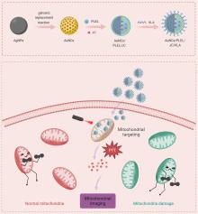
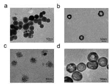
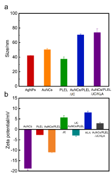
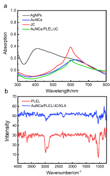
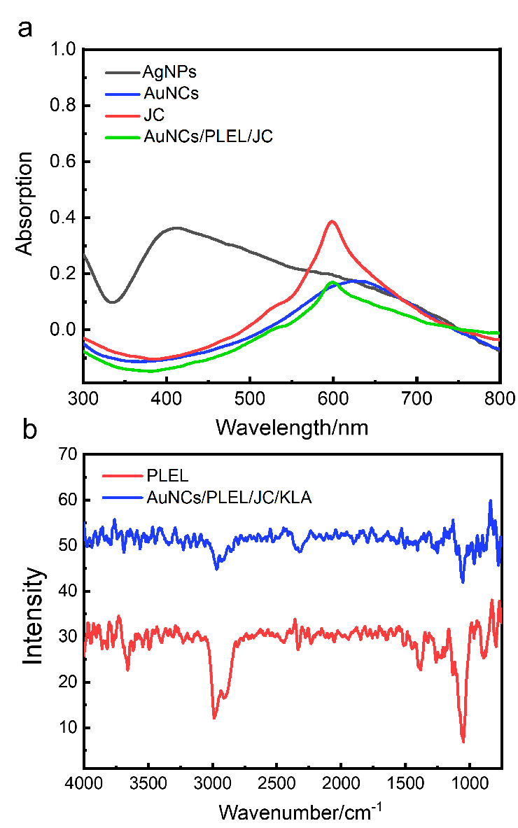
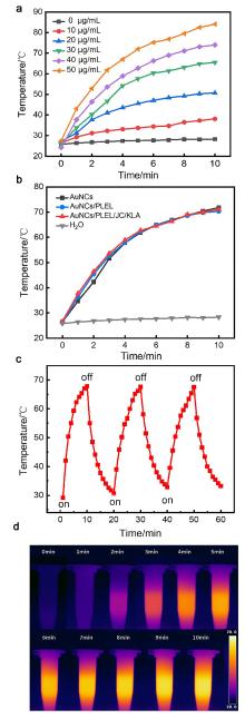
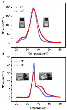
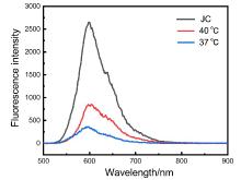
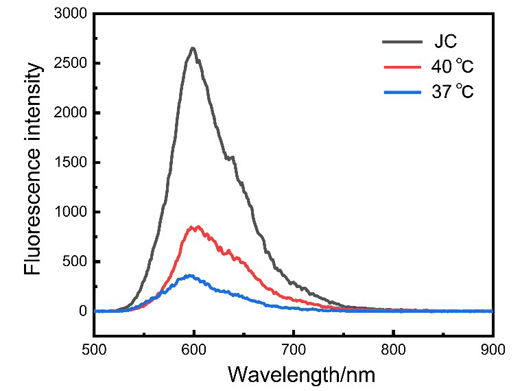
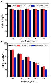
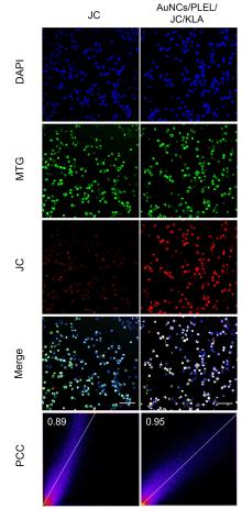
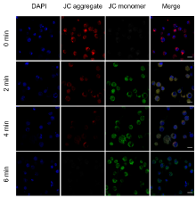
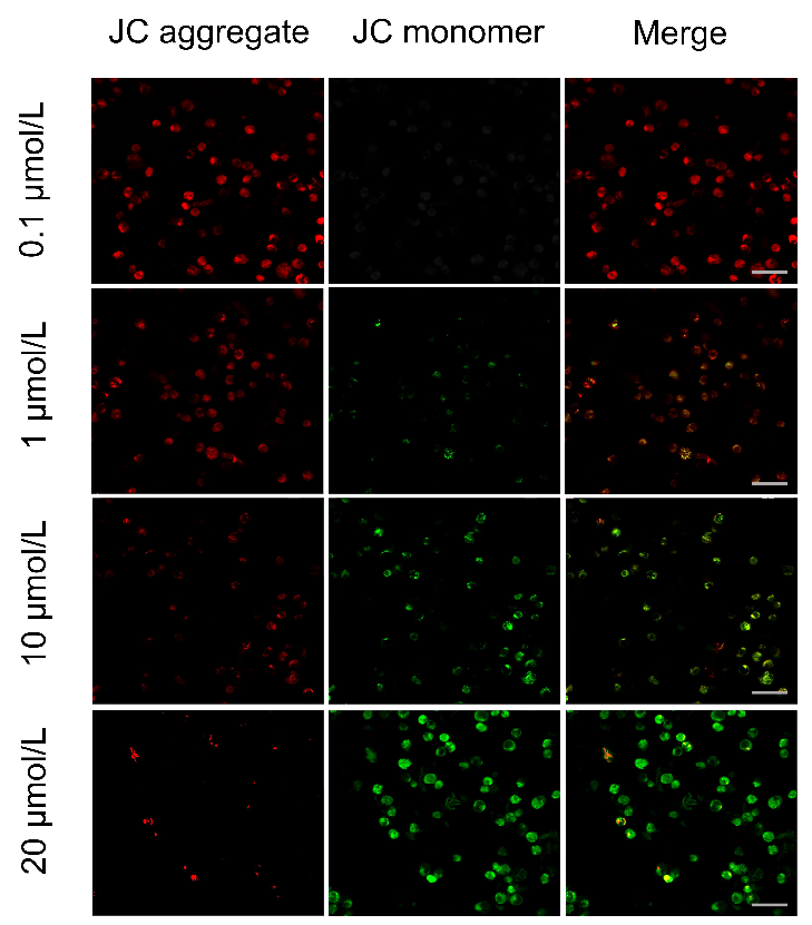
线粒体是许多细胞行为的关键调节细胞器, 线粒体膜电位降低被认为是细胞凋亡所发生的最早事件之一, 因此线粒体成像及其膜电位的检测分析, 对疾病的检测与治疗有重要的科学意义. 采用金纳米笼(Au nanocages, AuNCs)介导的光热损伤与温度敏感的药物释放相结合, 开发了一种线粒体靶向的荧光纳米探针AuNCs/PLEL/JC/KLA. 引入一种线粒体靶向肽(KLAKLAKKLAKLAK, KLA), 作为纳米探针的“指向标”, 指引着探针特异地靶向到细胞线粒体部位, 随后在近红外光的照射下, AuNCs吸收光能转化为热量, 实现光热介导的细胞损伤. 同时, 高温促使外层温敏水凝胶发生凝胶-溶胶转变, 实现荧光染料(JC-10)的释放. 所释放的JC-10荧光染料可根据线粒体的活力表现出两种荧光信号, 用于监测线粒体膜电位的变化. 总之, 该荧光纳米探针不仅实现了线粒体靶向的荧光成像与损伤细胞, 同时还可以监测线粒体膜电位的变化.
孙丽, 王亚静, 李涛, 郭英姝, 张书圣. 金纳米笼探针用于线粒体成像和光热损伤细胞★[J]. 化学学报, 2023, 81(10): 1301-1310.
Li Sun, Yajing Wang, Tao Li, Yingshu Guo, Shusheng Zhang. Au Nanocages Probes for Mitochondrial Imaging and Photothermal Damage Cells★[J]. Acta Chimica Sinica, 2023, 81(10): 1301-1310.


| [1] |
Al Kelabi, D.; Dey, A.; Alimi, L. O.; Piwonski, H.; Habuchi, S.; Khashab, N. M. Chem. Sci. 2022, 13, 7341.
doi: 10.1039/D2SC00836J |
| [2] |
Anishiya Chella Daisy, E. R.; Rajendran, N. K.; Jeyaraj, M.; Ramu, A.; Rajan, M. J. Liposome Res. 2021, 31, 203.
doi: 10.1080/08982104.2020.1768111 |
| [3] |
Asadian-Birjand, M.; Bergueiro, J.; Wedepohl, S.; Calderon, M. Macromol. Biosci. 2016, 16, 1432.
doi: 10.1002/mabi.v16.10 |
| [4] |
Cao, M. X.; Dai, X. G.; Chen, B. B.; Zhao, N. N.; Xu, F. J. Acta Chim. Sinica 2020, 78, 1054 (in Chinese).
doi: 10.6023/A20070295 |
|
(曹萌轩, 代晓光, 陈贝贝, 赵娜娜, 徐福建, 化学学报, 2020, 78, 1054.)
|
|
| [5] |
Bae, Y.; Joo, C.; Kim, G. Y.; Ko, K. S.; Huh, K. M.; Han, J.; Choi, J. S. Macromol. Res. 2019, 27, 1071.
doi: 10.1007/s13233-019-7153-x |
| [6] |
Bouchaala, R.; Anton, N.; Anton, H.; Vandamme, T.; Vermot, J.; Smail, D.; Mely, Y.; Klymchenko, A. S. Colloid Surface B 2017, 156, 414.
doi: 10.1016/j.colsurfb.2017.05.035 |
| [7] |
Wu, D.; Wang, Y. J.; Wang, Q. Q.; Li, T.; Guo, Y. S. Chin. J. Analysis Laboratory 2023, DOI: 10.13595/j.cnki.issn1000-0720.2023.032703 (in Chinese).
|
|
(吴谛, 王亚静, 王倩倩, 李涛, 郭英姝, 分析试验室, 2023, DOI: 10.13595/j.cnki.issn1000-0720.2023.032703.)
|
|
| [8] |
Cai, Y.; Zhu, H. S.; Zhou, W. C.; Qiu, Z. Y.; Chen, C. C.; Qileng, A.; Li, K. S.; Liu, Y. J. Anal. Chem. 2021, 93, 7275.
doi: 10.1021/acs.analchem.1c00616 |
| [9] |
Chen, H. C.; Wang, Y. R.; Yao, Y. Y.; Qiao, S. L.; Wang, H.; Tan, N. H. Theranostics 2017, 7, 3781.
doi: 10.7150/thno.20892 |
| [10] |
Chen, M. M.; Li, R. Y.; Liu, Y.; Song, X. R.; Tian, J.; Fu, Y. L.; Yang, Y. J.; Liu, C.; Zhang, Q. Q. Chem. Eng. J. 2021, 406, 126745.
doi: 10.1016/j.cej.2020.126745 |
| [11] |
Chen, S.; Lei, Q.; Qiu, W. X.; Liu, L. H.; Zheng, D. W.; Fan, J. X.; Rong, L.; Sun, Y. X.; Zhang, X. Z. Biomaterials 2017, 117, 92.
doi: 10.1016/j.biomaterials.2016.11.056 |
| [12] |
Chen, S. B.; Zhong, H.; Gu, B.; Wang, Y. Z.; Li, X. M.; Cheng, Z. P.; Zhang, L. L.; Yao, C. Mat. Sci. Eng. C-Mater. 2012, 32, 2199.
doi: 10.1016/j.msec.2012.05.052 |
| [13] |
Chen, W.; Shi, K.; Liu, J.; Yang, P. P.; Han, R. X.; Pan, M.; Yuan, L. P.; Fang, C.; Yu, Y. Y.; Qian, Z. Y. Bioact. Mater. 2023, 23, 1.
|
| [14] |
Zhou, H. M.; Tang, Y. H.; Lu, H. X.; Zhang, Q.; Lin, W. Y. Chin. J. Org. Chem. 2022, 42, 1687 (in Chinese).
doi: 10.6023/cjoc202112012 |
|
(周红梅, 唐永和, 卢辉旭, 张倩, 林伟英, 有机化学, 2022, 42, 1687.)
|
|
| [15] |
Chen, X. M.; Chen, D. R.; Liu, H. M.; Yang, L.; Zhang, Y. T.; Bu, L. L.; Sun, Z. J.; Cai, L. L. J. Control. Release 2022, 351, 381.
doi: 10.1016/j.jconrel.2022.09.010 |
| [16] |
Chi, J. N.; Ma, Q. M.; Shen, Z. J.; Ma, C. Y.; Zhu, W.; Han, S. C.; Liang, Y.; Cao, J.; Sun, Y. Nanoscale 2020, 12, 11008.
doi: 10.1039/C9NR10674J |
| [17] |
Chuang, C. H.; Chen, W. Y.; Tseng, W. B.; Lin, A. Y.; Lu, C. Y.; Tseng, W. L. ACS Sustainable Chem. Eng. 2022, 10, 2461.
doi: 10.1021/acssuschemeng.1c07440 |
| [18] |
Guo, Y. S.; Cao, X. P.; Zhang, S. S. Chem. Commun. 2021, 57, 10504.
doi: 10.1039/D1CC03454E |
| [19] |
Guo, Y. S.; Zheng, X. F.; Gai, T. T.; Wei, Z. Y.; Zhang, S. S. Chem. Commun. 2021, 57, 5754.
doi: 10.1039/D1CC01375K |
| [20] |
Guo, Y. S.; Wang, Y. J.; Li, S.; Niu, L.; Wei, D.; Zhang, S. S. Chem. Commun. 2017, 53, 4826.
doi: 10.1039/C7CC00310B |
| [21] |
Liu, F.; Guo, Y. S.; Hu, Y. H.; Zhang, X. R.; Zheng, X. J. Anal. Bioanal. Chem. 2019, 411, 5845.
doi: 10.1007/s00216-019-01966-0 |
| [22] |
Guo, Y. S.; Zheng, X. F.; Cao, X. P.; Li, W. X.; Wu, D.; Zhang, S. S. Chin. J. Chem. 2020, 38, 1793.
doi: 10.1002/cjoc.v38.12 |
| [23] |
Hu, X. J.; Gao, G. B.; Zhang, M. X. Acta Phys.-Chim. Sin. 2017, 33, 1324 (in Chinese).
doi: 10.3866/PKU.WHXB201704112 |
|
(胡雪娇, 高冠斌, 张明曦, 物理化学学报, 2017, 33, 1324.)
|
|
| [24] |
Li, Y. R.; Wang, Z. G.; Tang, C. H. Acta Chim. Sinica 2022, 80, 291 (in Chinese).
doi: 10.6023/A21120544 |
|
(李嫣然, 王子贵, 汤朝晖, 化学学报, 2022, 80, 291.)
|
|
| [25] |
Zheng, B. X.; Bi, C. F.; Hou, W. B.; Li, W. L. Acta Pharm. Sin. 2022, 57, 681 (in Chinese).
|
|
(郑宝鑫, 毕常芬, 侯文彬, 李祎亮, 药学学报, 2022, 57, 681.)
|
|
| [26] |
Guo, X. L.; Yang, N. D.; Ji, W. H.; Zhang, H.; Dong, X.; Zhou, Z. Q.; Li, L.; Shen, H. M.; Yao, S. Q.; Huang, W. Adv. Mater. 2021, 33, 2007778.
doi: 10.1002/adma.v33.43 |
| [27] |
Zhang, W.; Du, X. F.; Liu, B.; Li, C. R.; Long, J.; Zhao, M. X.; Yao, Z. Y.; Liang, X. J.; Lai, Y. X. ACS Nano 2022, 16, 1421.
doi: 10.1021/acsnano.1c09555 |
| [28] |
Hu, X. W.; Zhang, J.; Yu, Z.; Xie, Y. C.; He, H. S.; Qi, J. P.; Dong, X. C.; Lu, Y.; Zhao, W. L.; Wu, W. Nanomedicine 2015, 11, 1939.
|
| [29] |
Huang, W. Q.; Wang, F.; Nie, X.; Zhang, Z.; Chen, G.; Xia, L.; Wang, L. H.; Ding, S. G.; Hao, Z. Y.; Zhang, W. J.; Hong, C. Y.; You, Y. Z. ACS Appl. Bio. Mater. 2020, 3, 1176.
doi: 10.1021/acsabm.9b01052 |
| [30] |
Jeevarathinam, A. S.; Lemaster, J. E.; Chen, F.; Zhao, E.; Jokerst, J. V. Angew. Chem., Int. Ed. 2020, 59, 4678.
doi: 10.1002/anie.v59.12 |
| [31] |
He, H. M.; Zhao, F.; Zhong, W. K.; Yang, Y. L.; Lin, Y. H.; Ding, Y. H.; Yang, J. J.; Lu, C. H.; Tu, X. K. Mater. Design 2022, 219, 110722.
|
| [32] |
Horton, K. L.; Stewart, K. M.; Fonseca, S. B.; Guo, Q.; Kelley, S. O. Chem. Biol. 2008, 15, 375.
doi: 10.1016/j.chembiol.2008.03.015 |
| [33] |
Fan, X. H.; Wang, Y.; Yang, Y. Y.; Zhang, Y. H. Chem. J. Chin. Univ. 2022, 43, 168 (in Chinese).
|
|
(樊晓慧, 汪洋, 杨园园, 张玉红, 高等学校化学学报, 2022, 43, 168.)
|
|
| [34] |
Hu, W. Y.; Zhou, C. T.; Jing, Q. G.; Li, Y. C.; Yang, J.; Yang, C.; Wang, L. Y.; Hu, J. Y.; Li, H. J.; Wang, H. R.; Yuan, C.; Zhou, Y.; Ren, X. Y.; Tong, X. M.; Du, J.; Wang, Y. Cancer Cell Int. 2021, 21, 709.
doi: 10.1186/s12935-021-02420-x |
| [35] |
Zhang, Z. P.; Xu, S. H.; Wang, Y.; Yu, Y. N.; Li, F. Z.; Zhu, H.; Shen, Y. Y.; Huang, S. T.; Guo, S. R. J. Colloid Inter. Sci. 2018, 509, 47.
doi: 10.1016/j.jcis.2017.08.097 |
| [36] |
Jia, Y. P.; Shi, K.; Yang, F.; Liao, J. F.; Han, R. X.; Yuan, L. P.; Hao, Y.; Pan, M.; Xiao, Y.; Qian, Z. Y.; Wei, X. W. Adv. Funct. Mater. 2020, 30, 2001059.
doi: 10.1002/adfm.v30.25 |
| [37] |
Qiu, M. L.; Chen, D. Y.; Shen, C. Y.; Shen, J.; Zhao, H. K.; He, Y. H. Int. J. Mol. Sci. 2016, 17, 2001442.
|
| [38] |
Jeong, C.; Uthaman, S.; Bagheri, B.; Kim, J.; Pillarisetti, S.; Park, I. K.; Kim, Y. C. J. Control. Release 2021, 329, 50.
doi: 10.1016/j.jconrel.2020.11.046 |
| [39] |
Jia, H. Y.; Yang, L.; Fan, D. W.; Kuang, X.; Sun, X.; Wei, Q.; Ju, H. X. Sensor. Actuat. B-Chem. 2022, 367, 132034.
doi: 10.1016/j.snb.2022.132034 |
| [40] |
Jiang, L.; Zhou, S. S.; Zhang, X. K.; Li, C.; Ji, S. L.; Mao, H.; Jiang, X. Q. Nat. Commun. 2021, 12, 2390.
doi: 10.1038/s41467-021-22594-2 |
| [41] |
Jiao, Y.; Guo, Y. M.; Fan, Y. C.; Wang, R.; Li, X.; Wu, H.; Meng, Z. C.; Yang, X.; Cui, Y. P.; Liu, H.; Pan, L. P.; Maimaitijuma, T.; Zhang, J. Z.; Wang, Y. H.; Cao, Y. P.; Zhang, T. Bio. Med. Res. Int. 2020, 2020, 2846297.
|
| [42] |
Kadkhoda, J.; Tarighatnia, A.; Nader, N. D.; Aghanejad, A. Life Sci. 2022, 307, 120898.
doi: 10.1016/j.lfs.2022.120898 |
| [43] |
Ke, L. B.; Zhang, C.; Liao, X. X.; Qiu, K. Q.; Rees, T. W.; Chen, Y.; Zhao, Z. Z.; Ji, L. N.; Chao, H. Chem. Commun. 2019, 55, 10273.
doi: 10.1039/C9CC05610F |
| [44] |
Kolygina, D. V.; Siek, M.; Borkowska, M.; Ahumada, G.; Barski, P.; Witt, D.; Jee, A. Y.; Miao, H.; Ahumada, J. C.; Granick, S.; Kandere-Grzybowska, K.; Grzybowski, B. A. ACS Nano 2021, 15, 11470.
doi: 10.1021/acsnano.1c01232 |
| [45] |
Skrabalak, S. E.; Chen, J. Y.; Au, L. L.; Lu, X. M.; Li, X. D.; Xia, Y. N. Adv. Mater. 2007, 19, 3177.
doi: 10.1002/adma.v19:20 |
| [46] |
Skrabalak, S. E.; Au, L. L.; Lu, X. M.; Li, X. D.; Xia, Y. N. Nanomedicine 2007, 2, 657.
doi: 10.2217/17435889.2.5.657 |
| [47] |
Zhou, X. H.; He, X. L.; Shi, K.; Yuan, L. P.; Yang, Y.; Liu, Q. Y.; Ming, Y.; Yi, C.; Qian, Z. Y. Adv. Sci. 2020, 7, 2001442.
doi: 10.1002/advs.v7.23 |
| [48] |
Luo, Y.; Yang, L.; Feng, P. P.; Qiu, H. F.; Wu, X. J.; Lu, S. W.; Zhou, M.; Xu, L.; Zhu, Y. B. Front. Bioeng. Biotech. 2020, 8, 581621.
doi: 10.3389/fbioe.2020.581621 |
| [49] |
Zhang, Z. J.; Zhang, X. L.; Wang, C. G.; Teng, W. S. Y.; Xing, H. Y.; Wang, F. Q.; Yinwang, E.; Sun, H. X.; Wu, Y.; Yu, C. C.; Chai, X. P.; Qian, Z. Q.; Yu, X. H.; Ye, Z. M.; Wang, X. Y. Chem. Eng. J. 2022, 433, 134465.
doi: 10.1016/j.cej.2021.134465 |
| [50] |
Xu, Z.; Liu, Y. J.; Ma, R.; Chen, J.; Qiu, J. M.; Du, S.; Li, C. C.; Wu, Z. H.; Yang, X. F.; Chen, Z. B.; Chen, T. K. ACS Appl. Mater. Interfaces 2022, 14, 14059.
doi: 10.1021/acsami.1c24569 |
| [51] |
Zheng, Z. Q.; Bian, S. Q.; Li, Z. Q.; Zhang, Z. Y.; Liu, Y.; Zhai, X. Y.; Pan, H. B.; Zhao, X. L. Carbohydr. Polym. 2020, 249, 116826.
doi: 10.1016/j.carbpol.2020.116826 |
| [52] |
Wan, J. S.; Geng, S. N.; Zhao, H.; Peng, X. L.; Xu, J. B.; Wei, M. H.; Mao, J. X.; Zhou, Y.; Zhu, Q.; Zhao, Y. B.; Yang, X. L. Nanoscale 2018, 10, 20020.
doi: 10.1039/C8NR06851H |
| [53] |
Wang, X. H.; Wang, K. X.; Liu, J. P.; Hong, X. Chem. J. Chin. Univ. 2019, 40, 1586 (in Chinese).
|
|
(王晓慧, 王可心, 刘俊平, 洪霞, 高等学校化学学报, 2019, 40, 1586.)
|
|
| [54] |
Shao, J. D.; Ruan, C. S.; Xie, H. H.; Li, Z. B.; Wang, H. Y.; Chu, P. K.; Yu, X. F. Adv. Sci. 2018, 5, 1700848.
doi: 10.1002/advs.v5.5 |
| [55] |
Tang, Q.; Lim, T.; Shen, L. Y.; Zheng, G.; Wei, X. J.; Zhang, C. Q.; Zhu, Z. Z. Biomaterials 2021, 268, 120605.
doi: 10.1016/j.biomaterials.2020.120605 |
| [56] |
Li, W. X.; Wang, Y. J.; Sun, L.; Li, T.; Guo, Y. S. Chin. J. Analysis Laboratory 2023, DOI: 10.13595/j.cnki.issn1000-0720.2023.041602 (in Chinese).
|
|
(李文鑫, 王亚静, 孙丽, 李涛, 郭英姝, 分析试验室, 2023, DOI: 10.13595/j.cnki.issn1000-0720.2023.041602.)
|
| [1] | 车飞达, 赵晓茗, 张馨, 丁琪, 王昕, 李平, 唐波. 抑郁症相关活性分子的荧光成像★[J]. 化学学报, 2023, 81(9): 1255-1264. |
| [2] | 武虹乐, 郭锐, 迟涵文, 唐永和, 宋思睿, 葛恩香, 林伟英. 喹啉基粘度荧光探针的合成及其检测应用[J]. 化学学报, 2023, 81(8): 905-911. |
| [3] | 吕鑫, 吴仪, 张勃然, 郭炜. 过氧化氢激活型近红外氟硼二吡咯光敏剂的设计、合成及光动力治疗研究[J]. 化学学报, 2023, 81(4): 359-370. |
| [4] | 黄艳琴, 栗丽君, 杨书培, 张瑞, 刘兴奋, 范曲立, 黄维. HA-AuNPs/FDF用于透明质酸酶的高灵敏检测、肿瘤靶向细胞荧光成像和光疗[J]. 化学学报, 2023, 81(12): 1687-1694. |
| [5] | 贺晓梦, 袁方, 张素雅, 张健健. 基于尼罗红类ONOO–近红外荧光探针的开发及其成像应用[J]. 化学学报, 2023, 81(11): 1515-1521. |
| [6] | 宋思睿, 唐永和, 孙良广, 郭锐, 姜冠帆, 林伟英. 基于香豆素荧光团的新型极性检测荧光探针的开发及其成像应用[J]. 化学学报, 2022, 80(9): 1217-1222. |
| [7] | 刘巴蒂, 王承俊, 钱鹰. 噻吩基氟硼二吡咯近红外光敏染料的合成、双光子荧光成像及光动力治疗研究[J]. 化学学报, 2022, 80(8): 1071-1083. |
| [8] | 吴志芬, 柯建熙, 刘永升, 孙蓬明, 洪茂椿. 稀土近红外二区纳米荧光影像探针及其生物医学应用※[J]. 化学学报, 2022, 80(4): 542-552. |
| [9] | 李嫣然, 王子贵, 汤朝晖. 水溶性IR-780聚合物用于线粒体靶向的光动力治疗※[J]. 化学学报, 2022, 80(3): 291-296. |
| [10] | 王其, 夏辉, 熊炎威, 张新敏, 蔡杰, 陈冲, 高逸聪, 陆峰, 范曲立. 调控供电子策略简易制备近红外二区有机小分子光学诊疗试剂[J]. 化学学报, 2022, 80(11): 1485-1493. |
| [11] | 潘立祥, 黄艳琴, 盛况, 张瑞, 范曲立, 黄维. 透明质酸纳米材料在荧光/光声成像和光疗中的应用[J]. 化学学报, 2021, 79(9): 1097-1106. |
| [12] | 任江波, 王蕾, 郭锐, 唐永和, 周红梅, 林伟英. 一种基于萘酰亚胺的检测细胞内pH值的荧光探针及其生物成像应用[J]. 化学学报, 2021, 79(1): 87-92. |
| [13] | 李勇, 王栩, 解希雷, 张建, 唐波. 一氧化碳有机荧光探针和光控释放剂研究进展[J]. 化学学报, 2021, 79(1): 36-44. |
| [14] | 魏廷文, 江龙, 陈亚辉, 陈小强. 光笼分子与材料研究进展[J]. 化学学报, 2021, 79(1): 58-70. |
| [15] | 桑若愚, 许兴鹏, 王其, 范曲立, 黄维. 近红外二区有机小分子荧光探针[J]. 化学学报, 2020, 78(9): 901-915. |
| 阅读次数 | ||||||
|
全文 |
|
|||||
|
摘要 |
|
|||||
