有机化学 ›› 2025, Vol. 45 ›› Issue (4): 1137-1152.DOI: 10.6023/cjoc202407041 上一篇 下一篇
综述与进展
相韩悦a,b, 魏少荫a, 王玉记a,*( ), 肖猱a,*()
收稿日期:2024-07-27
修回日期:2024-10-09
发布日期:2024-11-08
基金资助:
Hanyue Xianga,b, Shaoyin Weia, Yuji Wanga(), Nao Xiaoa()
Received:2024-07-27
Revised:2024-10-09
Published:2024-11-08
Contact:
* E-mail: Supported by:文章分享
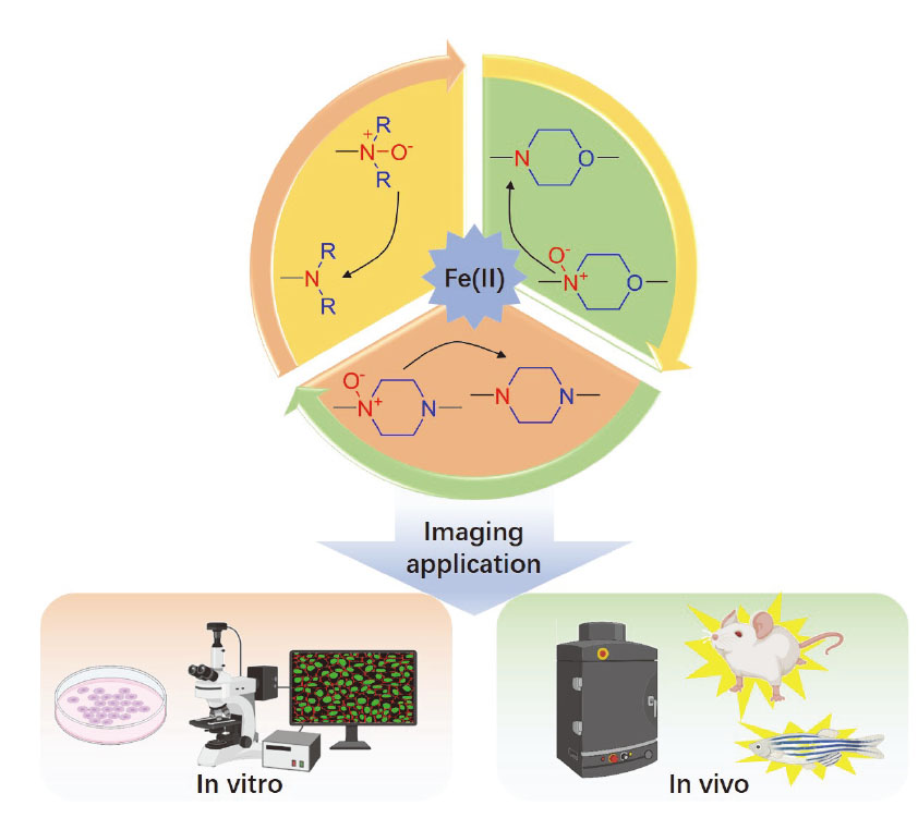
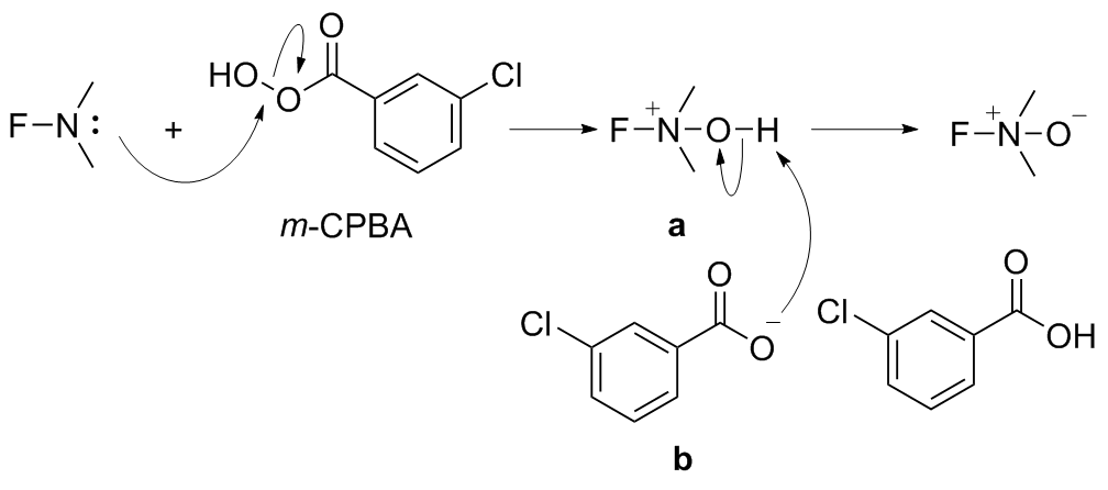
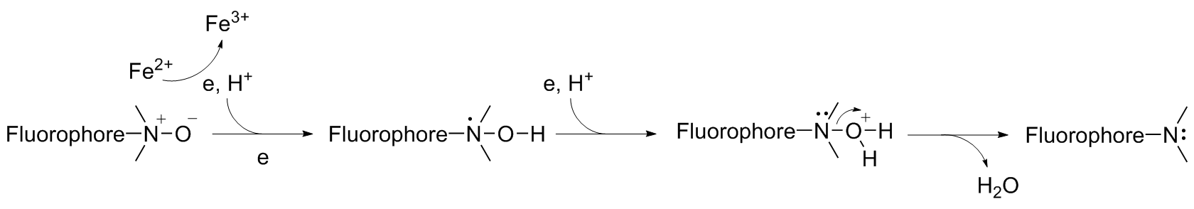
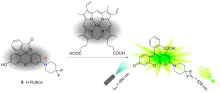
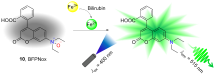
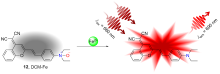
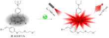
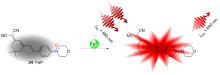
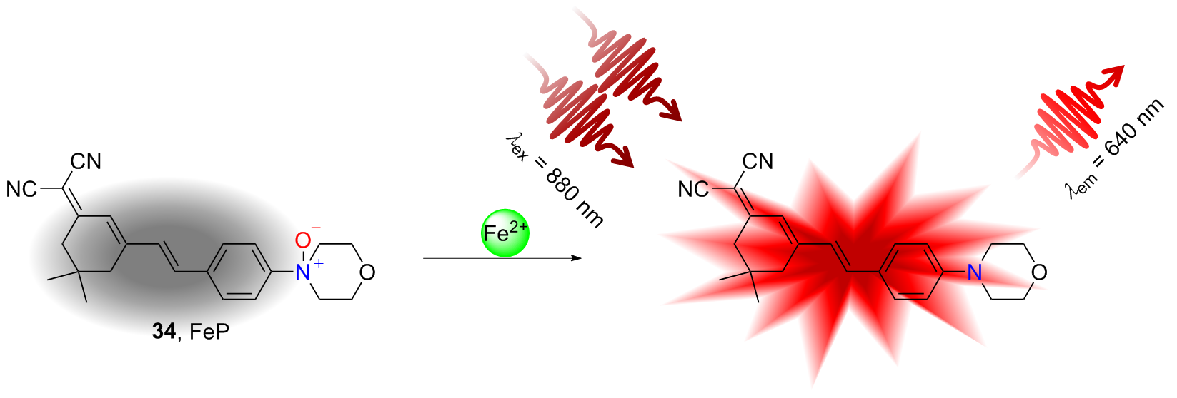
铁离子在人体内分布广泛, 参与一系列生化反应. 细胞中的铁离子主要以Fe2+与Fe3+的形式存在, 对维持细胞正常功能发挥着重要作用. 在活细胞还原环境中, 细胞内游离铁主要以Fe2+离子形式存在. 游离Fe2+可作为芬顿反应催化剂, 使细胞内产生活性氧物种(ROS), 对细胞造成严重损伤, 引起一系列与之相关的疾病. 因此, 在生理环境中对Fe2+的可视化监测具有必要性, 有助于进一步了解疾病的发病机制和进展. 近年来, 已有一些研究人员致力于设计并合成具有优异光学性能并可用于体内外监测Fe2+的荧光探针. 其中, N-氧化物结构的荧光探针凭借其选择性强、灵敏度高及可进行原位实时成像等优势受到了研究人员的广泛关注. 针对N-氧化物类Fe2+荧光探针的结构、设计思路、光学性质与应用等方面进行了介绍.
相韩悦, 魏少荫, 王玉记, 肖猱. 基于N-氧化物结构Fe2+荧光探针的研究进展[J]. 有机化学, 2025, 45(4): 1137-1152.
Hanyue Xiang, Shaoyin Wei, Yuji Wang, Nao Xiao. Recent Advances of Fe(II) Fluorescence Probes Based on N-Oxide Structure[J]. Chinese Journal of Organic Chemistry, 2025, 45(4): 1137-1152.


| Probe | Ex/Em/nm | Time/min | Solvent | Response mode | Co-localization |
|---|---|---|---|---|---|
| 1 | 540/575 | 60 | HEPES buffer | Turn-On | — |
| 2 | 550/575 | 20 | HEPES buffer (0.2% DMF) | Turn-On | Lysosome |
| 3 | 550/578 | 60 | HEPES buffer (0.2% DMF) | Turn-On | Lysosome |
| 4 | 515/535 | 60 | HEPES buffer (0.2% DMF) | Turn-On | — |
| 5 | 515/535 | 60 | HEPES buffer (0.2% DMF) | Turn-On | — |
| 6 | 575/660 | 30 | HEPES buffer | Turn-On | Endoplasmic reticulum |
| 7 | 580/630 | 10 | HEPES buffer (5% MeOH) | Turn-On | — |
| 8 | 630/665 | 30 | HEPES buffer (30% dioxane) | Turn-On | Golgi |
| 9 | 490/535 | 30 | HEPES buffer (0.2% DMSO) | Turn-On | — |
| 10 | 400/515 | — | HEPES buffer (5% DMSO) | Turn-On | — |
| 11 | 390/460 | 60 | HEPES buffer (0.2% DMSO) | Turn-On | — |
| 12 | 540/690 | 15 | PBS buffer (50% DMSO) | Turn-On Two-photon | — |
| 13 | 480/630 | 5 | Sodium borate buffer (pH=7.4) | Turn-On | — |
| 14 | 540/700 | 5 | HEPES buffer (40% DMSO) | Turn-On | Lipid droplet |
| 15 | 400/675 | 5 | PBS buffer (40% DMSO) | Turn-On | — |
| 16 | 467/625 | 20 | PBS buffer (30% EtOH) | Turn-On | — |
| 17 | 540/720 | 5 | PBS buffer (2.5% EtOH, 1 mmol/L CTAB) | Turn-On | Lysosome |
| 18 | 362/519 | 10 | PBS buffer | Turn-On | — |
| 19 | 365/520 | 10 | PBS buffer | Turn-On | — |
| 20 | 360/515 | 10 | PBS buffer | Turn-On | — |
| 21 | 350/489 | 20 | Tris-HCl buffer (20% DMSO) | Turn-On | — |
| 22 | 295/495 | 30 | HEPES buffer | Turn-On | Endoplasmic reticulum |
| 23 | 425/603 | 0.67 | PBS buffer (20% EtOH) | Turn-On | — |
| 24 | 380/495 | 1 | PBS buffer (20% EtOH) | Turn-On | — |
| 25 | 640/671 | 2 | DMSO (1% TFA) | Turn-Off | Lysosome |
| 26 | 370/486 | 0.42 | PBS solution (90% ACN, 10 mmol/L, v/v) | Turn-On | — |
| 27 | 400/518 | 0.58 | PBS solution (90% ACN, 10 mmol/L, v/v) | Turn-On | — |
| 28 | 510/535 | 30 | HEPES buffer | Turn-On | — |
| 29 | 540/575 | 60 | HEPES buffer (0.2% DMSO) | Turn-On | — |
| 30 | 540/575 | 60 | HEPES buffer (0.2% DMSO) | Turn-On | Cell membrane |
| 31 | 430/675 | 30 | Tris-HCl buffer (30% DMSO) | Ratiometric | — |
| 32 | 395/540 | 30 | Aqueous solution (0.5% CH3CN) | Turn-On | — |
| 33 | 450/530 | 50 | HEPES buffer | Turn-On | — |
| 34 | 456/640 | 10 | PBS buffer (1% DMSO, 1 mmol/L CTAB) | Turn-On Two-photon | Lysosome |
| [1] |
Siri-Angkul, N.; Xie, L. H.; Chattipakorn, S. C.; Chattipakorn, N. Front. Physiol. 2018, 9, 1615.
doi: 10.3389/fphys.2018.01615 pmid: 30498456 |
| [2] |
Andreini, C.; Putignano, V.; Rosato, A.; Banci, L. Metallomics 2018, 10, 1223.
doi: 10.1039/c8mt00146d pmid: 30095136 |
| [3] |
Kamal, A.; Kumar, N.; Bhalla, V.; Kumar, M.; Mahajan, R. K. Sens. Actuators, B 2014, 190, 127.
|
| [4] |
Jiang, T.; Huang, J.; Ran, G.; Song, Q.; Wang, C. Anal. Sci. 2023, 39, 325.
|
| [5] |
Wu, Y.; Zhang, S.; Gong, X.; Tam, S.; Xiao, D.; Liu, S.; Tao, Y. Mol. Cancer 2020, 19, 39.
|
| [6] |
Anita, P. K.; Alireza, B.; Zohreh, S. G.; Jahan, B. G. J. Mol. Struct. 2022, 1252, 131978.
|
| [7] |
Liu, H.; Mo, T.; Zhou, Y.; Gong, H.; Zhao, D. Microchem. J. 2022, 183, 108106.
|
| [8] |
Siahcheshm, P.; Heiden, P. J. Photochem. Photobiol., A 2023, 435, 114284.
|
| [9] |
Goetze, O.; Schmitt, J.; Spliethoff, K.; Theurl, I.; Weiss, G.; Swinkels, D. W.; Tjalsma, H.; Magginorini, M.; Krayenbuhl, P.; Rau, M.; Fruehauf, H.; Wojtal, K. A.; Mullhaupt, B.; Fried, M.; Gass- mann, M.; Lutz, T.; Geier, A. Hepatology 2013, 58, 2153.
|
| [10] |
Hirayama, T. Acta Histochem. Cytochem. 2018, 51, 137.
|
| [11] |
Zhang, S.; Xin, W.; Anderson, G. J.; Li, R.; Gao, L.; Chen, S.; Zhao, J.; Liu, S. Cell Death Dis. 2022, 13, 40.
|
| [12] |
Dong, X.; Cheng, X.; Mills, E.; Delling, M.; Wang, F.; Kurz, T.; Xu, H. Biophys. J. 2009, 96, 266a.
|
| [13] |
Ji, R.; Jia, F.; Chen, X.; Wang, Z.; Jin, W.; Yang, J. Arch. Biochem. Biophys. 2022, 715, 109094.
|
| [14] |
Li, J.; He, R.; Duan, S.; Li, J.; Han, X.; Ye, Y. Chin. J. Org. Chem. 2022, 42, 2428 (in Chinese).
|
|
(李嘉欣, 贺如艳, 段森林, 黎锦华, 韩校净, 叶勇, 有机化学, 2022, 42, 2428.)
doi: 10.6023/cjoc202203023 |
|
| [15] |
Zi, Y.; Wang, X.; Zi, Y.; Yu, H.; Lan, Y.; Fan, Y.; Ren, C; Liao, K.; Chen, H. Free Radical Biol. Med. 2023, 200, 73.
|
| [16] |
Li, D.; Zhang, G.; Wang, Z.; Guo, J.; Liu, Y.; Lu, Y.; Qin, Z.; Xu, Y.; Cao, C.; Wang, B.; Guo, Q.; Wang, Y.; Liu, G.; Cui, X.; Zhang, J.; Tang, J. Eur. J. Pharmacol. 2023, 943, 175569.
|
| [17] |
Wang, Y.; Wang, M.; Liu, Y.; Tao, H.; Banerjee, S.; Srinivasan, S.; Nemeth, E.; Czaja, M. J.; He, P. Redox Biol. 2022, 55, 102407.
|
| [18] |
Dixon, S. J.; Lemberg, K. M.; Lamprecht, M. R.; Skouta, R.; Zaitsev, E. M.; Gleason, C. E.; Patel, D. N.; Bauer, A. J.; Cantley, A. M.; Yang, W. S.; Morrison, B.; Stockwell, B. R. Cell 2012, 149, 1060.
|
| [19] |
Li, J.; Xiao, W.; Wei, W.; Wu, M.; Xiong, K.; Lyu, J.; Li, Y. Mol. Immunol. 2023, 158, 79.
|
| [20] |
Lee, J.; Roh, J. L. Cancer Lett. 2023, 559, 216119.
|
| [21] |
Yan, J.; Lee, S.; Zhang, A.; Yoon, J. Chem. Soc. Rev. 2018, 47, 6900.
|
| [22] |
Singh, H.; Tiwari, K.; Tiwari, R.; Pramanik, S. K.; Das, A. Chem. Rev. 2019, 119, 11718.
|
| [23] |
Zhu, J.; Miao, C.; Wang, X. J. Photochem. Photobiol.,A 2023, 440, 114659.
|
| [24] |
Zhu, J.; Miao, C.; Wang, X. Spectrochim. Acta, Part A 2023, 294, 122546.
|
| [25] |
Wei, X.; Zhu, T.; Ma, Y.; Sun, J.; Zheng, G.; Ma, T.; Yang, X.; Song, X.; Lv, Y.; Zhang, J.; Yan, M. Sens. Actuators, B 2023, 380, 133392.
|
| [26] |
Xiang, H.; Wang, T.; Tang, S.; Wang, Y.; Xiao, N. Spectrochim. Acta, Part A 2022, 267, 1386.
|
| [27] |
Li, H.; An, Y.; Gao, J.; Yang, M.; Luo, J.; Li, X.; Lv, J.; Li, X.; Yuan, Z.; Ma, H. Chemosensors 2022, 10, 233.
|
| [28] |
Guo, L.; Chen, X.; Xie, R.; Han, L. J. Mol. Struct. 2023, 1275, 134615.
|
| [29] |
Tanaka, H.; Kurokawa, Y.; Takechi, M.; Ohbuchi, A. Talanta Open 2021, 3, 100031.
|
| [30] |
Patel, M.; Bisht, N.; Prabhakar, P.; Sen, R. K.; Kumar, P.; Dwivedi, N.; Ashiq, M.; Mondal, D. P.; Strivastava, A. K.; Dhand, C. Environ. Res. 2023, 221, 115317.
|
| [31] |
Siyal, P.; Nafady, A.; Sirajuddin.; Memon, R.; Sherazi, S. T. H.; Nisar, J.; Siyal, A. A.; Shah, M. R.; Mahesar, S. A.; Bhagat, S. Spectrochim. Acta, Part A 2021, 254, 119645.
|
| [32] |
El-Bindary, M. A.; Shahat, A.; El-Deen, I. M.; Khalil, M. A.; Hassan, N. J. Mol. Liq. 2023, 382, 121946.
|
| [33] |
Toit, J. P.; Krieg, H. M.; Mans, N. J. Power Sources 2023, 533, 232178.
|
| [34] |
Wu, D.; Sedgwick, A. C.; Gunnlaugsson, T.; Akkaya, E. U.; Yoon, J.; James, T. D. Chem. Soc. Rev. 2017, 46, 7105.
|
| [35] |
Yin, J.; Zhan, J.; Hu, Q.; Huang, S.; Lin, W. Chem. Soc. Rev. 2023, 52, 2011.
|
| [36] |
La, D. D.; Bhosale, S. V.; Jones, L. A.; Bhosale, S. V. ACS Appl. Mater. Interfaces 2018, 10, 12189.
|
| [37] |
Guo, L.; Li, L.; Wang, X.; Zhang, Y.; Cui, F. ACS Omega 2023, 8, 37098.
|
| [38] |
Lu, Y.; Ruan, G.; Du, W.; Li, J.; Yang, N.; Wu, Q.; Lu, L.; Zhang, C.; Li, L. Dyes Pigm. 2021, 190, 109337.
|
| [39] |
Kumar, N.; Bhalla, V.; Kumar, M. Analyst 2014, 139, 543.
|
| [40] |
Wang, X.; Zhang, S.; Zhao, B. Spectrochim. Acta, Part A 2020, 227, 117699.
|
| [41] |
Yan, H.; Xu, X.; Li, J.; Xie, P.; Cao, W.; Yang, X.; Ye, Y. J. Photochem. Photobiol., A 2023, 440, 114638.
|
| [42] |
Duangkamol, C.; Muangsopa, P.; Rattanopas, S.; Wongsuwan, P.; Khrrotkaew, T.; Chueakwon, P.; Niamnont, N.; Chansaenpak, K.; Kamkaew, A. Dyes Pigm. 2023, 216, 111365.
|
| [43] |
Meng, X.; Zhao, J.; Ma, W. Chin. J. Org. Chem. 2020, 40, 276 (in Chinese).
|
|
(孟宪娇, 赵晋忠, 马文兵, 有机化学, 2020, 40, 276.)
doi: 10.6023/cjoc201908039 |
|
| [44] |
Guo, L.; Chen, X.; Xie, R.; Han, L.; Zhu, N. J. Mol. Struct. 2023, 1275, 134615.
|
| [45] |
Grabowski, Z. R.; Rotkiewicz, K.; Rettig, W. Chem. Rev. 2003, 103, 3899.
doi: 10.1021/cr940745l pmid: 14531716 |
| [46] |
Sedgwick, A. C.; Wu, L.; Han, H. H.; Bull, S. D.; He, X. P.; James, T. D.; Sessler, J. L.; Tang, B. Z.; Tian, H.; Yoon, J. Chem. Soc. Rev. 2018, 47, 8842.
doi: 10.1039/c8cs00185e pmid: 30361725 |
| [47] |
Wu, L.; Huang, C.; Emery, B. P.; Sedgwick, A. C.; Bull, S. D.; He, X. P.; Tian, H.; Yoon, J.; Sessler, J. L.; James, T. D. Chem. Soc. Rev. 2020, 49, 5110.
|
| [48] |
Xing, W.; Xu, H.; Ma, H.; Abedi, S. A. A.; Wang, S.; Zhang, X.; Liu, X.; Xu, H.; Wang, W.; Lou, K. Chem. Commun. 2022, 58, 2979.
|
| [49] |
Li, Z.; Xiao, M.; Zheng, Y.; Zhao, B. Spectrochim. Acta, Part A 2022, 279, 121397.
|
| [50] |
Li, K.; Xu, S.; Xiong, M.; Huan, S. Y.; Yuan, L.; Zhang, X. B. Chem. Soc. Rev. 2021, 50, 11766.
|
| [51] |
Huang, W.; Yang, Y.; Ye, D. Chin. J. Chem. 2023, 41, 2382.
|
| [52] |
Yin, J.; Huang, L.; Wu, L.; Li, J.; James, T. D.; Lin, W. Chem. Soc. Rev. 2021, 50, 12098.
|
| [53] |
Wang, X.; Ding, Q.; Groleau, R. R.; Wu, L.; Mao, Y.; Che, F.; Kotova, O.; Scanlan, E. M.; Lewis, S. E.; Li, P.; Tang, B.; James, T. D.; Gunnlaugsson, T. Chem. Rev. 2024, 124, 7106.
|
| [54] |
Owens, E. A.; Henary, M.; Fakhri, G. E.; Choi, H. S. Acc. Chem. Res. 2016, 49, 1731.
|
| [55] |
Li, H.; Kim, Y.; Jung, H.; Hyun, J. Y.; Shin, I. Chem. Soc. Rev. 2022, 51, 8957.
|
| [56] |
Zhang, Y.; Zhang, G.; Zeng, Z.; Pu, K. Chem. Soc. Rev. 2022, 51, 566.
|
| [57] |
Fujita, K.; Urano, Y. Chem. Rev. 2024, 124, 4021.
|
| [58] |
Liu, Y.; Li, Y.; Koo, S.; Sun, Y.; Liu, Y.; Liu, X.; Pan, Y.; Zhang, Z.; Du, M.; Lu, S.; Qiao, X.; Gao, J.; Wang, X.; Deng, Z.; Meng, X.; Xiao, Y.; Kim, J.S.; Hong, X. Chem. Rev. 2022, 122, 209.
|
| [59] |
Xu, G.; Li, N.; Sun, Y.; Gao, C.; Ma, L.; Song, P.; Xia, L. Chem. Eng. J. 2021, 414, 128741.
|
| [60] |
Kouser, R.; Zehra, S.; Khan, R. A.; Alsalme, A.; Arjmand, F.; Tabassum, S. Spectrochim. Acta, Part A 2021, 247, 119156.
|
| [61] |
Guan, J.; Tu, Q.; Chen, L.; Yuan, M.; Wang, J. Spectrochim. Acta, Part A 2019, 220, 117114.
|
| [62] |
Zhang, J.; Yang, Y.; Zhang, J.; She, W. Chin. J. Org. Chem. 2024, 44, 1337 (in Chinese).
|
|
(张继东, 杨垚, 张杰, 厍伟, 有机化学, 2024, 44, 1337.)
doi: 10.6023/cjoc202307021 |
|
| [63] |
Pinheiro, G. E. G.; Ihmels, H. J. Fluoresc. 2021, 31, 269.
doi: 10.1007/s10895-020-02666-0 pmid: 33340065 |
| [64] |
Sarkar, D.; Chowdhury, M.; Das, P. K. J. Mater. Chem. B 2021, 9, 494.
|
| [65] |
Hou, X.; Song, Y.; Lv, Y.; Wang, P.; Chen, K.; Li, G.; Guo, L. Spectrochim. Acta, Part A 2023, 290, 122254.
|
| [66] |
Khomami, A. P.; Badiei, A.; Ghavami, Z. S.; Ghasemi, B. J. J. Mol. Struct. 2022, 1252, 131978.
|
| [67] |
Khunkhong, N.; Kitchawengkul, N.; Wongnongwa, Y.; Jungsuttiwong, S.; Keawin, T.; Promarak, V.; Nalaoh, P.; Suttisintong, K.; Chansaenpak, K.; Jarujamrus, P. Dyes Pigm. 2022, 208, 110869.
|
| [68] |
Yun, L.; Cheng, X. Cellulose 2023, 30, 933.
|
| [69] |
Zhang, S.; Cheng, X. Mater. Today Commun. 2023, 36, 106696.
|
| [70] |
Chao, M.; Zhang, H.; Hu, Q.; Ma, S.; Cui, X.; Zhu, X.; Wang, H.; Yu, X.; Han, B. Anal. Bioanal. Chem. 2024, 416, 6473.
|
| [71] |
Nan, X.; Huyan, Y.; Li, H.; Sun, S.; Xu, Y. Coord. Chem. Rev. 2021, 426, 213580.
|
| [72] |
Xu, G.; Li, N.; Sun, Y.; Gao, C.; Ma, L.; Song, P.; Xia, L. J. Lumin. 2022, 250, 119069.
|
| [73] |
Grimm, J. B.; Tkachuk, A. N.; Patel, R.; Hennigan, T. S.; Gutu, A.; Dong, P.; Gandin, V.; Osowski, A. M.; Holland, K. L.; Liu, Z. J.; Brown, T. A.; Lavis, L. D. J. Am. Chem. Soc. 2023, 145, 23000.
|
| [74] |
Liu, D.; He, Z.; Zhao Y.; Yang, Y.; Shi, W.; Li, X.; Ma, H. J. Am. Chem. Soc. 2021, 143, 17136.
|
| [75] |
Hirayama, T.; Okuda, K.; Nagasawa, H. Chem. Sci. 2013, 4, 1250.
|
| [76] |
Niwa, M.; Hirayama, T.; Okuda, K.; Nagasawa, H. Org. Biomol. Chem. 2014, 12, 6590.
|
| [77] |
Hirayama, T.; Tsuboi, H.; Niwa, M.; Miki, A.; Kadota, S.; Ikeshita, Y.; Okuda, K.; Nagasawa, H. Chem. Sci. 2017, 8, 4858.
doi: 10.1039/c6sc05457a pmid: 28959409 |
| [78] |
Dong, B.; Song W.; Lu, Y.; Tian, M.; Kong, X.; Mehmood, A. H.; Lin, W. Sens. Actuators, B 2020, 305, 127470.
|
| [79] |
Hirayama, T.; Inden, M.; Tsuboi, H.; Niwa, M.; Uchida, Y.; Naka, Y.; Hozumib, I.; Nagasawa, H. Chem. Sci. 2019, 10, 1514.
doi: 10.1039/c8sc04386h pmid: 30809369 |
| [80] |
Kawai, K.; Hirayama, T.; Imai, H. Murakami, T.; Inden, M.; Hozumi, I.; Nagasawa, H. J. Am. Chem. Soc. 2022, 144, 3793.
|
| [81] |
Huang, X. C.; Tang, H.; Wei, X.; He, Y.; Hu, S.; Wu, J.; Xu, D.; Qiao, F.; Xue, J.; Zhao, Y. Nat. Commun. 2024, 15, 6864.
|
| [82] |
Yan, L.; Li, X.; Li, J. ChemistrySelect 2018, 3, 10157.
|
| [83] |
Chen, S.; Wang, H.; Wu, B.; Li, Q.; Gong, J.; Zhao, Y.; Zhao, Y.; Xiao, X.; Lam, J. W. Y.; Zhao, Z.; Luo, X.; Tang, B. Z. ACS Cent. Sci. 2023, 9, 883.
|
| [84] |
Ahmmed, E.; Mondal, A.; Sarkar, A.; Chakraborty, S.; Lohar, S.; Saha, N. C.; Dhara, K.; Chattopadhyay, P. ACS Appl. Bio Mater. 2020, 3, 4074.
doi: 10.1021/acsabm.0c00170 pmid: 35025482 |
| [85] |
Khatun, S.; Biswas, S.; Binoy, A.; Podder, A.; Mishra, N.; Bhuniya, S. J. Photochem. Photobiol., B 2020, 209, 111943.
|
| [86] |
Yang, X.; Wang, Y.; Liu, R.; Zhang, Y.; Tang, J.; Yang, E.; Zhang, D.; Zhao, Y.; Ye, Y. Sens. Actuators, B 2019, 288, 217.
|
| [87] |
Yang, L.; Chen, Q.; Gan, S.; Guo, Q.; Zhang, J.; Zhang, H.; Xie, Y.; Xiao, H.; Wang, W.; Sun, H. Dyes Pigm. 2021, 190, 109271.
|
| [88] |
Zheng, J.; Feng, S.; Gong, S.; Xia, Q.; Feng, G. Sens. Actuators, B 2020, 309, 127796.
|
| [89] |
Zhu, M.; Zhao, Z.; Liu, X.; Chen, P.; Fan, F.; Wu, X.; Hua, R.; Wang, Y. J. Hazard. Mater. 2021, 406, 124767.
|
| [90] |
Zeng, X.; Chen, J.; Yu, S.; Liu, Z.; Ma, M. J. Lumin. 2022, 250, 119069.
|
| [91] |
Xiang, H.; Song, Y.; Wang, Y.; Xiao, N. Mater. Adv. 2024, 5, 5624.
|
| [92] |
Xu, C.; Zou, H.; Zhao, Z.; Zhang, P.; Kwok, R. T. K.; Lam, J. W. Y.; Sung, H. H. Y.; Williams, I. D.; Tang, B. Z. Adv. Funct. Mater. 2019, 29, 1903278.
|
| [93] |
Ga, G.; Wang, X.; Wang, Z.; Jin, X.; Ou, L.; Zhou, J.; Xie, P. Talanta 2020, 215, 120908.
|
| [94] |
Zhang, Y.; Gong, S.; Song, J.; Li, M.; Qin, A.; Gao, Y.; Wang, Z.; Wang, S. Dyes Pigm. 2021, 194, 109590.
|
| [95] |
Liang, Y.; Zhang, Y.; Li, M.; Meng, Z.; Gao, Y.; Yin, J.; Yang, Y.; Wang, Z.; Wang, S. Anal. Bioanal. Chem. 2021, 413, 6267.
|
| [96] |
Xu, W.; Wu, P.; Li, X.; Liu, S.; Feng, L.; Xiong, H. Talanta 2021, 233, 122601.
|
| [97] |
Shen, Z.; Yang, Y.; Gong, S.; Meng, Z.; Liang, Y.; Zhang, Y.; Wang, Z.; Wang, S. Microchem. J. 2023, 195, 109519.
|
| [98] |
Hirayama, T.; Miki, A.; Nagasawa, H. Metallomics, 2019, 11, 111.
doi: 10.1039/c8mt00212f pmid: 30215439 |
| [99] |
Hirayama, T.; Niwa, M.; Hirosawa, S.; Nagasawa, H. ACS Sens. 2020, 5, 2950.
|
| [100] |
Niwa, M.; Hirayama, T.; Oomoto, I.; Wang, D.O.; Nagasawa, H. ACS Chem. Biol. 2018, 13, 1853.
|
| [101] |
Feng, S.; Zheng, J.; Zhang, J.; Gui, Z.; Feng, G. Sens. Actuators, B 2022, 371, 132512.
|
| [102] |
Lee, Y. H.; Verwilst, P.; Kim, H. S.; Ju, J.; Kim, J. S.; Kim, K. Chem. Commun. 2019, 55, 12136.
|
| [103] |
Shao, C.; Liu, Y.; Chen, Z.; Qin, Y.; Wang, X.; Wang, X.; Yan, C.; Zhu, H.; Zhao, J.; Qian, Y. Cell Chem. Biol. 2022, 29, 43.
|
| [1] | 李兆周, 谢亚芳, 陶健, 魏雪冰, 黎乐乐, 李亚娟, 唐嘉敏, 牛华伟, 陈秀金, 高红丽, 李芳, 于慧春, 袁云霞, 古绍彬, 康怀彬, 孙晓菲, 任国艳, 吴影. 半胱氨酸荧光探针的构建及其在病理学检验中的应用[J]. 有机化学, 2025, 45(7): 2335-2349. |
| [2] | 张文博, 苏思铭, 杨鹏. 新型杯[3]咔唑衍生物的合成及其对羟喜树碱识别与检测研究[J]. 有机化学, 2025, 45(4): 1395-1401. |
| [3] | 沈晴, 曾玉, 黎忠昊, 曹西颖, 郭玉婷, 叶蕴仪, 汪朝阳. 基于共价键形成与切断的反应型小分子荧光探针的研究进展[J]. 有机化学, 2025, 45(4): 1194-1205. |
| [4] | 曾玉, 黎忠昊, 邓思威, 陈祖佳, 陈璧瑜, 宇世伟, 沈晴, 汪朝阳. 氰基乙烯类化合物的制备与应用研究进展[J]. 有机化学, 2024, 44(9): 2722-2731. |
| [5] | 何春, 刘姝祺, 成奋民, 郝远强, 陈述, 张培盛, 曾荣今. 具有双态发光性质的双光子丹磺酰类荧光探针的合成及其在硫化氢检测中的应用研究[J]. 有机化学, 2024, 44(9): 2869-2875. |
| [6] | 黄凯航, 尹理, 姜青云, 汪乾, 石光, 许炳佳. 具有聚集诱导发光性质的高效热激活延迟荧光材料用于脂滴成像[J]. 有机化学, 2024, 44(8): 2479-2486. |
| [7] | 苏小龙, 李健鹏, 刘孟鑫, 邹莉, 杨得锁, 冯海涛. 四苯乙烯酰胺类化合物的合成及其高灵敏度、高选择性识别Cu2+[J]. 有机化学, 2024, 44(8): 2581-2587. |
| [8] | 胡亦然, 张巳亮, 罗海艳, 赵璐瑶, 郭旭东, 王双青, 胡睿, 杨国强. 新型查尔酮衍生物氨肽酶N荧光探针的设计及应用[J]. 有机化学, 2024, 44(7): 2257-2264. |
| [9] | 李非凡, 余康, 倪传志, 朱园园, 曾婕, 古双喜. 测定氨基酸浓度和对映体组成的手性荧光探针[J]. 有机化学, 2024, 44(6): 1862-1869. |
| [10] | 张文艳, 王丹, 罗仁洁, 刘会玲. 近红外荧光手术导航探针的研究进展[J]. 有机化学, 2024, 44(6): 1760-1776. |
| [11] | 刘琳, 陈琳, 胡晓玲, 钟克利, 张璟琳, 汤立军. 基于苯并吡喃点亮型硫化氢荧光探针在食品检测中的应用与细胞成像[J]. 有机化学, 2024, 44(6): 2027-2032. |
| [12] | 程晓红, 刘发龙, 孙进博, 张锐. 一种用于次氯酸根实时高灵敏度检测的络合物基荧光探针[J]. 有机化学, 2024, 44(4): 1284-1292. |
| [13] | 徐冬青, 童海姗, 沈杰, 邱万伟, 钱立生. 脂滴特异性荧光探针的构建及可视化肝肿瘤细胞[J]. 有机化学, 2024, 44(4): 1240-1246. |
| [14] | 张继东, 杨垚, 张杰, 厍伟. 基于聚集诱导效应(AIE)-激发态分子内质子转移(ESIPT)效应的四苯乙烯荧光探针对Zn(II)检测研究[J]. 有机化学, 2024, 44(4): 1337-1342. |
| [15] | 张飞, 梅煜, 丁雪峰, 李再峰, 张鹏, 丁彩凤. 激活型近红外荧光探针用于肝癌中二肽基肽酶活性检测[J]. 有机化学, 2024, 44(11): 3417-3426. |
| 阅读次数 | ||||||
|
全文 |
|
|||||
|
摘要 |
|
|||||