
|
Molecular Probes and Nanobiology and Bioanalytical Chemistry

|
| Default Latest Most Read |
|
Please wait a minute...
|
ImageJ software and then reconstruct STORM images with a Falcon algorithm to show marked imaging resolution enhancement, compared with wide-field images, which provide a new protocol for biomedical imaging.

Review
Progress in Analysis and Detection of Salivary Tumor Biomarkers Associated with Oral Cancer
Acta Chim. Sinica
2019, 77 (4):
340-350.
DOI: 10.6023/A18100414
Published: 05 December 2018 Oral cancer is head and neck cancer, and cancer tissue is located in the oral cavity. The non-invasive early diagnosis is an effective method to reduce the death of the disease. The oral cancer-related substances are first released into the saliva, which is convenient, safe and non-invasive, and is the first choice for screening and early diagnosis of oral cancer. In this paper, the specific types and the commonly used detection methods of the salivary tumor biomarkers at home and abroad were summarized and compared. Specifically, the latest application of new electrochemical biosensor in the detection of the salivary tumor biomarkers associated with oral cancer was mainly described. Futhermore, the summary of its future directions and the potential applications was proposed, which provided reference for the further research and application of the salivary tumor biomarkers in oral cancer. 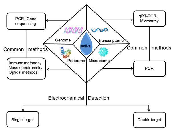
Review
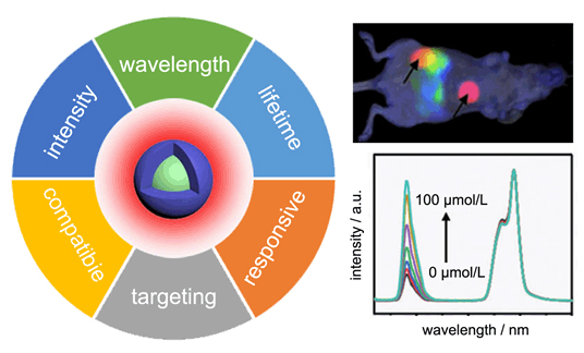
Research Progress on Rare Earth Nanocrystals for In Vivo Imaging and Sensing in Near Infrared Region
Acta Chimica Sinica
2019, 77 (12):
1239-1249.
DOI: 10.6023/A19080305
Published: 18 September 2019
In vivo imaging and sensing play a critical role in modern biological and medical research. Compared with other techniques such as computed tomography (CT), positron emission tomography (PET) and nuclear magnetic resonance (NMR), fluorescence imaging and analysis are featured by fast feedback, high sensitivity, and high spatiotemporal resolution. Especially, the application of near infrared (NIR) light as both excitation and emission signals provides increased tissue penetration and improved imaging quality and sensitivity due to reduced light scattering and auto-fluorescence. Among various materials investigated for in vivo imaging and bio-sensing, lanthanide-based nanocrystals display rich excitation/emission wavelengths in the NIR range, good photo and chemical stability, large Stokes shifts. In recent years, the research on lanthanide-based nanocrystals for in vivo imaging and sensing has seen rapid progress. Through nanoscale material design and synthesis, it is possible to fine tune the optical properties of lanthanide-based nanocrystals. By properly choosing different lanthanide ions as activators and sensitizers, multiple excitation/emission wavelengths can be obtained. The careful design of core-shell structure of nanocrystals enables improved fluorescence efficiency and tailorable fluorescence life time through controlled energy transfer. On the other side, the surface of lanthanide-based nanocrystals can be modified through coating, absorption or ligand exchange to enhance the biocompatibility, targeting capability, and bio-responsiveness. Taking advantage of this high flexibility and versatility, there are great opportunities for these lanthanide-based nanocrystals in various in vivo imaging and sensing applications. This review first outlines the general technique requirements for in vivo imaging and sensing. Then, the composition, synthesis and basic properties of lanthanide-based nanocrystals are briefly introduced. Subsequently, the routes for tailoring the optical and biochemical properties of lanthanide-based nanocrystals are discussed in detail, with an emphasis on the material designs and surface modifications for in vivo imaging and analysis. It is expected that this work will inspire new ideas for accelerating the clinic translation of rare earth nanocrystals-based imaging and sensing techniques.
Article
Colorimetric Sensing of Prostate Specific Membrane Antigen Based on Gold Nanoparticles
Acta Chim. Sinica
2019, 77 (5):
422-426.
DOI: 10.6023/A19010018
Published: 26 March 2019 Cancer is a major cause of death and its early diagnosis has been a research goal for many decades. For males, prostatic carcinoma has become the second leading cause of cancer death worldwide. Prostate specific membrane antigen (PSMA) has been widely recognized as a prostate cancer marker. Thus, measurement of PSMA would be more valuable for the early diagnosis of prostate cancer. Nanomaterials have the characteristics of small size effect, quantum size effect, macroscopic quantum tunneling effect and surface effect, and have been widely used in various fields, such as cell imaging, analysis and detection, drug release and treatment. Gold nanoparticles have been widely used in biosensing and medical diagnosis due to their simple preparation, high stability and unique photoelectric properties. In this paper, a new colorimetric approach is proposed for simple detection of PSMA based on gold nanoparticles. In the experiment, we synthesized gold nanoparticles with positive charges, and the polyanionic peptide as the substrate of PSMA. The detection of PSMA was based on the property that different aggregation states of gold nanoparticles can lead to the change of color and the specific recognition of PSMA for its substrate. The positively charged gold nanoparticles interact electrostatically with polyanionic peptide, resulting in aggregation of gold nanoparticles. In the presence of PSMA, however, the polyanionic peptide are hydrolyzed into glutamic acid fragment due to the reaction between the PSMA and the polyanionic peptide, resulting in the dispersion of gold nanoparticles. This behaviour leads to the development of a rapid and simple colorimetric method for assaying PSMA activity, with a detection limit of 0.5 nmol/L and the linear range of 2~10 nmol/L. This approach is simple compared to the existing ones since the gold nanoparticles-peptide based sensor is easy to be assembled and the detection can be achieved without the involvement of complicated procedures. Moreover, the applicability of the method has been demonstrated by detecting PSMA spiked into urine samples. 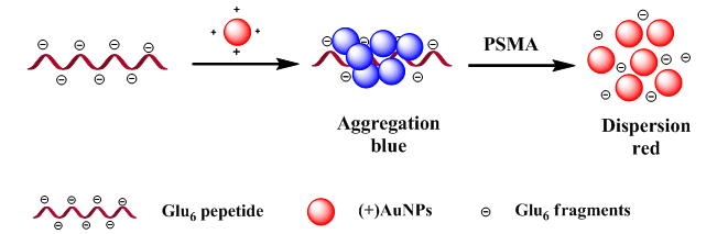
Article
DNA Interaction and Antitumor Activity of A Copper(II) Complex Containing Sparfloxacin and Triazine Derivatives
Acta Chimica Sinica
2020, 78 (3):
263-270.
DOI: 10.6023/A19110403
Published: 24 February 2020
DNA is an important target for antitumor drugs, hence investigation of the interaction between drug molecules and DNA can help to design targeted DNA antitumor drugs. New ternary copper(II) complex[Cu(Sf)(PyTA)(H2O)]·ClO4· 3.5H2O[Sf=sparfloxacin, 5-amino-1-cyclopropyl-7-(cis-3,5-dimethyl-1-piperazinyl)-6,8-difluoro-1,4-dihydro-4-oxoquino-line-3-carboxylic acid, PyTA=2,4-diamino-6-(2'-pyridyl)-1,3,5-triazine] was synthesized and characterized by elemental analyses, molar conductivity measurement and various spectroscopic techniques such as infrared, ultraviolet-visible, and electrospray ionization mass spectra. The interaction of the complex with DNA was investigated using electronic absorption spectroscopy, KI fluorescence quench, viscosity measurement and molecular docking techniques. It was found that the complex could bind to DNA through an intercalation mode being related with the quinoline ring of ligand Sf, and the corresponding binding constant Kb is 1.23×10⁴ L/mol. Moreover, the antitumor activity of the complex was evaluated using the MTT[3-(4,5-dimethyl-2-thiazolyl)-2,5-diphenyl-2-H-tetrazolium bromide] method, revealing that the complex displayed favorable cytotoxic effects[IC50=(57.0±1.6)~(77.6±1.4) μmol/L] toward cancer cells (A549, Bel-7402 and Eca-109) and less toxic towards normal cells (3T3). Most importantly, the cytotoxic mechanism of the complex towards Eca-109 cells was explored by single cell gel electrophoresis assay, Hoechst 33342 staining, Annexin V-FITC/PI double dye flow cytometry, measurement of mitochondrial membrane potential change, detection of intracellular cytochrome C and Ca2+ levels, and test of cell cycle arrest. Single cell gel electrophoresis assay (comet assays) demonstrated that the complex could damage DNA and cause apoptosis. Double staining analysis showed that the complex could induce apoptosis in Eca-109 cells. Cell cycle arrest studies revealed the cell growth arrest at S and G2/M phases. The complex also could induce a reduction in the mitochondrial membrane potential and release of the cytochrome C, and increase the intracellular Ca2+ level. The results demonstrated that the complex could induce apoptosis in Eca-109 cells through DNA-binding mitochondrial dysfunctional pathways, which was accompanied by the cell growth arrest at S and G2/M phases and damage of DNA.

|