|
Molecular Probes and Nanobiology and Bioanalytical Chemistry

|
| Default Latest Most Read |
|
Please wait a minute...
|
| ImageJ software and then reconstruct STORM images with a Falcon algorithm to show marked imaging resolution enhancement, compared with wide-field images, which provide a new protocol for biomedical imaging.
Article
Construction of Nitric Oxide (NO)-Responsive Fluorescent Polymer and Its Application in Cell Imaging
Zheng Bin, Cheng Sheng, Dong Huaze, Zhu Jinmiao, Han Yu, Yang Liang, Hu Jinming
Acta Chimica Sinica
2020, 78 (10):
1089-1095.
DOI: 10.6023/A20060280
Published: 21 August 2020
Nitric oxide (NO) is a ubiquitous physiological signal messenger, but the use of NO as a trigger event to delicately tune the self-assembly behaviors of biomimetic polymers has been far less exploited. In this work, a single primary amine-containing 2-(3-(2-aminophenyl)ureido)ethyl methacrylate (APUEMA) monomer was first synthesized by the reaction between o-phenylenediamine and 2-isocyanatoethyl methacrylate. Then, the well-defined double hydrophilic block copolymer (DHBC), poly[oligo(ethylene glycol)methyl ether methacrylate]-b-poly[2-(3-(2-aminophenyl)ureido)ethyl methacrylate-co-4-(2-methylacryloyloxyethylamino)-7-nitro-2,1,3-benzoxadiazole)] (POEGMA-b-P(APUEMA-co-NBD)), was synthesized via sequential reversible addition-fragmentation chain transfer (RAFT) polymerization. Since there is a free amine group in the APUEMA monomer, it can be competent to quench the fluorescence of dyes and react with NO showing NO-responsiveness property. The reaction product of APUEMA and NO was purified by column chromatography, and 1H and 13C NMR results displayed the formation of urea-functionalized benzotriazole residual. The pKa values of APUEMA monomer and POEGMA-b-P(APUEMA-co-NBD) block polymer were measured to be 3.36 and 2.15, respectively, indicating that APUEMA monomer and PAPUEMA moieties of POEGMA-b-P(APUEMA-co-NBD) showed hydrophilic ability at acidic medium and hydrophobic ability at neutral medium. The aqueous solution of POEGMA-b-P(APUEMA-co-NBD) block copolymer exhibited a small diameter with about 5.0 nm at pH 2.0, which illustrates that block copolymer can dissolve into water with a unimer state. After changing the solution pH value to 7, the solution diameter increased to about 10 nm recorded by dynamic light scattering (DLS). Transmission electron microscope (TEM) results displayed micelles of POEGMA-b-P(APUEMA-co-NBD) block copolymer aqueous solution with spherical structures at pH 7.4. Furthermore, the fluorescence intensity of the block copolymer solution was decreased quickly after the pH value increased from 2 to 7. The NO-responsive property of block copolymer POEGMA-b-P(APUEMA-co-NBD) was also detected by DLS and fluorescent spectrometry methods. At pH 2.0, the diameter of the block copolymer aqueous solution increased from 5 nm to about 150 nm upon sparging with NO for 24 h. At pH 7.0, the diameter of block copolymer micelles increased from 10 nm to about 100 nm after exposure to NO for 24 h. The transmittance of POEGMA-b-P(APUEMA-co-NBD) block copolymer aqueous solution at pH 2.0 or pH 7.0 decreased upon NO addition, which were in accorded with DLS results. Moreover, the fluorescence intensity of the block copolymer solution at pH 2.0 improved rapidly upon sparging with NO for 0.5 h, implying that the NO-triggered self-assembly of micelles decreased environmental polarity. The fluorescence intensity decreased with further addition. The fluorescence intensity of block copolymer micelles at pH 7.0 exhibited 15-fold increased after addition with NO for 24 h. The in vitro study of block copolymer POEGMA-b-P(APUEMA-co-NBD) was conducted in normal MRC-5 cells. The block copolymer showed negligible cytotoxicity even at the block copolymer concentration of 100 g/mL. We herein report on a novel pH-responsive DHBC with unique NO-reactive feature, where NO can spontaneously trigger the self-assembly and morphological transformation in acidic and neutral milieus, respectively. After the introduction of fluorophores, these transitions are also associated with significant fluorescence turn-on due to eliminations of photoinduced electron transfer (PET) process in the presence of NO, imparting the opportunities to visualize intracellular NO.
Article
A Cell Membrane-Anchored DNA Tetrahedral Sensor for Real-time Monitoring of Exosome Secretion
Zhao Li-Dong, Zuo Peng, Yin Bin-Cheng, Hong Chenglin, Ye Bang-Ce
Acta Chimica Sinica
2020, 78 (10):
1076-1081.
DOI: 10.6023/A20060235
Published: 28 July 2020
Exosomes are nanoscale bilayer membrane vesicles actively secreted by cells, which carry abundant cell-specific substances. They can directly reflect the physiological and functional status of the secreting cells and play important roles in intercellular communication, physiological and pathological processes. In this work, we combined membrane modification technique with fluorescence imaging technique and blended CD63 aptamers into a highly stable and universal DNA tetrahedral structure to construct a cell membrane-anchored DNA sensor for real-time monitoring the secretion of exosomes. We designed four functional toes on each vertex of the tetrahedral sensor, respectively. A signal report toe on the top vertex consisted of fluorophore-modified CD63 aptamer, quencher-modified quencher probe(QP) binding part of the CD63 aptamer, and block probe (BP) binding the rest of the CD63 aptamer. The other three extended toes on the vertices were immobilized to the cell membrane by hybridizing with cholesterol-modified anchor probes(AP), which spontaneously incorporated to a lipid bilayer via hydrophobic interaction between the cholesterol moieties and the cellular membrane. In the initial state, the proposed DNA tetrahedral sensor was tethered to membrane with fluorophores quenched by QP and CD63 aptamer blocked by QP and BP. Trigger probes (TP) were add to bind to BP, resulting in the activation of the sensor. Subsequently, CD63 aptamers were specifically bound to the secreted exosomes, leading to the release of QP and concurrent fluorescence restoration of fluorophore. The intensity of the fluorescent signal in cell membrane was proportional to the amount of exosomes captured, thus realizing the real-time monitoring of the exosomes by analysis the changes of the fluorescence intensity. The experimental results showed that the sensor exhibited a good stability and a high capture efficiency for secreted exosomes. This strategy would provide a potentially useful tool for a variety of applications in biomedical research, drug discovery and tissue engineering.
Article
Rapid Synthesis of Bi@ZIF-8 Composite Nanomaterials for the Second Near-infrarad Window Photothermal Therapy and Controlled Drug Release
Wang Yingmei, Zhu Daoming, Yang Yang, Zhang Ke, Zhang Xiuke, Lv Mingshan, Hu Li, Ding Shuaijie, Wang Liang
Acta Chimica Sinica
2020, 78 (1):
76-81.
DOI: 10.6023/A19100371
With the development of nanotechnology and its penetration into the field of medicine, nanotechnology has opened a new way for the treatment of tumors. Building an effective nanocarrier system is significant for the treatment of tumors. Compared with the traditional drug therapy, the drug which uses the nanomaterial as the carrier can greatly improve the treatment effect of the medicine and the side effect caused by the medicine in the in-vivo circulation process is extremely reduced simultaneously. At the same time, due to the protective effect of the carrier, the stability of the drug can be improved obviously. In this paper, we report a composite nanomaterial Bi@ZIF-8@TPZ (BZT) which is the formation of Bi nanoparticles and tirapazamine (TPZ) embedded in ZIF-8, this novel nanomaterial combines chemotherapy with photothermal therapy in the second near-infrared region (NIR-II), and achieves a good therapeutic effect. First, we prepared a Bi@ZIF-8 (BZ) nanoparticle by a simple one-step reduction method. The morphology and microstructure of the nanoparticle were analyzed by transmission electron microscopy (TEM) and X-ray diffraction (XRD). Next, the anticancer drug tirapazamine (TPZ) was efficiently loaded into the BZ nanomaterial by physical mixing. The UV absorption spectrum proved that it could be successfully loaded, and the loading efficiency (LE) was 30%. Furthermore, the embedded Bi nanoparticles make the composite nanomaterials have good photothermal properties in NIR-II area, and the photothermal conversion efficiency is about 31.75%. Because ZIF-8 has a good pH response ability, the material can achieve controllable drug release under weak acid (pH=5.5) and light conditions. In vitro results show that BZ loaded with the chemotherapeutic drug TPZ can achieve a good therapeutic effect. The composite materials reported in this article realize the synergistic treatment of chemotherapy and NIR-II photothermal treatment, which makes it highly clinically useful.
Article
Self-Assembly of a Highly Fluorescent Three-Dimensional Supramolecular Organic Framework and Selective Sensing for Picric Acid
Wu Yi-Peng, Wang Ze-Kun, Wang Hui, Zhang Dan-Wei, Zhao Xin, Li Zhan-Ting
Acta Chimica Sinica
2019, 77 (8):
735-740.
DOI: 10.6023/A19060214
Published: 28 June 2019 Cucurbit[8]uril (CB[8])-encapsulation-based host-guest chemistry has been utilized to construct supramolecular organic frameworks, a family of water-soluble, self-assembled periodic porous structures, from multi-armed preorganized building blocks. The tetrahedral prototype building block has been incorporated with four CH2 units to connect the central tetraphenylmethane and appended aromatic arms. Herein we designed and prepared a new fully conjugated tetrahedral building block T-1, which possesses four N-methyl 4-(4-styrylphenyl)pyridinium (SPP) arms. The 1:2 mixture of T-1 with CB[8] in water leads to the formation of a new three-dimensional homogeneous diamondoid supramolecular organic framework SOF-r-SPP through CB[8] encapsulation for intermolecular dimers of the appended SPP units. 1H NMR, absorption and fluorescence experiments conformed strong binding between the two components at diluted concentrations and 1:2 binding stoichiometry. Isothermal calorimetric (ITC) experiments established that the three-component (SPP)2ÌCB[8] complexes formed between the SPP units of T-1 and CB[8] had an apparent binding constant of 5.5×10¹³ M-2, which was 5.5×10⁴ times as high as that of the complex of a SPP control. ITC experiments also revealed that the self-assembly of SOF-r-SPP are driven both enthalpically and entropically, but the enthalpic contribution was overwhelmingly higher. Dynamic light scattering experiments revealed that within the concentration range of 0.031 mmol/L to 1.0 mmol/L of T-1, the framework possessed a hydrodynamic diameter of 41 nm to 68 nm. Molecular modelling study indicated that the new regular framework formed an aperture of 2.3 nm. Although T-1 has nearly no fluorescence, SOF-r-SPP exhibits strong fluorescence in water probably due to the encapsulation of the SPP dimers by CB[8] that suppresses the relative rotation of the aromatic rings. Adding nitrobenzene or naphthalene derivatives to the solution of SOF-r-SPP remarkably quenched the fluorescence of the framework. Among other sixteen nitro-bearing aromatic molecules, picric acid (2,4,6-trinitrophenol) exhibited the largest quenching ability. At the low concentration of 1.0 μmol/L for T-1 of SOF-r-SPP, 0.1 μmol/L of 2,4,6-tirnitrophenol could cause 16% quenching of the fluorescence of SOF-r-SPP and 0.1 mmol/L of 2,4,6-tirnitrophenol could realize nearly complete quench (>97%). Following a reported method, the limit of detection of SOF-r-SPP for picric acid was as low as 0.024 μmol/L.
Article
Colorimetric Sensing of Prostate Specific Membrane Antigen Based on Gold Nanoparticles
Feng Tingting, Gao Shouqin, Wang Kun
Acta Chim. Sinica
2019, 77 (5):
422-426.
DOI: 10.6023/A19010018
Published: 26 March 2019 Cancer is a major cause of death and its early diagnosis has been a research goal for many decades. For males, prostatic carcinoma has become the second leading cause of cancer death worldwide. Prostate specific membrane antigen (PSMA) has been widely recognized as a prostate cancer marker. Thus, measurement of PSMA would be more valuable for the early diagnosis of prostate cancer. Nanomaterials have the characteristics of small size effect, quantum size effect, macroscopic quantum tunneling effect and surface effect, and have been widely used in various fields, such as cell imaging, analysis and detection, drug release and treatment. Gold nanoparticles have been widely used in biosensing and medical diagnosis due to their simple preparation, high stability and unique photoelectric properties. In this paper, a new colorimetric approach is proposed for simple detection of PSMA based on gold nanoparticles. In the experiment, we synthesized gold nanoparticles with positive charges, and the polyanionic peptide as the substrate of PSMA. The detection of PSMA was based on the property that different aggregation states of gold nanoparticles can lead to the change of color and the specific recognition of PSMA for its substrate. The positively charged gold nanoparticles interact electrostatically with polyanionic peptide, resulting in aggregation of gold nanoparticles. In the presence of PSMA, however, the polyanionic peptide are hydrolyzed into glutamic acid fragment due to the reaction between the PSMA and the polyanionic peptide, resulting in the dispersion of gold nanoparticles. This behaviour leads to the development of a rapid and simple colorimetric method for assaying PSMA activity, with a detection limit of 0.5 nmol/L and the linear range of 2~10 nmol/L. This approach is simple compared to the existing ones since the gold nanoparticles-peptide based sensor is easy to be assembled and the detection can be achieved without the involvement of complicated procedures. Moreover, the applicability of the method has been demonstrated by detecting PSMA spiked into urine samples.
Review
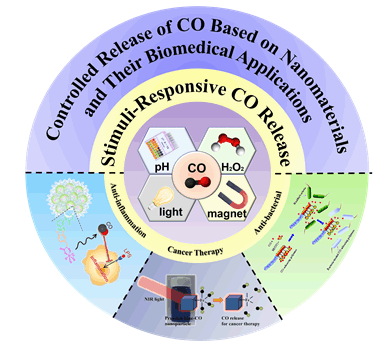
Controlled Release of Carbon Monoxide Based on Nanomaterials and Their Biomedical Applications
Zhang Xiaolei, Tian Gan, Zhang Xia, Wang Qing, Gu Zhanjun
Acta Chim. Sinica
2019, 77 (5):
406-417.
DOI: 10.6023/A18120504
Published: 14 February 2019 In recent years, the use of gas therapy has been more and more concerned by researchers in biomedical applications. Carbon monoxide (CO) is a diatomic gas messenger molecule with the function of transmitting intercellular information and regulating cellular signals. CO is found to play an extremely important physiological role in multiple systems, including cardiovascular system, nervous system, immune system, endocrine system and respiratory system, cancer therapy, coagulation and fibrinolysis system, organ transplantation and preservation, and so on. The biological functions of carbon monoxide molecule greatly depend on the its concentration, position, and duration. However, the existing carbon monoxide donors including Mn2(CO)10, Ru2Cl4(CO)6, Ru(CO)3Cl(glycinato), CORM-F, CORM-A1 have some disadvantages, such as poor stability, difficulties in dose control, lack of targeting, potential toxic and side effects on normal cells and tissues, which limited their further applications. How to control the concentration of carbon monoxide in the specific region has always been a big challenge in the field of biomedical applications. With the rapid development of nanoscience and technology, researchers have constructed a series of multifunctional carbon monoxide releasing nanomaterials, provided a new idea for CO controlled release, and applied them in the field of biomedicine. In this paper, several kinds of endogenous/exogenous stimulus-responsive CO releasing nanomaterials with the unique advantages are introduced based on the stimuli source. Then, the applications of these controlled CO releasing nanomaterials in biomedical fields, such as inhibiting inflammation, anti-bacte- rial and cancer therapy, are summarized. Finally, the challenges and prospects of CO releasing nanomaterials are discussed.
Article
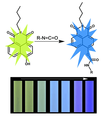
A Ratiometric Fluorescence Probe for Detecting Gaseous Isocyanates Directly
Chen Kai, Han Baichuan, Ji Sixin, Sun Jin, Gao Zhenzhong, Hou Xianfeng
Acta Chimica Sinica
2019, 77 (4):
365-370.
DOI: 10.6023/A18120484
Published: 31 January 2019 Isocyanates is a widely-used chemical in many manufacturing industries, such as polymer industry, pharmaceutical production and production of a variety of agricultural chemicals. However, it is harmful to human health due to the volatility. Therefore, it is necessary to develop methods to detect isocyanates quickly and conveniently, especially to gaseous isocyanates. In this work, a novel fluorescent probe, N-buty-4-hydroxy-1,8naphthalimide, was developed for detection of isocyanates. This fluorescence probe can be synthesized by a simple three-steps synthetic route, and the overall yield of the whole synthesizing process reached 75%. In the absence of isocyanate, the probe solution displays an emission centering at 596 nm when excited at 370 nm, which is yellow to the naked eye. Once isocyanate is added, the fluorescence of solution changes from yellow to blue, and the process finishes in 4 min. The detecting limit of this probe to isocyanates is calculated to be 112 nmol·L-1. It is also proved that this probe possesses excellent selectivity for isocyanate and distinct anti-interference to common organic volatilized compounds. In addition, the reaction mechanism between the probe and isocyanate were proved by HPLC, NMR and ESI-MASS. Results show that the hydroxy group on the 4th position of naphthalene ring of probe reacts with isocyanate group (-NCO) of isocyanate, and resulting in carbamates, which alter 4th substituent group of probe molecule and lead to change of fluorescence. In order to detect the gaseous isocyanates directly, test paper are developed based on N-buty-4-hydroxy-1,8naphthalimide. When the test paper exposed to isocyanates vapor, the yellow fluorescence fade away gradually and a blue fluorescence appear in 6 min. And the test paper possesses excellent selectivity for gaseous isocyanate and distinct anti-interference to common VOCs. In conclusion, this strategy is an efficient way to detect gaseous isocyanates, and it may provide a referable approach for directly monitoring the volatile organic compounds in air.
Review
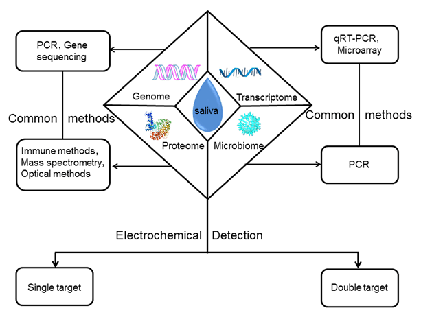
Progress in Analysis and Detection of Salivary Tumor Biomarkers Associated with Oral Cancer
Jin Xin, Wang XiaoYing
Acta Chim. Sinica
2019, 77 (4):
340-350.
DOI: 10.6023/A18100414
Published: 05 December 2018 Oral cancer is head and neck cancer, and cancer tissue is located in the oral cavity. The non-invasive early diagnosis is an effective method to reduce the death of the disease. The oral cancer-related substances are first released into the saliva, which is convenient, safe and non-invasive, and is the first choice for screening and early diagnosis of oral cancer. In this paper, the specific types and the commonly used detection methods of the salivary tumor biomarkers at home and abroad were summarized and compared. Specifically, the latest application of new electrochemical biosensor in the detection of the salivary tumor biomarkers associated with oral cancer was mainly described. Futhermore, the summary of its future directions and the potential applications was proposed, which provided reference for the further research and application of the salivary tumor biomarkers in oral cancer.
Communication
Fluorescent Aptamer-functionalized Graphene Oxide Biosensor for Rapid Detection of Chloramphenicol
Lu Jinghe, Tan Shuzhen, Zhu Yuqing, Li Wei, Chen Tianxiao, Wang Yao, Liu Chen
Acta Chim. Sinica
2019, 77 (3):
253-256.
DOI: 10.6023/A18100433
Published: 17 January 2019 A label free and rapid fluorescent method for quantitative detection of chloramphenicol (CAP) based on graphene oxide (GO) fluorescence functional G-quadruplex probe (FGP) was developed. The FGP consisted of a choramphenicol aptamer and a G-rich sequence. The aptamer was used to bind CAP and the G-quadruplex formed by G-rich sequence was employed as a signal reporter after binding to Thioflavin T (ThT). In the absence of CAP, the FGP was absorbed onto the surface of GO through π-π stacking interactions, which restrained the G-rich sequence to form a G-quadruplex structure. Thus, the fluorescent intensity of background was low. In the addition of the CAP, the aptamer part of FGP could recognize and bind CAP to form a target-FGP complex, which led to the desorption of the complex from GO. Therefore, the free G-rich sequence could form G-quadruplex structure and bind to ThT, resulting a increase in the fluorescence intensity of the solution. We observed that the fluorescence increasement of the sensing platform had a linear relationship with the concentrations of CAP in the range of 2~20 nmol/L, and the limit of detection was 1.45 nmol/L. Besides, this detection system was applied for detecting CAP in the spiked milk, the recovery rate was between 93.2%~103.3%. These results indicated that this developed method can be used to efficiently recognize CAP in real samples.
Article
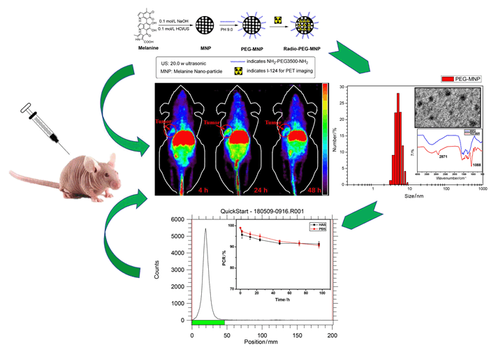
Preparation and Preliminary Molecular Imaging Study of 124I in-situ Labeled Organic Melanin Nanoparticles
Xia Lei, Cheng Zhen, Zhu Hua, Yang Zhi
Acta Chimica Sinica
2019, 77 (2):
172-178.
DOI: 10.6023/A18090410
Published: 01 November 2018 Developing biocompatible, multifunctional and in-situ labeling nanoplatform is high challenging for molecular imaging. Organic derivates melanin nanoparticles (MNPs) holds great potential to be multimodal contrast agents, and have been used for photoacoustic imaging, magnetic resonance imaging, and 64Cu PET imaging with simple modifications. In order to extend MNPs application in molecular imaging, here a novel radio-nuclide was applied to in-situ labeling of MNPs. Large numbers of active dihydroxyindole/indolequinone groups and natural binding ability of MNPs enabled them to have the ability to label different types of radionuclides which have unique half-life and functions, especially long-life elemental nuclide. This project explored the in-situ labeling methods of organic melanin nanoparticles with a promising diagnostic radionuclides named Iodine-124 (124I), and using this novel multifunctional organic nanoparticles for preliminary molecular imaging studies. Generally, ultrafine particle size melanin nanoparticles (5.5 nm in diameter) were prepared by ultrasonication method using naturally occurring melanin, then PEG3500 which had amino group at both ends was used as a stabilizer agent to obtain PEG-MNP nanocarriers (7.5 nm in diameter) with better water solubility and stability. The nanoparticles were full characterized by dynamic light scattering (DLS), transmission electron microscope (TEM) and 1H NMR, respectively. Then, one kind of elemental nuclide was labeled. Classic iodine labeled method with N-Bromo Succinimide (NBS) was used as oxidant to oxidize active dihydroxyindole/indolequinone ring of PEG-MNP for electrophilic substitution reaction labeling 124I (100.8 h). This reaction rate is extremely fast (60 s reaction time) and high labelling yield (>99%). The 124I was labeled successfully and in-situ labeled PEG-MNP nanocarriers were obtained. After that, 124I and 124I-PEG-MNP were used to further preclinical evaluation by micro-PET imaging. Micro-PET images were collected at 2 h, 24 h and 48 h after intravenous injection 7.4 MBq 124I and 124I-PEG-MNP in normal Kunming mice (n=3). The ROI target area of heart, liver and thyroid were delineated for semi-quantitative analysis. Then, in order to verify the imaging ability of 124I-PEG-MNP in solid tumor. We built human pancreatic cancer BxPC3 xenograft model (n=3), and Micro-PET scans were performed at different time points. Results showed that the labeling rate of 124I on PEG-MNP was 99.9%. And the radiochemical purity in vitro stability of 124I-PEG-MNP in 96 h was more than 90%. Micro-PET images showed that 124I-PEG-MNP had no obvious thyroid uptake which indicated no de-marking in mice. The radio-distribution of 124I and 124I-PEG-MNP was substantially different in liver and thyroid (P<0.001). In vivo semi-quantitative analysis showed that the radio uptakes of organs were consistent with the distribution of nanoparticles. And the PET imaging of xenograft mice showed that 124I-PEG-MNP can utilize the enhanced permeability and retention effect (EPR) to be significantly enriched at the tumor and retained in the tumor site for more than 48 h. PEG-MNP has the ability to label long half-life nuclide 124I. This research provides an experimental basis for further construction of long-circulation multimodal imaging probes.
Article
Self-Assembly of Cyclic Dipeptides and Their Fluorescent Properties
Yang Jingge, Li Yang, Wang Xiaoai, Wang Dong, Sun Yawei, Wang Jiqian, Xu Hai
Acta Chimica Sinica
2019, 77 (12):
1279-1286.
DOI: 10.6023/A19090331
Published: 21 October 2019
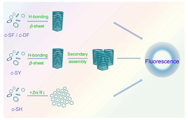
Cyclic dipeptide (CDP) is a kind of the smallest cyclic peptide with two amino acids cyclization through amide bonds. The two amide bonds with four hydrogen bonding sites give CDPs a high self-assembly propensity, mainly driven by the hydrogen bonding interactions. In this paper, we have designed four CDPs, c-SF, c-SY, c-SH and c-DF, and studied their self-assembly performance in aqueous solution with circular dichroism spectroscopy (CD) and atomic force microscopy (AFM), including the effects of pH and zinc ion coordination on self-assembly. The fluorescence properties of CDP self-assemblies have also been studied. CD results showed that c-SF, c-SY and c-DF adopted a β-sheet conformation, while c-SH was random coil secondary structure at the concentration of 2.0 mmol/L and pH 5.0. AFM results showed that c-SF, c-SY and c-DF could form nanofibers with different diameters ranged from 1.0 to 3.0 nm. In addition, c-SY self-assembled hierarchically over time. Not only the nanofiber diameter gradually increased, but also the nanofibers entangled into 3D networks. Although c-SH did not self-assemble at the concentration of 3.0 mmol/L and pH 7.0, it could form monolayers with the induction of zinc ion at pH 9.0. The self-assemblies of each CDP had different multiple fluorescent emission peaks with excitation of different wavelengths. Especially, c-SF emitted green fluorescent light under UV light of 365 nm. The fluorescent emission intensity of CDPs was much stronger than their corresponding linear dipeptides. It was assumed that the diketopiperazine structure contributed to the fluorescence enhancement. Moreover, the fluorescent emission intensity of CDP self-assemblies was much higher than that of their free molecules, which meant that the ordered aggregation made a significant contribution to the fluorescent properties. Both the coordination of zinc ions with the imidazole groups on histidine and the oxidation of phenolic hydroxyl groups in tyrosine could enhance the fluorescent emission intensity of CDPs. It was assumed that CDP molecules stacked one by one to form nanofibers during self-assembly. The diketopiperazine ring of CDPs and its self-assembly endowed CDPs with special fluorescent properties.
Article
Preparation of AIE Polymer Dots (Pdots) Based on Poly(N-vinyl-2-pyrrolidone)-Eu(III) Complex and Dual-color Live Cell Imaging
Guan Xiaolin, Li Zhifei, Wang Lin, Liu Meina, Wang Kailong, Yang Xueqin, Li Yali, Hu Lili, Zhao Xiaolong, Lai Shoujun, Lei Ziqiang
Acta Chimica Sinica
2019, 77 (12):
1268-1278.
DOI: 10.6023/A19090349
In recent years, polymer dots (Pdots) have been developed as an excellent organic fluorescent nanoparticles due to its excellent optical properties, diverse structures, easy surface modification and good biocompatibility. So, they have important application potential in biological imaging, sensing and detection, drug delivery and therapeutic diagnosis. However, the fluorescence quenching of semiconducting Pdots with large conjugated structure due to aggregation-caused quenching (ACQ) effect limits its applications for bioimaging in aggregated states. The ACQ phenomenon of Pdots could been eliminated by introducing aggregation-induced emission (AIE)-active molecules in Pdots. In this paper, a kind of responsive AIE-active Pdots, which were composed of tetraphenylethylene (TPE) with blue fluorescent light emission and poly(N-vinyl-2-pyrrolidone)-Eu(III) complex (PVP-Eu(III)) with red fluorescent light emission, were constructed. Firstly, a TPE derivative initiator (TPE-tetraAZO) containing four arms was synthesized by using 4,4'-azobis-(4-cyanovaleric acid) to modify TPE, and a multi-stimuli-responsive amphiphilic polymer of tetraphenylethene-graft-poly(N-vinyl-2-pyrrolidone) (TPE-tetraPVP) was then successfully synthesized by using TPE-tetraAZO as initiator. Finally, the complex TPE-tetraPVP-Eu(III) with AIE characteristic and dual fluorescence was obtained through the coordination between TPE-tetraPVP and rare earth element Eu(III). The amphiphilic 4-arm star polymer TPE-tetraPVP-Eu(III) formed Pdots consisted of hydrophobic AIEgens TPE core and hydrophilic PVP shell by a self-assembling process. The morphology and particle size of Pdots were investigated by transmission electron microscope (TEM). Results showed that Pdots was a relatively uniform diameter around 20 nm and exhibited regular sphere morphology. The results of fluorescence experiments showed that TPE-tetraPVP-Eu(III) Pdots had two emission bands centered at about 435 (blue) and 615 nm (red) with a wavelength difference of 180 nm, which were obtained under optimum excitation at 360 and 395 nm, respectively. Among them, the blue emission showed typical AIE property. Moreover, the lower critical solution temperature (LCST) of TPE-tetraPVP-Eu(III) in aqueous solution was about 37℃, which was close to normal body temperature. Meanwhile, at different temperatures from 10 to 60℃, photoluminescence (PL) intensities of TPE-tetraPVP-Eu(III) Pdots firstly decreased with increasing temperature from 10 to 36℃, and then increased with increasing temperature from 37 to 60℃. It was interesting that the fluorescent response of Pdots could be caused by the phase transfer of PVP. Besides, the PL intensity of Pdots in aqueous solution changed at different pH. Therefore, TPE-tetraPVP-Eu(III) Pdots might be used as multi-functional and smart fluorescent sensors. Furthermore, the results of cellular imaging indicated the efficient cellular uptake and low cytotoxicity of Pdots in HeLa, HepG2 and A549 cells. And, the photoswitchable dual-emission could be easily realized in three cells by simply tuning the excitation wavelength. Thus, the non-conjugated Pdots is an ideal dual-color live cell imaging probe.
Review
Research Progress on Rare Earth Nanocrystals for In Vivo Imaging and Sensing in Near Infrared Region
Xiong Lin, Fan Yong, Zhang Fan
Acta Chimica Sinica
2019, 77 (12):
1239-1249.
DOI: 10.6023/A19080305
Published: 18 September 2019
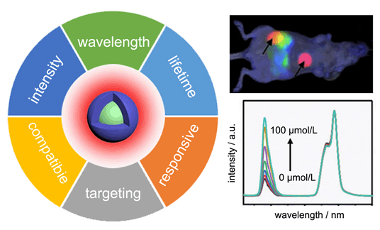
In vivo imaging and sensing play a critical role in modern biological and medical research. Compared with other techniques such as computed tomography (CT), positron emission tomography (PET) and nuclear magnetic resonance (NMR), fluorescence imaging and analysis are featured by fast feedback, high sensitivity, and high spatiotemporal resolution. Especially, the application of near infrared (NIR) light as both excitation and emission signals provides increased tissue penetration and improved imaging quality and sensitivity due to reduced light scattering and auto-fluorescence. Among various materials investigated for in vivo imaging and bio-sensing, lanthanide-based nanocrystals display rich excitation/emission wavelengths in the NIR range, good photo and chemical stability, large Stokes shifts. In recent years, the research on lanthanide-based nanocrystals for in vivo imaging and sensing has seen rapid progress. Through nanoscale material design and synthesis, it is possible to fine tune the optical properties of lanthanide-based nanocrystals. By properly choosing different lanthanide ions as activators and sensitizers, multiple excitation/emission wavelengths can be obtained. The careful design of core-shell structure of nanocrystals enables improved fluorescence efficiency and tailorable fluorescence life time through controlled energy transfer. On the other side, the surface of lanthanide-based nanocrystals can be modified through coating, absorption or ligand exchange to enhance the biocompatibility, targeting capability, and bio-responsiveness. Taking advantage of this high flexibility and versatility, there are great opportunities for these lanthanide-based nanocrystals in various in vivo imaging and sensing applications. This review first outlines the general technique requirements for in vivo imaging and sensing. Then, the composition, synthesis and basic properties of lanthanide-based nanocrystals are briefly introduced. Subsequently, the routes for tailoring the optical and biochemical properties of lanthanide-based nanocrystals are discussed in detail, with an emphasis on the material designs and surface modifications for in vivo imaging and analysis. It is expected that this work will inspire new ideas for accelerating the clinic translation of rare earth nanocrystals-based imaging and sensing techniques.
Review
Construction and Application of DNA-two-dimensional Layered Nanomaterials Sensing Platform
Chi Jingyuan, Li Jing, Ren Shaokang, Su Shao, Wang Lianhui
Acta Chimica Sinica
2019, 77 (12):
1230-1238.
DOI: 10.6023/A19070262
Published: 18 September 2019
Combining the specific recognition ability of DNA molecules with the superior physical and chemical properties of two-dimensional (2D) layered materials, a DNA-2D layered nanomaterial sensing platform was constructed. More and more researchers are devoted to develop this sensing platform, which has become one of the important research directions in the field of chemical/biological sensors. In view of the rapid development of the 2D layered materials, this paper firstly introduces the construction principle of the DNA-2D layered material sensing platform. Then we mainly review the application of the sensing platform in the analysis of chemical and biological molecules, including metal ions, mycotoxins, ATP, amino acid, antibiotics, nucleic acids, proteins and cancer cells. Finally, the future of this sensing platform is prospected.
Article
Preparation of Multi-stimulus Responsive Polymer Nanospheres Based on AIE Effect and Its Cell Tracing Application
Guan, Xiaolin, Wang, Lin, Li, Zhifei, Liu, Meina, Wang, Kailong, Lin, Bin, Yang, Xueqing, Lai, Shoujun, Lei, Ziqiang
Acta Chimica Sinica
2019, 77 (10):
1036-1044.
DOI: 10.6023/A19060226
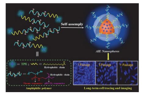
Published: 13 August 2019 In recent years, fluorescent bioimaging technology has great advantages in the fields of life science research and medical diagnosis because of its advantages of fast and effective, high sensitivity, easy realization of multi-channel imaging and economic efficiency. Organic fluorescent dyes have been widely used as biological imaging reagents due to their excellent photoelectric properties, functional modification, adjustable optical properties, and good biocompatibility. However, conventional organic fluorescent molecules cause aggregation-caused quenching (ACQ) due to π-π stacking in the aggregated state, limiting their bioimaging applications in aggregated or high concentrations. Since the discovery of the unique luminescence phenomenon of aggregation-induced emission (AIE), the ACQ phenomenon of traditional fluorescent materials has been eliminated. Stimulating responsive polymer nanoparticles have been widely used in the life sciences due to their combination of nanoparticle and polymer advantages and their ability to respond intelligently with environmental changes. Therefore, nanomaterials with excellent aggregation-induced emission (AIE) property, environmental stimuli responsiveness and biocompatibility based on AIE molecules and smart responsive polymers have shown attractive application prospects in the life sciences. A kind of multi-responsive AIE-active polymer nanospheres, which were composed of tetraphenylethylene (TPE) and stimuli-responsive poly[N]-2-(diethylamino)-ethyl]acrylamide (PDEAEAM), were constructed in this study. Firstly, a multi-stimulation responsive monomer N-[2-(diethylamino)ethyl]acrylamide (DEAEAM) and TPE derivative tetraphenylethene-4-(12-hydroxydodecyl-2-methylpropionyl) (TPE-BIB) with propionyl bromide were synthesized, respectively, and a multi-stimuli-responsive amphiphilic polymer of tetraphenylethene-graft-poly[N-[2-(diethylamino)ethyl]acrylamide] (TPE-g-PDEAEAM) was then successfully synthesized by atom transfer radical polymerization (ATRP) using TPE-BIB as initiator. Lastly, polymer nanospheres TPE-g-PDEAEAM of approximately 200 nm were formed by a self-assembling pro-cess. The results of the performed experiments showed that the LCST of TPE-g-PDEAEAM in aqueous solution is about 60 ℃. Meanwhile, the luminescence change of TPE-g-PDEAEAM at different temperatures from 20 to 66 ℃ was observed. The fluorescence intensity of TPE-g-PDEAEAM firstly decreased with increasing temperature from 20 to 58 ℃, and the fluorescence intensity increased with increasing temperature from 58 to 66 ℃. The phase transfer of PDEAEAM in TPE-g-PDEAEAM may be the reason of luminescence change which may lead to the fluorescent temperature response. Moreover, the fluorescence intensity of TPE-g-PDEAEAM nanospheres in aqueous solution increased with increasing temperature pH. Besides, the fluorescence intensity of TPE-g-PDEAEAM decreased dramatically when the volume of CO2 increased from 0.0 to 1.2 mL. Therefore, TPE-g-PDEAEAM was a new temperature and pH/CO2 responsive materials and might be used as multi-functional smart fluorescent sensors. More importantly, the fluorescent signals were significantly strong in HeLa cells after cells were incubated with TPE-g-PDEAEAM for 24 h based on the characteristic of AIE fluorescence and low cytotoxicity. The resultant nanospheres were able to be internalized by the cancer cells and effectively track the HeLa cells for as long as 11 passages. So, the polymer nanomaterial is an ideal living cell fluorescent tracer probe, which is expected to be applied as biosensors, long-term cell traces and medical biomaterials.
Article
Trace Detection of Rhodamine B in Infant Candy by g-C3N4/Ag Nanocomposite as Surface-Enhanced Raman Scattering Substrate
Ma, Chao, Wu, Jiawei, Zhu, Lin, Han, Xiaoxia, Ruan, Weidong, Song, Wei, Wang, Xu, Zhao, Bing
Acta Chimica Sinica
2019, 77 (10):
1024-1030.
DOI: 10.6023/A19050191
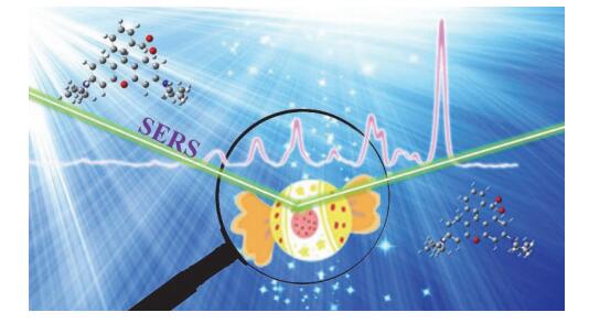
Published: 22 July 2019 In recent years, food safety problems caused by illegal additions in infant foods have received widespread attention. Surface-enhanced Raman scattering (SERS) technique is used to rapidly and non-destructively detect the banned RhB that is usually added in food. In this study, we have prepared g-C3N4/Ag composites via a simple method successfully, their morphology and structure were characterized by transmission electron microscope (TEM), ultraviolet-visible (UV-Vis), X-ray diffraction (XRD), fluorescence spectrophotometer and confocal micro-Raman spectrometer (Raman). The g-C3N4 nanosheet possesses good adsorption performance due to its highly delocalized π-conjugated system, which acts as a carrier for Ag nanoparticles. Therefore, Ag nanoparticles are more uniformly and stably distributed on the surface of g-C3N4 nanosheets to form g-C3N4/Ag nanocomposite, which can be used for rapid adsorption and trace detection of RhB. In the experiment, the pH of the test and the absorbed time between the substrate and RhB were optimized. The influence of pH on the SPR of the substrate and the SERS intensity of the probe molecule were investigated in detail. As g-C3N4/Ag nanocomposite shows a significant higher absorption in the visible region around 500 nm than Ag nanoparticles, g-C3N4/Ag nanocomposite is more favorable for SPR absorption. A wide SPR absorption range is achieved due to the synergy between g-C3N4 and Ag nanoparticles, providing an improved SERS enhancement performance. Under the optimal experimental conditions by using RhB as probe molecule, an enhancement factor of 7.6×10 5 is achieved. Due to the electrostatic interaction and π-π interaction between the substrate and the probe molecules, the substrate can enrich in a large amount of cationic dyes, offering a detection of RhB. The g-C3N4/Ag SERS substrate can be used to detect RhB with a linear relationship from 1.0×10 –9 to 1.0×10 –6 mol/L and a detection limit as low as 0.39 nmol/L. In addition, the g-C3N4/Ag nanocomposite SERS substrate can also detect trace amounts of RhB molecules in the commercially available rainbow lollipops with a high sensitivity, and the recovery were 93.6%~95.04%. In summary, the g-C3N4/Ag nanocomposite is not only a SERS substrate with high sensitivity, uniformity and stability, but also can be used as a rapid trace detection method of Rhodamine B in real food and environment. |
Special Issue
|
Molecular Probes and Nanobiology and Bioanalytical Chemistry

|
| Default Latest Most Read |
|
Please wait a minute...
|
| ImageJ software and then reconstruct STORM images with a Falcon algorithm to show marked imaging resolution enhancement, compared with wide-field images, which provide a new protocol for biomedical imaging.
|


