有机化学 ›› 2025, Vol. 45 ›› Issue (11): 4082-4107.DOI: 10.6023/cjoc202503034 上一篇 下一篇
综述与进展
陈冰燕a, 孙洁a, 熊玲红b, 何学文a,*( )
)
收稿日期:2025-03-31
修回日期:2025-05-23
发布日期:2025-06-30
基金资助:
Bingyan Chena, Jie Suna, Linghong Xiongb, Xuewen Hea,*()
Received:2025-03-31
Revised:2025-05-23
Published:2025-06-30
Contact:
*E-mail: xheao@suda.edu.cn
Supported by:文章分享

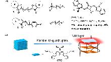
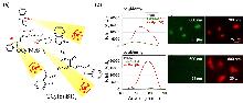
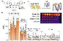
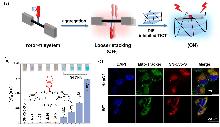
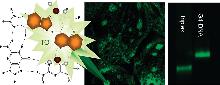
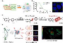
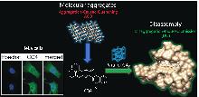
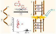
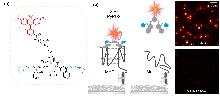
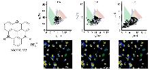
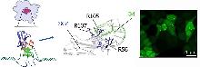
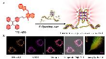
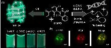
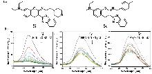
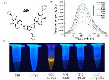
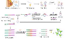
相比于经典的单链和双链型经典核酸结构, 非典型核酸结构(如G4s、i-motif、Triplex及环形核酸)因其被发现的重要生物功能和在生理环境下的动态平衡异常与多种重大疾病的密切关联性而逐渐成为生物医学研究的热点. 传统凝胶电泳、核磁共振、圆二色性检测技术存在空间分辨率低、破坏性大、缺乏实时动态监测能力等不足. 近年来, 荧光探针材料因其高灵敏度、快速响应性以及动态实时观测性能等逐渐成为非经典核酸结构检测的前沿工具. 综述了非经典核酸结构的荧光点亮材料, 包括传统荧光小分子、聚集诱导发光原(AIEgens)等, 并详述了设计原理、检测机制及应用场景. 当前探针技术通过优化分子构效关系提升识别性与信噪比, 但仍面临选择性不足、活体穿透性差等挑战. 未来需融合多模态成像、人工智能辅助设计及靶向递送系统, 构建高灵敏、多通道响应的检测平台, 以解析核酸动态构象与疾病关联性, 推动开发精准诊断与新型治疗策略.
陈冰燕, 孙洁, 熊玲红, 何学文. 非典型核酸结构的荧光点亮成像检测[J]. 有机化学, 2025, 45(11): 4082-4107.
Bingyan Chen, Jie Sun, Linghong Xiong, Xuewen He. Fluorescence Light-Up Detection and Imaging of Atypical Nucleic Acid Structures[J]. Chinese Journal of Organic Chemistry, 2025, 45(11): 4082-4107.


| Probe | Types of nucleic acid | Key features | Ref. |
|---|---|---|---|
| CSTS | G4s | Disaggregation-Induced Emission (DIE), V-shaped rigid plane π bracket combined with the parallel G4s through the | [ |
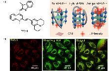
| QCy(BnBT)3 | G4s | High specificity recognition of G4s and 500 times fluorescence enhancement | [ |
| SCY-5 | G4s | J-Aggregates dissociate into monomers, high specificity recognition of parallel G4s | [ |
| SN-Cy5-S | G4s | Disaggregation-Induced Emission (DIE), Y-shaped plane is combined with the top plane at the 5' end of G4s | [ |
| TO | G4s and Triplex | Distinguish between Triplex and G4s, strong fluorescence enhancement | [ |
| TOVJ | G4s | High specificity recognition of antiparallel G4s, near-infrared emission | [ |
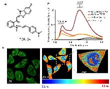
| TOR-G4 | G4s | Two-photon excitation, fluorescence lifetime has increased significantly | [ |
| ThT | G4s | High cell membrane permeability, specific recognition ability for G4s | [ |
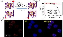
| IMT | G4s | Stack on the 3'-G4 end plane, low background and high signal-to-noise ratio emissions | [ |
| ThT-NE | G4s | Intramolecular rotation is restricted and fluorescence is activated, high selectivity and sensitivity | [ |
| ThT-NA | G4s | Red light emission, large Stokes shift, high fluorescence turn-on ratio and high selectivity for G4s | [ |
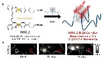
| NIRG-2 | G4s | Near-infrared Region II emission, hydrogen bonds and π-π stacking combine with G4s | [ |
| CQ4 | G4s | Disaggregation-Induced Emission (DIE), specifically bind parallel G4s | [ |
| P0 | G4s | The dimer G4/P0 system carried out highly selective detection of K+ | [ |
| SiR-PyPDS | G4s | The G4s ligand Pyridostatin (PyPDS) forms hydrogen bonds and hydrophobic interactions to specifically bind to G4s | [ |
| DAOTA-M2 | G4s | G4s were identified by fluorescence lifetime imaging microscopy (FLIM) technology | [ |
| SG4 | G4s | Antibodies against the structure of human c-MYC G4, bind to green fluorescent protein (GFP) | [ |
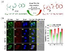
| CV2 | G4s | Peptide sequences specifically recognizing G4s (L-ARG-L-Gli-glutaric acid), Imaging of mitochondrial G4s | [ |
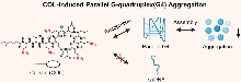
| COL | G4s | Induce parallel G4s to aggregate from the nucleic acid mixture | [ |
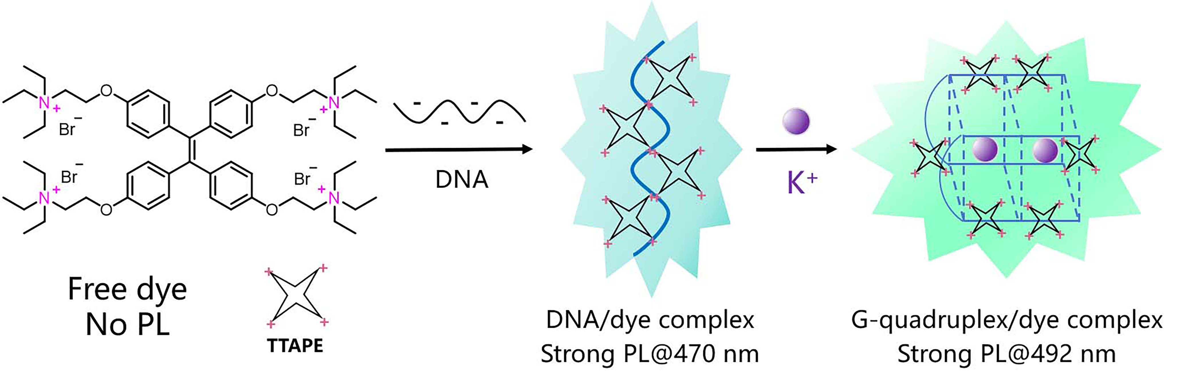
| TTAPE | G4s | Aggregation-induced emission detection (AIE), real-time monitoring of the folding process of G-rich DNA strands to form G4s | [ |
| PZ-1 | G4s | Aggregation-induced emission detection (AIE), G4s is combined through electrostatic interaction and π-π stacking | [ |
| TPE-mTO | G4s | Electron-deficient cation conjugated systems and crescent-shaped scaffolds, specifically locate G4s in mitochondria | [ |
| TPA-mTO | G4s | Near-infrared emission, ideal photostability, and high fluorescence contras | [ |
| HMPQ | G4s | AIEgens of biological origin, the π-π interaction binds to G4s, high selectivity and high sensitivity | [ |
| 5i and 5c | i-motif | Combining different types of I-motifs, the fluorescence intensity is enhanced and the fluorescence lifetime is increased | [ |
| G59 | i-motif | Specifically recognize and visually detect the i-motif structure of the c-MYC gene promoter | [ |
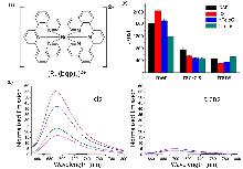
| [Ru(bqp)2]2+ | i-motif | The cis isomer combines with the DAP i-motif, resulting in fluorescence activation and increased fluorescence lifetime | [ |
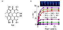
| Hyp | i-motif | The i-motif structures of different lengths were distinguished by fluorescence intensity | [ |
| NIAD-4 | Triplex | Uncharged near-infrared molecular rotor probe, topological match with the Triplex terminal triad | [ |
| FIS | Triplex | The quantity ratio of 2∶1 is combined with Triplex, the ESIPT process is triggered and the green fluorescence lights up | [ |
| Berberine | Triplex | Isoquinoline alkaloids, combined with the Triplex structure, it will show strong fluorescence at 530 nm | [ |
| AgNCs | Triplex | The dynamic changes of photophysical properties with the Triplex structure | [ |
| DFHBI | circRNA | Using circMTO1 as the template, the aptamers generated by RPA and transcriptional amplification techniques were combined with DFHBI | [ |
| LC | circDNA | Efficient intracellular uptake capacity, high-resolution visual imaging detection of mitochondrial DNA (mtDNA) | [ |
| YON | circDNA | Near-infrared twisted intramolecular charge transfer (TICT) fluorescent probe, high sensitivity and large Stokes displacement | [ |
| Probe | Types of nucleic acid | Key features | Ref. |
|---|---|---|---|
| CSTS | G4s | Disaggregation-Induced Emission (DIE), V-shaped rigid plane π bracket combined with the parallel G4s through the | [ |
| QCy(BnBT)3 | G4s | High specificity recognition of G4s and 500 times fluorescence enhancement | [ |
| SCY-5 | G4s | J-Aggregates dissociate into monomers, high specificity recognition of parallel G4s | [ |
| SN-Cy5-S | G4s | Disaggregation-Induced Emission (DIE), Y-shaped plane is combined with the top plane at the 5' end of G4s | [ |
| TO | G4s and Triplex | Distinguish between Triplex and G4s, strong fluorescence enhancement | [ |
| TOVJ | G4s | High specificity recognition of antiparallel G4s, near-infrared emission | [ |
| TOR-G4 | G4s | Two-photon excitation, fluorescence lifetime has increased significantly | [ |
| ThT | G4s | High cell membrane permeability, specific recognition ability for G4s | [ |
| IMT | G4s | Stack on the 3'-G4 end plane, low background and high signal-to-noise ratio emissions | [ |
| ThT-NE | G4s | Intramolecular rotation is restricted and fluorescence is activated, high selectivity and sensitivity | [ |
| ThT-NA | G4s | Red light emission, large Stokes shift, high fluorescence turn-on ratio and high selectivity for G4s | [ |
| NIRG-2 | G4s | Near-infrared Region II emission, hydrogen bonds and π-π stacking combine with G4s | [ |
| CQ4 | G4s | Disaggregation-Induced Emission (DIE), specifically bind parallel G4s | [ |
| P0 | G4s | The dimer G4/P0 system carried out highly selective detection of K+ | [ |
| SiR-PyPDS | G4s | The G4s ligand Pyridostatin (PyPDS) forms hydrogen bonds and hydrophobic interactions to specifically bind to G4s | [ |
| DAOTA-M2 | G4s | G4s were identified by fluorescence lifetime imaging microscopy (FLIM) technology | [ |
| SG4 | G4s | Antibodies against the structure of human c-MYC G4, bind to green fluorescent protein (GFP) | [ |
| CV2 | G4s | Peptide sequences specifically recognizing G4s (L-ARG-L-Gli-glutaric acid), Imaging of mitochondrial G4s | [ |
| COL | G4s | Induce parallel G4s to aggregate from the nucleic acid mixture | [ |
| TTAPE | G4s | Aggregation-induced emission detection (AIE), real-time monitoring of the folding process of G-rich DNA strands to form G4s | [ |
| PZ-1 | G4s | Aggregation-induced emission detection (AIE), G4s is combined through electrostatic interaction and π-π stacking | [ |
| TPE-mTO | G4s | Electron-deficient cation conjugated systems and crescent-shaped scaffolds, specifically locate G4s in mitochondria | [ |
| TPA-mTO | G4s | Near-infrared emission, ideal photostability, and high fluorescence contras | [ |
| HMPQ | G4s | AIEgens of biological origin, the π-π interaction binds to G4s, high selectivity and high sensitivity | [ |
| 5i and 5c | i-motif | Combining different types of I-motifs, the fluorescence intensity is enhanced and the fluorescence lifetime is increased | [ |
| G59 | i-motif | Specifically recognize and visually detect the i-motif structure of the c-MYC gene promoter | [ |
| [Ru(bqp)2]2+ | i-motif | The cis isomer combines with the DAP i-motif, resulting in fluorescence activation and increased fluorescence lifetime | [ |
| Hyp | i-motif | The i-motif structures of different lengths were distinguished by fluorescence intensity | [ |
| NIAD-4 | Triplex | Uncharged near-infrared molecular rotor probe, topological match with the Triplex terminal triad | [ |
| FIS | Triplex | The quantity ratio of 2∶1 is combined with Triplex, the ESIPT process is triggered and the green fluorescence lights up | [ |
| Berberine | Triplex | Isoquinoline alkaloids, combined with the Triplex structure, it will show strong fluorescence at 530 nm | [ |
| AgNCs | Triplex | The dynamic changes of photophysical properties with the Triplex structure | [ |
| DFHBI | circRNA | Using circMTO1 as the template, the aptamers generated by RPA and transcriptional amplification techniques were combined with DFHBI | [ |
| LC | circDNA | Efficient intracellular uptake capacity, high-resolution visual imaging detection of mitochondrial DNA (mtDNA) | [ |
| YON | circDNA | Near-infrared twisted intramolecular charge transfer (TICT) fluorescent probe, high sensitivity and large Stokes displacement | [ |
| [1] |
doi: 10.1038/nsmb1171 pmid: 17146462 |
| [2] |
doi: 10.1093/nar/gkab187 |
| [3] |
doi: 10.1021/acs.jmedchem.4c02518 |
| [4] |
doi: 10.1073/pnas.1810409116 |
| [5] |
doi: 10.1080/10409238.2021.1926417 |
| [6] |
doi: 10.3390/biomedicines10081871 |
| [7] |
|
|
(刘芷玥, 景海涛, 朱婷, 付文强, 张钠, 胡文萱, 生物学杂志, 2025, 42, 7.)
|
|
| [8] |
doi: 10.1021/ja2061984 |
| [9] |
doi: 10.1016/j.drup.2022.100907 |
| [10] |
doi: 10.1093/nar/gkab580 pmid: 34244785 |
| [11] |
doi: 10.1093/nar/gkaa615 |
| [12] |
doi: 10.1007/s00018-021-03921-8 |
| [13] |
doi: 10.1186/s12929-024-01041-6 pmid: 38741159 |
| [14] |
doi: 10.1021/jacs.4c02091 pmid: 38832857 |
| [15] |
doi: 10.1021/acssensors.2c00992 |
| [16] |
doi: 10.1021/jacs.3c11219 pmid: 38191993 |
| [17] |
doi: 10.1002/anie.202100280 pmid: 33539622 |
| [18] |
doi: 10.3390/molecules27051541 |
| [19] |
doi: 10.1021/acschembio.9b00475 pmid: 31532996 |
| [20] |
doi: 10.1016/j.cclet.2022.02.048 |
| [21] |
doi: 10.1016/j.saa.2023.122615 |
| [22] |
doi: 10.6023/A23040121 |
|
(李兰英, 陶晴, 闻艳丽, 王乐乐, 郭瑞妍, 刘刚, 左小磊, 化学学报, 2023, 81, 681.)
doi: 10.6023/A23040121 |
|
| [23] |
doi: 10.1002/anie.v59.25 |
| [24] |
doi: 10.1021/acsami.0c12482 |
| [25] |
doi: 10.1002/anie.v53.52 |
| [26] |
doi: 10.1021/acsnano.4c14792 |
| [27] |
doi: 10.1021/ac500590d |
| [28] |
doi: 10.1016/j.cej.2023.144022 |
| [29] |
doi: 10.1002/adfm.v34.23 |
| [30] |
doi: 10.1093/nar/gky729 pmid: 30107602 |
| [31] |
doi: 10.1016/j.ijbiomac.2025.139844 |
| [32] |
doi: 10.1093/nar/gkx201 |
| [33] |
doi: 10.1021/jacs.1c05468 pmid: 34554731 |
| [34] |
doi: 10.1002/advs.v12.8 |
| [1] |
doi: 10.1093/nar/gkac482 |
| [35] |
doi: 10.1016/j.ijbiomac.2023.126835 |
| [36] |
doi: 10.1016/j.compbiomed.2024.108683 |
| [37] |
|
|
(周鑫辰, 张卓, 董姝含, 靳茁, 战星彤, 王鹤霖, 杨舒惠, 刘丽梅, 化学通报, 2024, 87, 1098.)
|
|
| [38] |
doi: 10.1007/s00216-022-03995-8 |
| [39] |
doi: 10.3389/fchem.2021.707876 |
| [40] |
doi: 10.1002/chem.v21.39 |
| [41] |
doi: 10.1021/ac501619v |
| [42] |
doi: 10.1021/acs.analchem.3c04318 pmid: 37955574 |
| [43] |
doi: 10.1021/jacs.4c07698 |
| [44] |
doi: 10.1021/acs.analchem.3c01153 pmid: 37290004 |
| [45] |
doi: 10.3390/molecules26092828 |
| [46] |
doi: 10.1021/bi1000849 pmid: 20329708 |
| [47] |
doi: 10.1021/acsami.1c07101 |
| [48] |
doi: 10.1021/jacs.3c11908 pmid: 38151240 |
| [49] |
|
| [50] |
doi: 10.1093/nar/gky665 pmid: 30085206 |
| [51] |
doi: 10.1021/jacs.8b10265 |
| [52] |
doi: 10.1021/acs.analchem.2c02049 |
| [53] |
doi: 10.1021/jacs.3c13851 |
| [54] |
doi: 10.1002/anie.201912027 pmid: 31644837 |
| [55] |
doi: 10.1021/acs.analchem.4c02368 |
| [56] |
doi: 10.1038/s41557-020-0506-4 pmid: 32690897 |
| [57] |
doi: 10.1038/ncomms9178 pmid: 26350962 |
| [58] |
doi: 10.1021/jacs.2c10656 |
| [59] |
|
| [60] |
doi: 10.1021/jacs.5c01172 |
| [61] |
doi: 10.1039/c4cs00444b pmid: 25686761 |
| [62] |
|
| [63] |
|
| [64] |
doi: 10.1016/j.biomaterials.2020.119834 |
| [65] |
|
| [66] |
|
| [67] |
doi: 10.3390/molecules27123914 |
| [68] |
doi: 10.3390/molecules28062863 |
| [69] |
doi: 10.3390/bios12080667 |
| [70] |
doi: 10.7150/thno.31844 |
| [71] |
doi: 10.1021/jacs.9b11544 |
| [72] |
doi: 10.1021/acsnano.8b05270 |
| [73] |
doi: 10.1021/acs.analchem.4c01500 |
| [74] |
doi: 10.1021/acsnano.4c03879 |
| [75] |
doi: 10.1039/D5SC01072A |
| [76] |
doi: 10.3390/molecules23020419 |
| [77] |
doi: 10.1016/j.talanta.2023.124562 |
| [78] |
doi: 10.1021/jacs.9b12936 |
| [79] |
doi: 10.1002/anie.v59.18 |
| [80] |
doi: 10.1021/jacs.9b09239 |
| [81] |
doi: 10.3390/molecules29050983 |
| [82] |
|
| [83] |
doi: 10.1002/chem.v14:21 |
| [84] |
doi: 10.1016/j.snb.2020.128990 |
| [85] |
doi: 10.1016/j.snb.2020.128479 |
| [86] |
|
| [87] |
doi: 10.1016/j.cej.2024.154947 |
| [88] |
doi: 10.1002/anie.v57.8 |
| [89] |
doi: 10.1016/j.tig.2024.05.011 |
| [90] |
doi: 10.1093/nar/gky035 |
| [91] |
doi: 10.1038/ncomms2091 |
| [92] |
doi: 10.1016/j.bioorg.2025.108227 |
| [93] |
doi: 10.3390/ijms23073872 |
| [94] |
doi: 10.1021/jacs.0c04789 |
| [95] |
doi: 10.1093/nar/gkac158 |
| [96] |
doi: 10.1021/acs.analchem.2c02875 |
| [97] |
doi: 10.1039/D2SC01793H |
| [98] |
doi: 10.1016/j.cej.2025.159425 |
| [99] |
doi: 10.1021/acs.analchem.3c03147 |
| [100] |
doi: 10.1021/acs.analchem.5b02851 |
| [101] |
doi: S0039-9140(18)30656-8 pmid: 30086897 |
| [102] |
doi: 10.1021/acsnano.2c06631 |
| [103] |
doi: 10.1021/acs.analchem.3c01624 |
| [104] |
doi: 10.1038/s41568-021-00358-w pmid: 34045735 |
| [105] |
doi: 10.1039/C8CC06276E |
| [106] |
doi: 10.1021/acs.analchem.1c05582 |
| [1] | 彭澳霈, 李扬, 朱园园, 吴广文, 古双喜. 席夫碱型荧光探针对氨基酸化学选择性识别研究进展[J]. 有机化学, 2025, 45(9): 3301-3313. |
| [2] | 李兆周, 谢亚芳, 陶健, 魏雪冰, 黎乐乐, 李亚娟, 唐嘉敏, 牛华伟, 陈秀金, 高红丽, 李芳, 于慧春, 袁云霞, 古绍彬, 康怀彬, 孙晓菲, 任国艳, 吴影. 半胱氨酸荧光探针的构建及其在病理学检验中的应用[J]. 有机化学, 2025, 45(7): 2335-2349. |
| [3] | 沈晴, 曾玉, 黎忠昊, 曹西颖, 郭玉婷, 叶蕴仪, 汪朝阳. 基于共价键形成与切断的反应型小分子荧光探针的研究进展[J]. 有机化学, 2025, 45(4): 1194-1205. |
| [4] | 张文博, 苏思铭, 杨鹏. 新型杯[3]咔唑衍生物的合成及其对羟喜树碱识别与检测研究[J]. 有机化学, 2025, 45(4): 1395-1401. |
| [5] | 相韩悦, 魏少荫, 王玉记, 肖猱. 基于N-氧化物结构Fe2+荧光探针的研究进展[J]. 有机化学, 2025, 45(4): 1137-1152. |
| [6] | 程晓红, 张仁杰, 游军, 付文斌, 沈文洁, 罗运强, 王松. 基于催化反应的荧光探针及其在气体发生剂中铜含量的检测应用[J]. 有机化学, 2025, 45(11): 4195-4201. |
| [7] | 陈怡萱, 赵征. 聚集诱导发光光敏剂用于光动力抗菌治疗[J]. 有机化学, 2025, 45(11): 3998-4012. |
| [8] | 董子璇, 苏军军, 潘宏斐, 任相魁, 陈志坚. 基于三芳胺修饰苯并噻二唑单元偶联的近红外聚集诱导发光分子[J]. 有机化学, 2025, 45(1): 205-211. |
| [9] | 何春, 刘姝祺, 成奋民, 郝远强, 陈述, 张培盛, 曾荣今. 具有双态发光性质的双光子丹磺酰类荧光探针的合成及其在硫化氢检测中的应用研究[J]. 有机化学, 2024, 44(9): 2869-2875. |
| [10] | 曾玉, 黎忠昊, 邓思威, 陈祖佳, 陈璧瑜, 宇世伟, 沈晴, 汪朝阳. 氰基乙烯类化合物的制备与应用研究进展[J]. 有机化学, 2024, 44(9): 2722-2731. |
| [11] | 杨玉杰, 曹微, 于际凯, 张志霞, 徐莉, 王华. 给体-受体(D-A)型苯基环八四噻吩的合成及其聚集诱导发光与高压发光性能研究[J]. 有机化学, 2024, 44(8): 2495-2503. |
| [12] | 苏小龙, 李健鹏, 刘孟鑫, 邹莉, 杨得锁, 冯海涛. 四苯乙烯酰胺类化合物的合成及其高灵敏度、高选择性识别Cu2+[J]. 有机化学, 2024, 44(8): 2581-2587. |
| [13] | 胡亦然, 张巳亮, 罗海艳, 赵璐瑶, 郭旭东, 王双青, 胡睿, 杨国强. 新型查尔酮衍生物氨肽酶N荧光探针的设计及应用[J]. 有机化学, 2024, 44(7): 2257-2264. |
| [14] | 李非凡, 余康, 倪传志, 朱园园, 曾婕, 古双喜. 测定氨基酸浓度和对映体组成的手性荧光探针[J]. 有机化学, 2024, 44(6): 1862-1869. |
| [15] | 刘琳, 陈琳, 胡晓玲, 钟克利, 张璟琳, 汤立军. 基于苯并吡喃点亮型硫化氢荧光探针在食品检测中的应用与细胞成像[J]. 有机化学, 2024, 44(6): 2027-2032. |
| 阅读次数 | ||||||
|
全文 |
|
|||||
|
摘要 |
|
|||||